


Services on Demand
Journal
Article
 English (pdf)
 Article in xml format
 Article references
 Send this article by e-mail
Indicators
-
 Cited by SciELO
 -
 Access statistics
Related links
-
 Cited by Google
 -
 Similars in
SciELO
Similars in
SciELO -
 Similars in Google
Share

 Permalink
Colombia Médica
On-line version ISSN 1657-9534
Colomb. Med. vol.42 no.2 Cali Apr./June 2010
Bio-anthropology and paleopathology of the SO10-IX muisca mummy from Sátivanorte, Boyacá, Colombia
Abel Fernando Martinez, MD, MSc1, Bernardo Francisco Meléndez, MD, MSc2, Fred Gustavo Manrique, RN, PhD3
1. Associate Professor, School of Medicine, Universidad Pedagógica y Tecnológica de Colombia, Tunja, Colombia.
e-mail: abel.martinez@uptc.edu.co
2. Auxiliar Professor, School of Medicine, Universidad Pedagógica y Tecnológica de Colombia, Tunja, Colombia.
e-mail: bernardo.melendez@uptc.edu.co
3. Associate Professor, School of Nursing, Universidad Pedagógica y Tecnológica de Colombia, Tunja, and Universidad Nacional de Colombia, Bogotá, DC, Colombia. e-mail: fred.manrique@uptc.edu.co
Received for publication July 16, 2009 Accepted for publication January 26, 2010
SUMMARY
Introduction: The practice of artificial mummification of human corpses in America was common to most Andean pre-Hispanic societies to which the Muiscas belonged.
Objective: bio-anthropologically and paleopathologically characterize the SO10-IX mummy.
Materials and methods: Case and field history graphic study with invasive and non-invasive techniques.
Results: Pre-Hispanic individual from the 14th century, male, 25 to 30 years of age. Macroscopic dental and osseous characteristics suggest it fitting the American Indian pattern. Evident absence of lower left member and right thigh, and the vertebral column reveals marked angular kyphosis, a pre-vertebral abscess in spindle shape, a lesion of vertebrate bodies T7 and T8, compatible with tuberculous spondylitis known as Potts disease. Also noted is the good state of the dentoalveolar complex.
Conclusions: For the first time, there is evidence of tuberculosis, in pre-Hispanic times, in a Muisca mummy from the Andean plateau (high plains). Due to the multidisciplinary approach, the historical, geographic, paleopathologic, bio-anthropologic, cultural, and chronological contexts of the mummy were reconstructed, granting importance to this national heritage asset. Two facial reconstruction studies are proposed, preservation and manipulation, and a genetic analysis to confirm the presence of Mycobacterium tuberculosis DNA. The ritual position of mummification caused difficulties in the study and measurement techniques.
Keywords: Paleopathology; Physical anthropology; Spondylitis; Mycobacterium tuberculosis; Colombia; Mummy Muisca pre-Hispanic.
Bioantropología y paleopatología de la momia muisca SO10-IX de Sátivanorte, Boyacá, Colombia
RESUMEN
Introducción: La práctica de la momificación artificial de cuerpos humanos en América, fue común a la mayoría de las sociedades prehispánicas andinas, a las que pertenecen los muiscas.
Objetivo: Caracterizar bioantropológica y paleopatológicamente la momia S010-IX.
Materiales y métodos: Estudio historiográfico de campo y de caso, con uso de técnicas invasivas y no invasivas.
Resultados: Individuo prehispánico, del siglo XIV, sexo masculino, de 25-30 años de edad. Las características macroscópicas dentales y óseas sugieren que pertenece al patrón indígena americano. Presenta ausencia de miembro inferior izquierdo y muslo derecho. En la columna vertebral se observa una marcada cifosis angular, restos de un absceso prevertebral en forma de huso, una lesión de los cuerpos vertebrales T7 y T8, compatibles con una espondilitis tuberculosa conocida como Mal de Pott. Presenta un buen estado del complejo dentoalveolar.
Conclusiones: Por primera vez se presenta evidencia de la existencia de tuberculosis, en época prehispánica, en una momia muisca del altiplano. Gracias al enfoque multidisciplinario se reconstruyó el contexto histórico, geográfico, paleopatológico, bioantropológico, cultural y cronológico de la momia, dándole la importancia a este bien patrimonial de la nación. Se proponen estudios de reconstrucción facial, preservación y manejo, y un análisis genético para confirmar la presencia de ADN de Mycobacterium tuberculosis. La posición ritual de la momificación, dificultó el estudio y las técnicas de medición.
Palabras clave: Paleopatología; Antropología física; Espondilitis; Mycobacterium tuberculosis; Colombia; Momia muisca prehispánica.
In the field of human biology, paleopathology studies the illnesses affecting humans in the past1 and bio-archaeology permits discovering anthropometric characteristics, population of descent, age, and sex. Interest in this historical project stems from more than the curiosity to decipher the past, it comes about from the conception of the present as a movement and of the need to find the laws and sense of this movement2.
The study of ancient human remains permits knowing how or why these individuals died, as well as providing valuable information about their lives, bio-cultural adaptations, and their diseases, which like tuberculosis, still prevail.
The practice of artificial mummification of human bodies in America was a common characteristic in most Andean pre-Hispanic societies, belonging to diverse ethnic groups that occupied, during different historical moments, the territories corresponding to what is now Colombia, Perú, Chile, Bolivia, Argentina, and Ecuador. In South America, mummification was a much more common practice, although it is registered in some regions of Meso-America and the southern territory of the United States.
Muiscas, Guanes, Chitareros, and Laches, four important pre-Hispanic indigenous ethnic groups from the Chibcha linguistic group inhabited the eastern Andes of the territory that is now Colombia. Their presence is archaeologically registered after the occupation of Early Agro Potters or Ironsmiths, until the Spanish conquest and colonization, which had to be equally encountered by the four population groups that had occupied the Eastern Mountain range of the Colombian Andes for nearly eight centuries, conducting mummification practices.
Mummies are a link between human biology and cultural practices, making their study a relevant field of research3. Dating of this ritualistic human practice in South America goes back over 7,000 years, that is, two millennia prior to when the ancient Egyptians began thinking of mummification practices. The Chinchorro peoples, a culture from the Atacama desert, in northern Chile, practiced elaborate conservation methods of their dead4.
As a cultural practice, artificial mummification is the complex preservation of the corpse due to mortuary rituals, which include an important additional expense of energy by society, specializing the preparation and treatment of cadavers for mummification. Energy investment in mortuary practices indicates the status of the deceased, it reflects the interest in maintaining certain prestige by the social group to which the deceased belongs5.
Artificial mummification in Colombia is registered via radiocarbon from the 5th to the 17th centuries AC.6,7 It was essentially practiced in the Eastern Andes, in areas occupied by Muisca, Lache, Guane, and Chitarero ethnic groups. There are 70 mummies documented in Colombia, 54 belonging to the Chibcha macro group (Muiscas, Guanes, Chitareros, and Laches) who share characteristics in the mummification process, and 16 from the Yuko group from the Perijá mountain range5. Cárdenas Arroyo explains that mummies have been found in Chiscas, Gachantivá, Iguaque, Leiva, Moniquirá, Muzo, Socotá, Sogamoso, Boavita, Tasco, Tópaga, Tunja, and Pisba in Boyacá; Suesca, Gachancipá, and Ubaté in Cundinamarca; Santanderes in Ocaña, Silos, La Belleza, Bucaramanga, and Los Santos. Mummification was also practiced in the departments of Valle del Cauca and Cauca8.
The Eliecer Silva Celis Archaeological Museum at Universidad Pedagógica y Tecnológica de Colombia (UPTC) in Sogamoso keeps a mummy identified as SO10-IX in the inventory of the Colombian Institute of Anthropology and History [Instituto Colombiano de Antropología e Historia (ICANH)] since 1962, without registering any contextual information regarding the specific site of its finding, the general state, associated elements, surroundings, funerary trousseau, or related archaeological material. There is also no knowledge of any study done on the mummy in the last 48 years, until 2004 when the Group on the History of Health in Boyacá (Grupo de Historia de la Salud en Boyacá, UPTC), and the Museum of History of Medicine (Museo de Historia de la Medicina) began the research project «Bio-anthropological and Paleopathological Characterization of the SO10-IX mummy from the Eliécer Silva Celis Archaeological Museum in Sogamoso». This article is an advance on the initial results of the study mentioned.
The aim of this article is to present the results of the context, C14 dating, and the bio-anthropological and paleopathological characterization of the SO10-IX mummy.
MATERIALS AND METHODS
A history graphic study was conducted through interviews of Eliécer Silva Celis and of family members of Abraham López, who brought the mummy to the Archeological Museum in Sogamoso, as well as through documentary revision, field visits, and research.
The case study employed invasive methods (C14, histopathological methods) and non-invasive methods (anthropometry, description of the dentoalveolar complex, imaging studies) from a multidisciplinary perspective.
The project was approved and authorized by the Research and Extension Center [Centro de Investigación y Extensión (CIES)] of the Faculty of Health Sciences at UPTC and by the ICANH for the study of osseous remains.
NON-INVASIVE METHODS
Determination of sex. The Acsadi-Nemeskeri arthroscopic method was used, as well as the metric method of the width of the sacral base and wing according to Kimura9. The evaluation of cranial features was done through observation of the three-dimensional reconstruction of the cranium and the face carried out via computerized axial tomography.
Determination of age. The sequence of dental wear was evaluated by using the Murphy diagram as a reference; for the estimation of age, evaluation systems were used through the wear of the molar occlusal faces, proposed by Miles and Brothwell, along with the closing of the ossification centers9. The oral cavity was observed under direct vision with a mouth mirror and through X-rays (peryapical and coronal), this whole process with the difficulties implied in the observation of a mummified individual.
Determination of height. The Genovese formula10 for Meso-American Indians was used for tibial length and the Krogman-Iscan formula9 for Mongoloid males was used for the humerus, radius, cubitus, tibia, and fibula lengths. In taking the measurements, a metric tape was used and the maximum lengths of the bones mentioned were calculated by approximation, bearing in mind that it was impossible to disarticulate the bones and use the osteometric table.
Determination of population descent. The mummys descent was established through bibliographic revision, textile characterization, classical methods of physical anthropology, and field exploration of the probable site of the finding and geographic coordinates.
Description of the dentoalveolar complex. We registered the dental chart elaborated by the Bio-anthropology Laboratory at the Archaeological Museum in UPTC in Tunja, inventoried the type of dentition, teeth present and missing, presence of dental plaque; registry of the pathological profile using cavities and fistulas as indicators and, finally, establishing the partial odontometry of anterior teeth11. We collected a simple of the material found in the lower right retromolar gap, which was microscopically observed and subjected to toxicological examination for alkaloids and carbohydrates; this examination was done by the Technical Investigations Corps [Cuerpo Técnico de Investigación (CTI)].
Imaging studies. Simple X-ray studies were done (cranium, thorax, pelvis, long bones, dental, spinal cord, hands, and foot), periapical X-rays; computerized axial tomography (simple tomography and three-dimensional reconstruction of the thorax, cranium, face, and spinal cord).
INVASIVE METHODS
Having been licensed and authorized by the ICANH, we obtained a simple from the 12th left rib. Thereafter, 1 g of the material was sent for C14 analysis through Accelerator Mass Spectrometry (AMS), after cleaning and purifying the simple through the HCL method at the Tandem Laboratory in Uppsala University in Sweden. This technique is less destructive, faster, and more productive than the traditional technique. The traditional method measures the radiation emitted by carbon-14; while AMS measures the number of carbon-14 atoms present in the sample.
The Ruffer technique was used for the histo-pathological identification12. Biopsies were taken of the lung pleura, muscle, skin, and hair. These were labeled and placed in mixture of ethyl alcohol at 48%, water, and buffered formaldehyde at 5%, where they were kept for a week. They were set in formaldehyde at 10% for one day and the routine histological process was conducted with alcohol dehydration and paraffin inclusion. Crosscuts were stained with hematoxylin and eosin, and mounted on plates to be observed with conventional light microscopy.
Data analysis. For analysis and discussion of the results, a meeting of experts was held (geneticist, orthopedist, radiologist, neuro-surgeon, anthropologists, medicine historians, paleopathologist, microbiologist, odontologists, textile designer, entomologist, forensic technicians, artists, and photographer) to discuss the findings and reach conclusions.
Multidisciplinary work team. The work was done with the participation of the authors and of the following members from the Research Group of the History of Health in Boyacá at UPTC; Bibiana Bernal; Sandra Bello Rosas; María Eugenia Bohórquez; Andrés Otálora Cascante; Gisela Bernal Devia; Martha Liliana Álvarez A; Paola Gamboa Gamboa, and Derly Judasy Díaz.
RESULTS
The mummy is part of the Eliécer Silva Celis collection of the Archaeological Museum in Sogamoso. It was loaned to the Museum of the History of Medicine (Museo de Historia de la Medicina) of the Faculty of Health Sciences at UPTC in 2003 for the Exposition «Muisca Medicine».
The mummy had been in the museums warehouse, referenced and coming from Gámeza, according to the museums registry and to the ICANH. During the preparation for the exposition, a paper was found with the inscription: mommy from Sativanorte, contribution of Mr. Abraham López Ávila in 1962.
In interview to Silva Celis, there was no precision on the data of the context, or the finding; Silva Celis repeatedly insisted that this was a female mummy because of the shape of the cranium, without furnishing further data.
Through interviews with Lópezs family members and filed visits, it was established that the mummy was found by children somewhere within the limits of the municipalities of Sativasur, Sativanorte, and Socotá in 1962, on the western margin of the median basin of the Chicamocha River. It had a cone-shaped hat tied to the hands with a cotton twine; it remained outdoors for eight months; it was covered by a blanket strip and two strips of fine blanket in the anterior part of the ribs. We were unable to establish the exact location of the mummys finding, because of the rugged terrain in the region and because of the lack of specialized equipment.
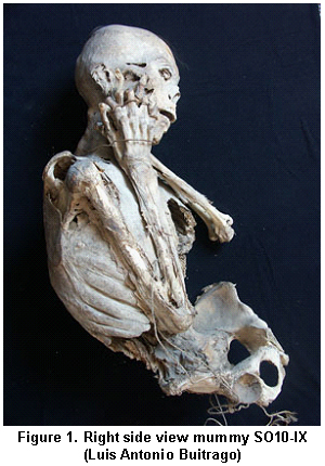
General morphoscopic description. The mummy is unwrapped in flexion, simulating the fetal position (Figure 1), missing the upper left limb, with partial loss of the right lower limb, conserving the leg and foot. There is loss of skin and soft tissue to the bone at the pelvis and the abdominal region. The upper limbs are flexed, the hands are interlaced and tied with a cotton lace; they are placed on the right side of the head. There is loss of skin and soft tissue in both upper limbs, in some regions to the bone. Three fragments of cotton blankets accompany the mummy.
The body shows evidence of perforations caused by cadaveric fauna, predominantly on the left hemi-trunk. Remains of coleoptera were identified and classified as: Class: Insect; Order: Coleoptera; Sub-order: Polyphaga; Super-family: Scarabaeoidea; Family: Melolonthidae; Subfamily: Melolonthinae; Genus: Ancognatha sp cf, Phylllophaga sp cf.
Inside the thoracic cavity, remains of the left lung were identified. Within the abdominal cavity, remains of the peritoneum were evident, without possible macroscopic identification of any specific organ.
ANATOMIC DESCRIPTION
Head. The cranium is symmetric dolichocephalic with size proportional to the rest of the body and no evidence of deformation. It abducted to the left. For the most part, there is good state of preservation of soft tissue and even skin, with loss of these to the bone in the left chin region and at the left fronto-temporal junction. Some small zones still preserve fine, straight black hair in the frontal and occipital parts with signs suggesting that has been cut.
The left auricular pavilion is strapped towards the front, obstructing the entrance orifice of the ear canal; the right auricular pavilion is in anatomic position with a perforation on the lobe. There is cotton inside the ear canal and inside the nostrils. On the right chin, maxillary, and malar region there are skin marks, corresponding to the type of pattern found on the blankets accompanying the mummy.
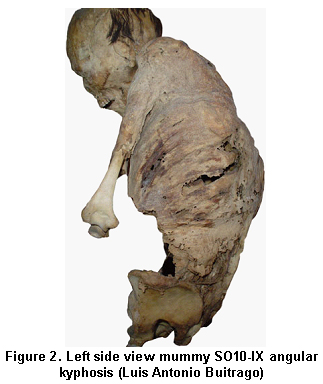
Trunk. There is marked increase of dorsal kyphosis (Figure 2). Perforations are present in the left dorsal region, attributable to taphonomic damage and to damage caused by cadaveric fauna.
On the anterior thoracic wall there is loss of soft tissue on the right side, from the union of the manubrium with the body of the sternum, continuing caudal-like and stemming from the sterna union with the second rib on the left side. Such permits visualizing the interior of the thoracic cavity, which reveals ribs, muscles, vertebrae, and the much descended left lung, which shows a zone with multiple miliar-like punctate nodules.
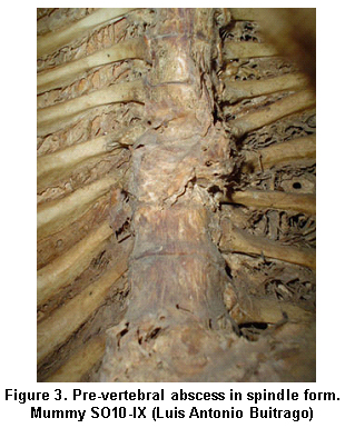
At the seventh thoracic vertebrate, in front of the anterior face of its vertebral body, we can observe the remains of a pre-vertebral abscess in spindle form, measuring 6 x 4 x 3 cm. Marked asymmetry is noted in the trajectory followed by the ribs on the left side in relation to the abscess described. The remaining vertebral bodies do not show signs of anomalies.
Angular kyphosis, the deviation of the ribs, miliary-type lesions on the lung, and the presence of the pre-vertebral abscess suggest the diagnosis of a tuberculous spondylitis or Potts disease (Figure 3).
Abdomen. There is total loss of soft tissue on the anterior wall of the abdomen, permitting a view of the inner abdominal cavity where some remains of the peritoneum are identified.
Pelvis. The pelvis lacks soft tissue, preserving small portions of such at iliac crest level where there is evidence of marks corresponding to the texture of the accompanying blankets. The left pubis shows loss of the descending limb.
Extremities. The right leg and foot are preserved; these are tied with a sisal rope at the pelvic waistline. The foot conserves soft tissue and skin, with the latter showing dark tonality in comparison to the appearance of the rest of the body; while the leg reveals a loss of skin and soft tissue to the bone. The upper limbs are flexed with the hands on the right side of the head, joined at the palms with fingers interlaced and tied with a dyed cotton lace.
Skin and muscles. Whitish 1-mm diameter friable spherical adhesions were noted, suggesting fungal contamination.
BASIC IDENTIFICATION QUATRAIN
Sex. Via cranium evaluation (glabella, mastoids, external occipital protuberance, supra-orbital arch, frontal inclination, orbit shape) and the general aspect of the mandible, features visible through TAC three-dimensional reconstruction, we obtained a weighted score of 1.4 indicative of male sex.
Through direct observation of the pelvis (greater sciatic notch, sub-pubic angle, obturator foramen, iliac fossa, acetabulum, greater and lesser pelvises), we obtained a weighted score of 1.54 indicative of a male individual. Through direct observation of the sacrum, we obtained a 55.55 index that places the mummy as male sex.
Age. The mummy shows dental wear corresponding to Murphys state of dental use number 4, which in the Brothwell age assignment corresponds to an adult individual from 25 to 35 years of age and through the Miles method indicates an individual from 20 to 24 years of age. Consultation with experts from the Biological Anthropology Group at Universidad Nacional de Colombia permitted establishing the age between 30 + 5 years, from the observation of ossification centers.
Height. The height obtained was 163.53 (IC 160.73-166.33) cm with the Genovese method; and with the Krogman-Iscan method, the average was 165.95 cm (DE 2.37).
Ancestry and descent. The mummy presents the dental formula consisting of 2 incisors, 1 canine, 2 pre-molars, and 3 molars completely erupted and in occlusion line corresponding to a young adult individual. Within the non-metric dental features, we noted blade morphology on the palatine surface grade 2 of 6 from the tables by the Arizona State University on the superior lateral incisors; while the reduction in the lateral incisor was zero, characteristic of American indigenous groups-indicating a Mongoloid origin.
Description of the dentoalveolar complex. No fistulae were observed compatible with infectious processes; the height of the alveolar bone in the premolar and molar zone was normal. The inferior incisors reveal a 6-mm loss of osseous height, compatible with localized periodontal disease. There are no cavities, dental enamel hypoplasia, erosions, or abfraction lesions, which reveals a good state of oral health. In samples taken from the retromolar space, it was possible to identify vegetable remains of diatoms and algae; the toxicological exam was negative for alkaloids and positive for carbohydrates.
Imaging studies. These describe the most significant findings from a series of imaging studies, without mentioning the X-ray numbering due to the impossibility of reproducing all of them in this work. The most remarkable aspect of the radiological study is localized at the mid level of the vertebral column. There is loss of intervertebral space between vertebrae T7 and T8. Selective osteolisis is identified on the body of the seventh dorsal vertebra, leading to the collapse and wedging of T7 causing marked spinal angulation (Figure 4).
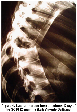
The posterior arches of the 7th and 8th left ribs have abnormal trajectories caused by the loss of the costovertebral joint due to the destruction of the T7 body. In T8, on the superior part of the body, there is evidence of a linear fracture, of oblique stroke, without displacement and with signs of periosteal reaction. It is clear that the tissues better withstanding the passage of time are the osseous structures and they are, in fact, the ones that can be radiologically described in the mummy. However, some of these structures like the spinal cord reveal osteopenia, present to a lesser degree in structures of the cranium.
No other images are observed suggesting articular degenerative and/or traumatic lesions of the spinal cord. In the intercranial tomography of the cranium, it is possible to observe intracranial laminar structures with the same density as the cortical bone extending from the cuts at the base to the highest. These structures could correspond to calcified meninges.
Both the simple X-ray and the tomography show punctate calcifications approximately 2 mm in diameter on the apex of the left lung.
INVASIVE METHODS
Carbon-14. It was established that the mummy dates to the year 1335 + 35 AC, pre-Hispanic Era, 202 years prior to the Spanish arrival in Muisca territory.
Histopathological description. Upon verifying the state of hydration of the samples, no changes were note don the first day. Minimal changes were observed on the third and fourth days, and on the seventh day a state of hydration was evidenced similar to the tissue set in formaldehyde. The histopathological samples showed muscular tissue and denatured collagen without evidence of epithelium. Through the sample taken from the thoracic cavity, we identified pleura in the presence of collagen and a simple flat epithelium of discontinuous coating. The visceral tissue was completely autolyzed with no identification of cell cytoplasm.
DISCUSSION
This is the first ever bio-anthropological characterization of a Muisca mummy in Boyacá. This work was accomplished with the aid of a multidisciplinary team, updating and correcting the ICAHN registry concerning the anthropological and pathological description, sex, age, place of finding, ethnics, textile description, and carbon-14 dating; leaving evidence for future studies on mummified human remains.
Interviews and field visits provided important knowledge to determine climatic, social, cultural, and geographic variables considered important in the analysis of Muisca ritual contexts.
In light of the results, the individual studied was most likely important within the social group. Evidence suggests disability due to illness with motor limitation that must have required care by the community13. The likelihood of this being a «Shaman» ritual figure in the Muisca society is evident through the perforation on the ear lobe, the cone-shaped hat, the very act of the mummification, the position, the funerary trousseau, and through its context.
For the Muiscas, mummies were a cult object and an example of valor. Historians indicate that they belonged to very important individuals, chiefs or sheiks, to whom tribute, homage, and reverence was offered, when mummified after death; draped in fine, hand-painted cotton blankets and bedecked with rich and varied adornments of fine gold. For their communities, mummies continued alive and performed a ritual, political, and social role. The mummification practice continued after the Conquest, as shown by Laches mummies with ovine skin7, animals introduced by the Spaniards.
Chronicler Friar Pedro Simón, states that the Muiscas practiced evisceration, employing vegetable resins to embalm the corpse, which they would then drape in cotton blankets and deposited with ritualistic offerings into a cave. Other times, the corpse would be dried by fire and smoke and deposited into a hut14.
In the 16th century, Friar Esteban de Asencio wrote that the viscera were removed from a chief in Bogotá and that he was then embalmed with a «balm powder» hut that after eight hours would make «the fats and blood expire from the human body leaving it as myrrh». The corpses «were preserved through ingredients»15,16. Likewise, Herodotus17, 25 centuries ago, claims that the Egyptians «spiced» their mummies treating them with salt and spices.
The remains of the funeral deposit elaborated in cotton and its textile characteristics reaffirm its Muisca origin. The presence of cadaveric fauna inside the mummy is associated to fauna belonging to Sogamoso and not to Sátivanorte; an indicator of probable contamination in the warehouse of the Sogamoso archaeological museum and suggesting inadequate conservation. This situation gave origin to a new project in 2006 on the conservation of mummies by the Casa Marqués de San Jorge Museum and the Sogamoso Archaeological Museum.
For the first time, tuberculous spondylitis (Potts disease) is documented in a pre-Hispanic Muisca mummy, in a high-ranking Muisca individual, ratifying the existence of this chronic infectious disease in the sedentary agricultural population of pre-Hispanic America, especially in the Andean high plains18; thus complementing the work of Romero19, Etxeberria20, and Sotomayor21 carried out in a Guane mummy.
All the methods employed to determine the sex confirmed the mummy as male, contrary to that stated by Silva Celis during the interview. The Miles method underestimated the age of the mummy, because it is mainly indicated for adults under 30 years of age, given that as wear is increased its level of precision is minor10.
The research group on Archaeology and History at UPTC evidences that the archaeological population of Tunja reveals average height of 152.7 + 3.6 cm. for women and 159.7 + 3.1 cm. for men22. The height of the mummy is above the measurement calculated regardless of the method.
The AMS method for C14 resulted less invasive given that it permits analysis with samples 20 times smaller than the standard, leaving evidence of remains for their conservation and future studies. The pre-Hispanic dating through carbon-14 corresponds to that found by other authors, placing it in Colombia from the 5th to the 18th century AC6,7.
The process of facial reconstruction of the mummy was begun in two and three dimensions from a three-dimensional model of the cranium via tomography, photography, and direct measurement for the purpose of presenting- for the first time- the face of a Muisca shaman.
Using current research techniques will permit conducting studies to prove the presence of tobacco metabolites, scopolamine, and coca in the hair and textiles. Furthermore, it is possible to determine some traces of the nutritional state and presence of parasitosis in the mummy.
The lesion suggesting Potts disease, anatomically and radiologically, added to pre-Hispanic dating, motivate further research with DNA identification and Mycobacerium typing of the tuberculosis complex. Additionally, we must stress the importance of these types of studies on the collections of human remains in our country, according to Legislation 1185 of 2008.
Conflict of interest. None of the authors has conflicts of interest related to this study.
REFERENCES
1. Jaramillo C, Guhl F, Gómez M, Yockteng R, Vallejo G. Hallazgo de Trypanosoma cruzi en momias de más de 4.000 años de antigüedad. Rev Med. 2000; 22: 53-8. [ Links ]
2. Zuleta E. Conferencias sobre historia económica de Colombia. Bogotá, DC: Hombre nuevo; 1964. [ Links ]
3. Rodríguez M, Cárdenas-Arroyo F. Historia de las investigaciones en momias. In: Rodríguez MC (ed.). Studies on ancient mummies and burial archaeology. Bogotá, DC: Fundación Erigai, Instituto Canario de Bioantropología, Universidad de los Andes; 2001. p. 13. [ Links ]
4. Langman J. Mudos testigos del desierto. Prácticas mortuorias de la civilización Chinchorra del área del desierto de Atacama en Chile. Americas (Spanish edition). 2001; 53: 1-4. [ Links ]
5. Valverde AM. Análisis funcional de la momificación prehispánica, el caso del altiplano cundi-boyacense. Bogotá, DC: Universidad de los Andes; 2002. [ Links ]
6. Cárdenas-Arroyo F. La momificación indígena en Colombia. Bol Museo del Oro. 1989; 25: 120-3. [ Links ]
7. Sotomayor HA, Correal G. Las calaveras enmascaradas de las momias Yuko-Yukpa (motilones). Rev Acad Colomb Cienc. 2003; 27: 5-14. [ Links ]
8. Cárdenas-Arroyo F. Bioantropología del pasado. Innovación y Ciencia. 1993; 11: 52-9. [ Links ]
9. Krogman W, Iscan M. The human skeleton in forensic medicine. Springfield: Charles C. Thomas; 1986. [ Links ]
10. Genovés S. La proporcionalidad entre los huesos largos y su relación con la estatura en restos mesoamericanos. México, D.F: Instituto de Investigaciones Históricas, UNAM; 1966. [ Links ]
11. Lozano-Ruiz M. Estudio del desgaste a nivel microscópico de los dientes anteriores de los homínidos del yacimiento pleistocénico de Sima de los Huesos (Sierra de Atapuerca, Burgos). Tarragona: Universitat Rovira i Virgili; 2005. [ Links ]
12. Ruffer M. Note on the presence of Bilharzia haematobia in Egyptian mummies of the Twentieth Dynasty (1250-1000 B.C.). BMJ. 1910; 1: 16. [ Links ]
13. Otálora-Cascante AR. Caracterización bioantropológica de una momia prehispánica: implicaciones metodológicas desde la antropología biológica. Rev Salud Hist Sanid. 2006; 1: 20-5. [ Links ]
14. Simón FP. Noticias historiales de las conquistas de tierra firme en las Indias Occidentales. Bogotá, DC: Biblioteca Banco Popular; 1981. [ Links ]
15. Silva-Celis E. Estudios sobre la cultura chibcha. Tunja: Academia Boyacense de Historia; 2005. [ Links ]
16. Pradilla H, Villate G, Ortiz F. Arqueología del Cercado Grande de los Santuarios. Bol Museo del Oro. 1992; 32: 21-148. [ Links ]
17. Herodoto. Los nueve libros de la historia. Bogotá, DC: Oveja Negra; 1983. [ Links ]
18. Rodríguez JV. Las enfermedades en las condiciones de vida prehispánica de Colombia. Bogotá: Universidad Nacional de Colombia; 2006. [ Links ]
19. Romero W. Estudio bioantropológico de las momias del Museo Arqueológico Marqués de San Jorge. Antropología. Bogotá, DC: Fondo de Promoción de la Cultura del Banco Popular, Universidad Nacional de Colombia; 1997. [ Links ]
20. Etxeberria F, Romero W, Herrasti L. Cifosis angular de la columna vertebral: identificación del Mal de Pott en una momia Guane prehispánica de Colombia. Chungara. 2000; 32: 41-8. [ Links ]
21. Sotomayor HA, Burgos J, Arango M. Demostración de tuberculosis en una momia prehispánica colombiana por la ribotipificación del ADN de Mycobacterium tuberculosis. Biomedica. 2004; 24 (supl): 18-26. [ Links ]
22. Rodríguez JV. La antropología forense en la identificación humana. Bogotá, DC: Universidad Nacional de Colombia; 2004. [ Links ]

