


 text in
  English (pdf)
 Article in xml format
 Article references
 Send this article by e-mail
 Cited by SciELO
  Cited by Google
  Similars in
SciELO
Similars in
SciELO  Similars in Google
 Permalink
Introduction
Scapulothoracic dissociation is a traumatic injury of the shoulder. The scapulothoracic joint is made up of the acromio-clavicular joint, glenohumeral joint, clavicle, scapula, and sternoclavicular joint. This type of injury can cause subclavian and axillary vascular injuries, partial or complete avulsion of the brachial plexus, and edema to the surrounding soft tissues (1. It is an uncommon injury with a mortality rate up to 10% and caused by motor vehicle accidents in 44% of cases. Above the elbow amputation has been noted to be in around 20% of cases due to low potential for neurological recovery 1-3.
The mechanism of injury is explained by the application of high energy traumatic forces, usually caused by high velocity mechanisms. Its management is challenging in all phases of care for the compromised patient 1.
Blunt trauma to the vertebral artery is a rare entity with an incidence of 0.24% to 2.0% in cervical 4, trauma cases and a mortality rate between 20-40%, which has decreased due to early diagnosis and identification of lesion characteristics 5.
This case report presents an unusual case of a patient where these two entities converge. We analyze the main points of intervention and decision-making that were effective in the management of the patient 6.
Case presentation
A 24-year-old male with no comorbidities presents due to a motorcycle accident that occured 20 minutes ago. At presentation, his vital signs are as follows: Glasgow 14/15 (confusion), BP: 85/45 mm Hg; MAP: 58 mm Hg; RR:22 respirations/minute, HR:130 beats/minute. Physical exam findings of a left transverse penetrating cervical trauma to zone I, left cervicothoracic edema and ecchymosis, left clavicle deformity, left upper extremity deformity with associated pulselessness, anesthesia, paralysis, and areflexia throughout. (Figure 1A).
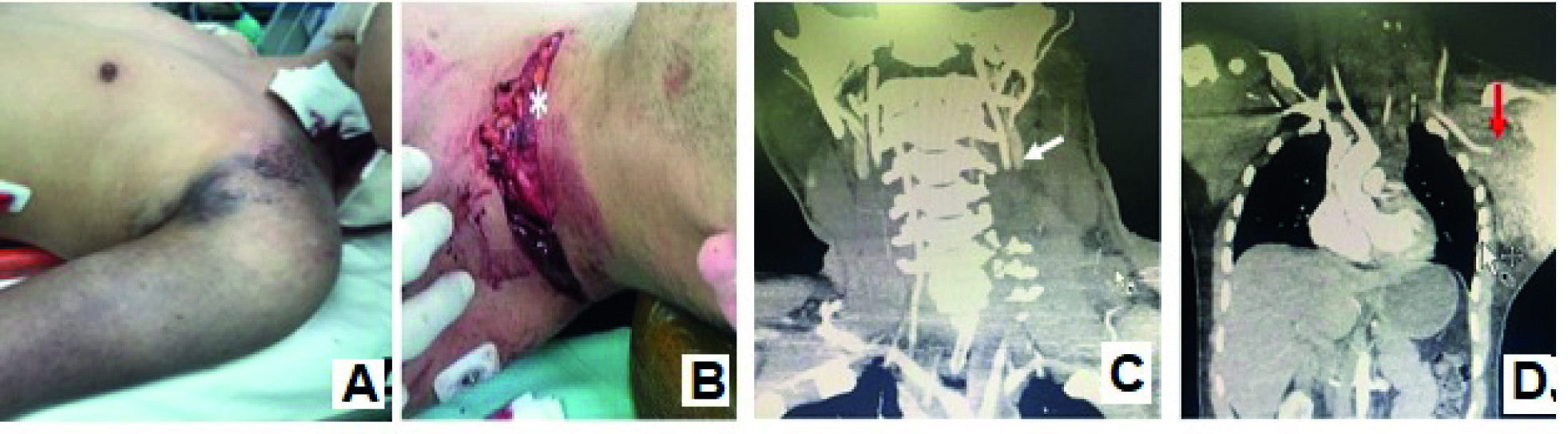
Figure 1 Scapulothoracic dissociation and associated blunt trauma of the vertebral artery. A. Inspection of the patient upon initiating resuscitation B. Identification of the left zone I cervical injury C. CT angiography of the neck, white arrow, thrombosis of segment V2 of the left vertebral artery. D. CT angiography of the neck, abdomen, and chest, red arrow, complete transection of the left axillary artery, increased soft tissue density in the axillary region.
The initial diagnosis was hypovolemic shock due to left zone I penetrating neck trauma, blunt trauma to the left axillary and subclavian vessels, and secondary neurovascular injury. Resuscitation was initiated with lactated ringers and oxygen therapy; packed red blood cells were reserved for the patient. Angiotomography was performed and reported a complete transection of the left axillary artery and vein in the distal third portion, ipsilateral scapular fracture, C4-C7 completely displaced transverse process fractures with compromise to the vertebral foramen and injury to the left vertebral artery. (Figure 1C and D)
The patient was taken into the operating room for surgical exploration of the penetrating neck injury where the left external jugular vein was localized and ligated in the setting of a partial transection. Infra-clavicular approach was used to drain 2,000 mL from a contained hematoma between the pectoralis and deltoid muscles, a complete transection of the axillary artery and vein were identified at its first segments, and avulsion of the humeral veins and secondary trunks of the brachial plexus were identified (Figure 2E ). At the intraoperative medical board with orthopedics, it was decided to continue with conservative management for the brachial plexus injury and a possible deferred neurological reconstruction was evaluated. For the time being, we performed an arterio-venous vascular reconstruction of the axillary vessels with internal saphenous vein grafting. The graft patency was confirmed with distal flow present on echo-Doppler intraoperatively and arteriography after surgery (Figure 2F). A concomitant bicompartmental fasciotomy of the forearm and Velpeau-type immobilization of the scapular fracture were also performed.
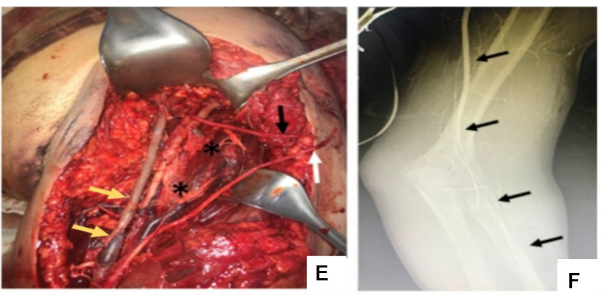
Figure 2 Scapulothoracic dissociation E. Yellow arrow, arterial reconstruction of the axillary vessels. White and black arrow, avulsed secondary trunks of the brachial plexus. F. Intraoperative arteriography with adequate contrast flow to the radial artery after vascular reconstruction.
Post-operatively the patient was transferred to the intensive care unit. Cervical spine and spinal cord MRI angiography and head CT showed a lesion in segments V1 and V2 of the left vertebral artery without signs of dissection, a spinal hematoma, and cerebral edema without ischemic lesions. A medical board of neurosurgery and interventional neuroradiology decided for conservative management to limit distal thrombosis of the vertebral artery using anticoagulation of unfractionated heparin at a dose of 18 units/kg/hour IV and use of thromboplastin time to perform dose adjustment. An MRI of the brachial plexus detected preganglionic lesions in the left C5 and C6 roots, with associated pseudomeningocele. Conservative management was initiated with deferred evaluation for the possibility of nerve reconstruction.
During the postoperative period, the patient experienced bleeding from the fasciotomy sites of the left upper limb that warranted a surgical revision with intraoperative findings of transection to the basilic vein. Ligation and exhaustive hemostasis were performed with subsequent control of bleeding.
The repeat CT scan revealed a localized thrombosis in the vertebral artery, which at that time had no major impact on postoperative course. Late secondary closure of the fasciotomies was performed.
The patient was discharged after 22 days of hospitalization managed by intensive care services, general surgery, plastic surgery, orthopedics, neurosurgery, and physical therapy. He demonstrated overall clinical improvement, even when there were minor cognitive memory deficits and the expected left brachial plexus injury. The patient was started on subcutaneous low molecular weight heparin 1 mg/kg every 12 hours for three months and subsequent anti-platelet therapy with 100 mg of oral acetylsalicylic acid every 24 hours and 100 mg of clopidogrel orally every 24 hours for six months.
Outpatient management was performed by general surgery, orthopedics, hand surgery, and physical therapy. Six months after the trauma, reconstruction of the C5-T1 left brachial plexus was performed, which resulted in a partial recovery of functionality up to the proximal third of the arm. After one year, the patient still has not considered amputation and continues with rehabilitation and follow up with hand surgery.
Discussion
The case presented combines two injuries of high morbidity and mortality, including scapulo-thoracic dissociation and blunt trauma to the vertebral artery. It highlights a scenario of high complexity, uncertainty, and with generally low consensus on diagnostic approaches, operative treatments, and prognosis, among others 5. The factors associated with high morbidity and mortality in this patient and described in cases of scapulo-thoracic dissociation include: hypovolemic shock, compromise to multiple vascular structures within the cervico-thoracic region including the vertebral artery, differing management options, the magnitude of these injuries, and the potential complications of the trauma and its management 1,2.
In regards to the complex vascular reconstructive procedure in this patient with the intention of preserving the affected limb, it could be considered a futile 6 task due to the severity of the brachial plexus injuries, unfavorable functional prognosis for the limb, and the uncertainty in the effectiveness of the treatments.
The discussion focuses on the points of controversy regarding the management of the two entities in a highly complex scenario.
Preoperative diagnostic approach vs immediate surgery
A medical board was held with the two attending surgeons, the emergency medicine doctor, and the radiologist present at the institution at the time of admission. Their analysis is described in Table 1.
Table 1 Considerations for the diagnostic and therapeutic approach to scapulo-thoracic dissociation and concomitant closed vertebral artery trauma
| Pro’s | Cons | |
|---|---|---|
| Immediate surgery | Control of bleeding | Uncertainty of injury severity upon admission |
| Prompt revascularization of ischemic limb. | Possibility of exsanguination and | |
| Theoretical increase in the probability of survival | Intraoperative death due to irreparable vascular approach. | |
| Preoperative radiological evaluation | Available resource of the institutional | Loss of time in the unstable patient |
| Anatomical and characterization imaging of the injury and evaluation for need of initial thoracotomy placement | Established indications for surgery according to trauma management protocols | |
| Establish an option for endovascular management | Complementary imaging studies are part of the secondary surgery of the polytrauma patient. | |
| Initial (balloon occlusion) |
The blunt trauma to the vertebral artery shown on imaging did not change the surgeon’s perspectives on the initial approach for the case, although it did darken the patient’s prognosis at the time.
CT angiography in patients with axillo-subclavian trauma is considered within the diagnostic approach of patients with injuries secondary to blunt trauma and in scenarios of hemodynamic stability. A multicenter evaluation carried out in the USA in patients with axillo-subclavian lesions documented a mechanism of penetrating trauma in 57% of the cases and the use of CT angiography in 50% of the patients as the initial diagnostic method. In regards to treatment, 17% of the cases received endovascular therapy regardless of the mechanism of the trauma, avoiding more complex surgical procedures (7-9). The authors conclude that its benefit lay in the possibility of offering concomitant endovascular management during the diagnostic procedure and thus represented an alternative to be considered in this group of patients (10).
The evaluation of the case as far as the surgical board in the emergency service is concerned determines that despite the presence of indications for surgery such as traumatic cervicotomy and signs of vascular trauma to the left upper limb in this patient, the performance of a radiological evaluation by means of CT angiography prior to surgical intervention helped to determine the order and operative approach according to the location of the lesions, thus contributing to the favorable outcome of the patient.
Immediate amputation vs preservation of limb
The preservation of the left upper limb in the setting of the brachial plexus injury is a topic to analyze in this case. According to the score on the MESS scale (Mangled Extremity Severity Score) 11, which assesses the prognosis of an injured limb, initial amputation would be considered in this case with 8/14 points (Table 2) 11-13. The intraoperative medical board discussion between surgery and orthopedics evaluated the situation and is described in Table 3.
Table 2 Modified from the Mangled Extremity Severity scale (MESS) 11.
| Mangled Extremity Severity | Points |
|---|---|
| Skeletal / Soft tissue injury | |
| Low energy (stab wound, simple fracture, low energy gunshot wound) | 1 |
| Medium energy (open or multiple fractures, dislocation) | 2 |
| High energy (high speed motor vehicle collision or rifle bullet wound, crush injury) | 3 |
| Very high energy (the above plus large contamination, soft tissue avulsion) | 4 |
| Limb ischemia* | |
| Reduced or absent pulse but normal perfusion | 1 |
| No pulse paresthesia, decreased capillary filling | 2 |
| Cold, paralyzed, senseless, rigid | 3 |
| Shock | |
| Systolic blood pressure >90 mmHg constant | 0 |
| Systolic blood pressure <90 mmHg transient | 1 |
| Systolic blood pressure <90 mmHg persistent | 2 |
| Age (years) | |
| Under 30 | 0 |
| Between 30-50 | 1 |
| Over 50 | 2 |
* Doubled score for ischemia time >6 hours
Scores less than or equal to 6 are associated with preservation of the limb. A MESS greater than or equal to 6 is associated with a higher rate of amputation
Table 3 Considerations for left upper limb amputation as an initial approach in scapulo-thoracic dissociation
| Pro’s | Con’s | |
|---|---|---|
| Immediate amputation | Control of bleeding in patients with shock and complex injuries | Technical capabilities of the professionals involved |
| Possibility of reperfusion syndrome | High-level institutional therapeutic support | |
| High MESS score indicating poor limb prognosis | Possibility of partial recovery from neurological injury, with a decrease in the level of a future amputation | |
| Highly complex surgery in a critical patient | Psychological impact on the patient |
The prognosis of a limb after a proximal neurologic injury has improved by the development of microsurgical techniques. Recent studies support conservative management even in cases of radical brachial plexus avulsion, with an early amputation rate close to 21% 2,3. These injuries have unpredictable responses and treatment options, including amputation 2 which must be individualized according to the type of injury. Post-ganglionic injuries are associated with a greater regenerative capacity when compared to pre-ganglionic injuries, which have a poor prognosis 1,3,10,14. Compromise to the brachial plexus in a scapulo-thoracic dissociation has a negative influence in limb prognosis (1. However, the partial effective response after reconstructive surgery and its rehabilitation, determined in this case report and according to the patient’s course presented thus far, warrants consideration of a more distal level of amputation in the patient's arm.
Similarly, the psychological impact derived from the process of acceptance and possible "programmed grief" after the exhaustion of therapeutic options, suggests better rehabilitation of an amputated limb 15.
Blunt trauma to the vertebral artery
Blunt cerebrovascular trauma includes traumas of the internal carotid artery and vertebral artery, both being entities that are difficult to diagnose in a polytrauma patient and are considered determinants of late mortality 5,16. Angiography technique in patients with a high risk for blunt cerebrovascular trauma (cervical hematoma, neurological deficit, high-energy trauma mechanism, subluxations or fractures of the cervical vertebrae) found an incidence of 34% of closed vertebral artery trauma (16. Therefore, a possible underreporting of cases exists due to the low index of suspicion of such injuries and the care of multiple injuries in a polytrauma patient 4. The importance of early detection along with early management has an impact on prognosis 1,4,16,17. For this reason, in 2017 the ATLS (Advaced Trauma Life Support) incorporated diagnostic strategies for recognition of such traumas in potential candidates 16. The diagnostic operative characteristics of CT angiography (sensitivity 97.7%, specificity 100%, positive predictive value 100% and negative predictive value 99.3%), although comparable to MRI angiography, make it the study of choice due to its greater availability 16.
A classification suggested by angiographic patterns, of blunt cerebrovascular trauma (trauma to the internal carotid artery and vertebral artery) considers their neurological prognosis and survival, 16,17 is presented in Table 4. In the present case, it corresponded to a grade IV/V, therapeutic anticoagulation was initiated in the immediate postoperative period after evaluating the risk/benefit in a complex vascular surgery with fasciotomies. The state of the art in the treatment of blunt trauma to the vertebral artery indicates use of this therapy in grade I-IV injuries, to procure control of distal thrombosis and prevention of late embolism, considering endovascular management, on a case-by-case basis.
Table 4 Classification of cerebrovascular blunt trauma (blunt trauma to the vertebral artery and blunt internal carotid artery trauma). Modified from 11.
| Grade of Injury | Characteristics | Rate of cerebrovascular event | Rate of mortality (%) |
|---|---|---|---|
| Grade I | Light compromise <25% | 3 | 11 |
| Grade II | Light compromise >25% | 11 | 11 |
| Intraluminal thrombus | |||
| Intimal tear | |||
| Grade III | Pseudoaneurysm | 33 | 11 |
| Grade IV | Arterial occlusion | 44 | 22 |
| Grade IV | Transection with extravasation | 100 | 100 |
The blunt trauma of the vertebral artery generates an additive interaction with respect to the scapulo-thoracic dissociation and the patient's prognosis 18, which contributes to a greater deleterious effect on the organic presentation, use of resources, concomitant therapies, and limits the possibilities of success. Its presence did not affect the final cerebral and neurological recovery of the patient, which is possibly attributed to a compensation from the circulation of the circle of Willis.
It is pertinent to note that during the course of the patient’s care, a permanent communication was established with the patient and his family, who expressed a feeling of gratitude and an attitude of collaboration with the medical and paramedical personnel involved in the case. Similarly, he illustrated the need to continue functional rehabilitation of the limb and about the possibility of a late amputation.
Conclusion
The case presented shows a context of high uncertainty and low probability of consensus, where decision-making must be carried out at a different level from that contemplated in daily clinical practice. Medical boards with various specialties are useful tools, in these complex scenarios, generate actions of shared responsibility, add experiences and knowledge in pursuit of a common final goal and implements teamwork as a determining factor in the outcome.
The generation of multidisciplinary cooperation in various phases of the patient's treatment contributed to the success of the case and reinforces the idea of its permanent execution for the benefit of patients, institutions, and health professionals.

