


 text in
  English (pdf)
 Article in xml format
 Article references
 Send this article by e-mail
 Cited by SciELO
  Cited by Google
  Similars in
SciELO
Similars in
SciELO  Similars in Google
 Permalink
Introduction
Accidental ingestion of foreign bodies is a common problem in pediatrics. Coins are the most frequently ingested object and cause 60% of esophageal impactions. Conventional plain radiography is the imaging modality of choice to locate most foreign bodies and guide treatment options 1-3. Various radiological findings that allow identifying the ingested object have been described, including the double rim or halo sign of the button battery (BB), a pathognomonic image that indicates the need for immediate removal 4. Treatment delay is associated with complications such as ulcers, perforation, stenosis, or fistulas 5. We report the first case of a child with three coins stacked and impacted in the esophagus and describe a new radiological finding associated with the superimposition of several foreign bodies that can help to improve diagnosis, treatment and outcomes.
Case description
A previously well 5-year-old girl presented to the emergency department with an acute onset of sialorrhea, odynophagia, and nausea. Vital signs were normal and no other abnormalities on physical examination were found. There was no significant medical, social, or family history. Given the child’s age group and symptoms, ingestion of a foreign body was suspected. An anteroposterior cervico-thoraco-abdominal radiograph (CTA X-ray) was obtained revealing a foreign body in the upper third of the esophagus which seemed to show a double rim sign, although an irregular layout was noted (Fig. 1a). Suspecting a button battery sign, immediate endoscopic removal was indicated. During the preoperative nasopharyngeal swab testing for SARS-CoV-2, the patient experienced an acute episode of emesis, expelling a 1-euro cent coin (cent €). After this, the child became asymptomatic. Despite the initial suspicion of a button battery sign and the patient is asymptomatic, a close inspection of the CTA X-ray with digital magnification demonstrated that the image of the foreign body was formed by the superimposition of three circumferential objects of different sizes (Fig. 1b).
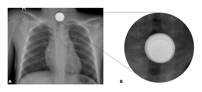
Figure 1 (A) Foreign body in the upper third of the esophagus with an apparent double-ring o halo image. (B) Radiological magnification shows the superimposition of 3 circumferential foreign bodies of different sizes.
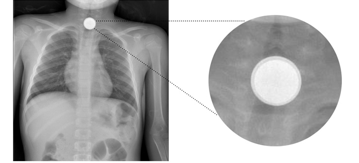
To discard multiple impacted objects, another anteroposterior CTA X-ray was obtained observing the persistence of the foreign body, but this time, only two superimposed objects were seen (Fig. 2). The patient was then taken to the operating room. During the flexible esophagoscopy, 2 coins (2 and 5 cents €) were found stacked at the level of the upper esophageal sphincter, both were removed without difficulty. The patient had an uneventful postoperative recovery and was discharged 12 hours after the procedure. There have been no long-term complications.
Discussion
The esophageal impaction of multiple coins is rare, accounting for 5-7% of all impactions and occurs most often in children under the age of 3, males, and patients with underlying esophageal abnormalities or other co-morbidities 2,6. X-ray imaging plays an important role in the workup and treatment of pediatric patients with suspected foreign-body ingestion. In the case of a radiopaque foreign body, X-ray imaging not only determines the anatomical location of the object, but the morphological features allow to suspect its nature and composition 6. The initial standard imaging protocol includes frontal and lateral radiographs of the neck, chest, and abdomen 7. Huyett et al. (8, state that a lateral X-ray has at least two purposes: to confirm that the foreign body is posterior to the airway and to differentiate multiple stacked coins from a button battery. In our case, no lateral X-ray was performed since a button battery was already suspected on the frontal radiograph thus, minimizing radiation dose following the ALARA radiation safety principle (As Low As Reasonably Achievable) and not delaying battery removal because of damage to the esophageal wall can occur within hours 4.
Unusual radiographic findings have been documented regarding esophageal foreign bodies, such as ingested coins with a sagittal orientation as if they were in the trachea 9-11. Ormeño et al. (9, reported a case of two coins impacted in the esophagus also simulating the double rim image of a button battery. In our case, the radiological finding was not a defined double ring but rather the superimposed silhouettes of three round objects of different sizes, arranged in the same way as celestial bodies do during a lunar eclipse (Fig. 3a). This finding (Fig. 1b*) created by the radiological arrangement of the coins in the patient's esophagus is a novel radiological finding that may alert the physician to the presence of multiple coins and distinguish them from a button battery, enabling the best therapeutic choice.
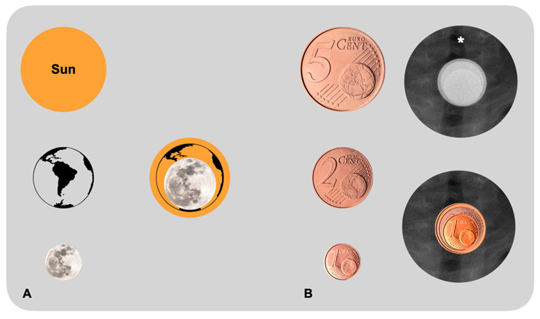
Figure 3 (A) Graphical representation of the alignment of the moon, the earth, and the sun during a lunar eclipse. (B*). Radiological arrangement of the coins in the patient's esophagus.
Informed consent and patient details
The authors declare that this report does not contain any personal information that could lead to the identification of the patient, however, written informed parental consent was obtained for publication of this case report and accompanying images. A copy of the written consent is available for review by the Editor-in-Chief of this journal.

