


Serviços Personalizados
Journal
Artigo
 Inglês (pdf)
 Artigo em XML
 Referências do artigo
 Enviar este artigo por email
Indicadores
-
 Citado por SciELO
 -
 Acessos
Links relacionados
-
 Citado por Google
 -
 Similares em
SciELO
Similares em
SciELO -
 Similares em Google
Compartilhar

 Permalink
Biosalud
versão impressa ISSN 1657-9550
Biosalud v.9 n.2 Manizales jul./dez. 2010
UPPER AERODIGESTIVE TRACT NEOPLASM (UATN): AN UPDATE
NEOPLASIA DEL TRACTO AERODIGESTIVO SUPERIOR (UATN): UNA ACTUALIZACIÓN
Anderson Rocha Buelvas1 y Cristian Rocha Buelvas2
1 Dentistry's Research System Coordinator. Faculty of Dentistry, Universidad Cooperativa de Colombia - Pasto (San Juan de Pasto, Nariño's Department - Colombia). E-mail: anderson.rocha@ucc.edu.co.
2 Student of Department of Biology, National University of Colombia -Bogotá. (Bogotá D.C.). crarochabu@unal.edu.co.
Recibido: julio 13 de 2010 - Aceptado: septiembre 10 de 2010
ABSTRACT
Introduction: according to the review carried out, while the majority of head and neck cancerous lesions are cutaneous and thyroid are associated with low lethality, there are others that are developed in the upper aerodigestive tract and are usually squamous cell carcinomas, which are usually diagnosed in advanced stages and are associated with very poor survival and functional prognosis. In Colombia, squamous cell carcinoma is the neoplasm of greatest occurrence in head and neck; it becomes evident as a detectable premalignant lesion and it registers between 100 and 120 new cases by year according to the Colombian National Cancer Institute in 1998. This update is important because of the new findings on the disease natural history and the characteristics of each location in the upper aerodigestive tract.
Methodology: the relevant biomedical literature was searched in several databases such as Medline, Proquest, Science Direct, Ovid and Cochrane, as well as available information in web sites of national scientific journals and Organizations.
KEY WORDS: neck and head, upper aerodigestive tract neoplasm, squamous cell carcinoma.
RESUMEN
Introducción: De acuerdo a la revisión llevada a cabo, mientras que la mayoría de las lesiones cancerosas de cabeza y cuello son cutáneas y asociadas con la tiroides y de baja letalidad, hay otras que se desarrollan en el tracto aerodigestivo superior y usualmente son carcinomas de células escamosas, las cuales son usualmente diagnosticadas en estados avanzados y asociadas con una muy pobre supervivencia y pronóstico funcional. En Colombia, el carcinoma de células escamosas es la neoplasia de mayor ocurrencia en la cabeza y el cuello, esta se manifiesta como una lesión premaligna detectable y registra entre 100 a 120 casos nuevos al año en Colombia de accauerdo al Instituto Nacional de Cancerología en 1998. Esta actualización es importante debido a los nuevos hallazgos en la historia natural de la enfermedad y a las características de cada localización en el tracto aerodigestivo superior.
Metodología: Fue buscada la literatura biomédica relevante en algunas bases de datos tales como Medline, Proquest, Science Direct, Ovid y Cochrane, así como también la información disponible en sitios Web de organizaciones y revistas científicas nacionales.
PALABRAS CLAVE: Cabeza y cuello, neoplasia del tracto aerodigestivo superior, carcinoma de células escamosas.
INTRODUCTION
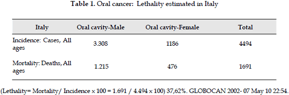
According to scientific literature, cancer is the second leading cause of mortality in developed countries and is a health problem of unquestionable importance (1). Squamous Cell Carcinoma of the Head and Neck affects 550,000 new patients worldwide annually (2). The head and neck cancer (HNC) accounts for 5-10% of all malignancies in Colombia (3, 4). In countries like Mexico, the prevalence of head and neck cancer ranges from 3-9%, according to a recent 24 years follow-up study (5). If it revise one localization of upper aerodigestive tract as oral cavity, the estimated lethality is 36.8% according to GLOBOCAN 2002 - 07 May 10/22:54 (Table 1). For example, the estimated lethality of oral cancer in Colombia is like seem the estimated lethality in Italy that is 37.62% according to GLOBOCAN 2002 - 22 Sep 08/23:38.
The frequency of head and neck cancer varies with the specific anatomical location, oral cancer is the most frequent with 40.6%, tongue 21%, gums 5%, floor of the mouth 2%, lips 2.5%, other oral mucosal locations 14.1%, in salivary glands is 15.3%; pharyngeal cancer accounts for 15% (5.7% oropharynx, 4.7% nasopharynx, 4.8% laryngopharynx) and unspecified sites cover the other 24.8%. The head and neck cancer includes some anatomical regions that because of their location and lymphatic dissemination have very different treatment. It is more frequent in men than in women and has its highest incidence in the fifth and sixth decade of life (6).
The cancer of the upper aerodigestive tract affects so important functions such as speech and swallowing. The cancer of the upper aerodigestive tract includes the following locations: nasal cavity and paranasal sinuses, nasopharynx or cavum, oral cavity, oropharynx, larynx, hypopharynx and salivary glands. The most common histological type is squamous cell carcinoma. There are premalignant lesions such as dysplasia and it is frequent the occurrence of second tumors. It may stay subclinical for a long time and symptoms appear when the disease has advanced. Its main feature is the easy dissemination to cervical lymph nodes but the metastatic effect is rare, however, when it occurs, is more common to the lung (7, 8).
Surgery and radiotherapy constitute the standard treatment for these tumors. In early stages they can be used exclusively with good results. In more advanced stages combination of surgery and radiation therapy produces better results (9).
The status of surgical margins, lymph node affectation and the degree of histological differentiation are critical to specify when the dose of radiation. Chemotherapy alone is not curative but should be taken into account in research protocols of advanced tumors, in order to try to preserve the phonation and swallowing functions since it is a conservative treatment and avoids mutilating surgery. In short, The cancer of the upper aerodigestive tract is a heterogeneous neoplasm which affects some anatomical regions that by their location and lymphatic dissemination, has a very different treatment (10).
Aetiology
This type of cancer is associated with a prolonged history of cigarette smoking and alcohol consumption. Cigarette is the major risk factor and alcohol enhances its carcinogenic effect. It has also been associated with deficits in the diet, such as vitamin A deficiency, and occupational exposure to certain products such as wood dust- related sinus cancer and asbestos with laryngeal cancer. Solar Ultraviolet radiation is associated with lip cancer. It may be of infectious origin, as it has been reported that nasopharyngeal cancer is closely related to Epstein-Barr virus, while the Plummer-Vinson syndrome is associated with tumors of the hypopharynx (11).
Regarding risk factors strongly associated with the development of these tumors, it is five times higher in smokers than in nonsmokers, increasing 17 times the risk in people who smoke 80 or more cigarettes, the risk increases just as twice in passive smokers. The emergence of second primary aerodigestive tract cancer in patients who stopped smoking after their first cancer cure is 18% while for those who continued to smoke is 30%, so the interruption of the habit of smoking decreases significantly the risk of oral premalignant and malignant lesions. The risk of precancerous lesions in the aerodigestive tract increases four times more than the cancerous ones in permanent smokers (12, 13).
Pipe or cigar smokers tend to develop more malignant lesions on lips and tongue. Cigarette smokers in addition to regular oral cavity have a higher risk of occurrence of these lesions in the larynx and pharynx. The type of tobacco affects the risk of cancer, for example, there are two types, the dark one is more alkaline, more irritating to mucous membranes and more linked to cancer of the larynx and supraglottic area because of its lower inhalation, whose habit and risk of cancer is geographically located in countries like Colombia, Brazil, Italy, Spain, Cuba, Uruguay, among others; and the consumption of light tobacco, which produces higher occurrence of cancer in glottal area due to its greater inhalation, it is most common in the United States (14, 15, 16, 17, 18).
Other cigarette elements linked to the increase of cancer are short cigarettes containing higher concentration of carcinogens and "Light" cigarettes that are consumed in greater numbers, it has also been reported a major increase with hand-rolled cigarettes. The habit of smoking inside the mouth or putting the cigarette in sublingual or cheek areas, is very common on the Pacific coast of southwest of Colombia. Thus the so called habit of inverted smokers (reverse smokers) according to the Anglo-Saxon literature increases the risk of oral cancer (lip, tongue and buccal mucosa) in more than 4-6 times , although other studies show a higher frequency on the palate (19, 20).
Histology
Squamous cell carcinoma is the predominant histological type (90%). The determination of the degree of differentiation is very important since well-differentiated tumors or Grade I (keratinization over 75%) are less aggressive than poorly differentiated tumors or Grade III, which are very aggressive tumors and although very responsive to treatment, relapse easily. In the histological diagnosis is very important that the pathologist specify the size of tumor, the status of surgical margins, the degree of differentiation and lymph node affection, specifying the status of the nodal capsule (21). In the nasopharynx predominates the undifferentiated carcinoma (lymphoepithelioma). Other varieties of rare occurrence are lymphomas, sarcomas and adenocarcinomas. The salivary gland tumors are mostly benign (pleomorphic adenona and Warthin's tumor). The more frequent malignant varieties are adenocarcinoma and mucoepidermoid tumor (22, 23).
Premalignant
Premalignant lesions exist in the upper aerodigestive tract which can lead to malignancy. They are leukoplakia, erythroplakia, hyperplasia and dysplasia ultimate precursor lesions. Dysplasia progresses to carcinoma in 15-30% of cases. It is therefore necessary to perform a successful exploration well in the first patient visit and in subsequent revisions. Patients with head and neck cancer are at high risk of developing second tumors. The etiology of oral cancer as both of precancerous lesions is multiple. The factors most commonly cited are the snuff, alcohol, genetics, nutrition, viruses, radiation and occupational hazards. Most of them have a cumulative effect over time, giving consistency to the epidemiological finding of higher prevalence of cancer in older people, this has led many authors claim that age is the main risk factor in developing cancer, oral cancer and specifically (24, 25).
The early detection of malignant lesions and their subsequent treatment are the cornerstone to provide the best prognosis in cancer. Despite the growing awareness among professionals and the population, the incidence and prevalence especially, oral cancer has not declined, on the contrary, it has been increasing in Western countries, even though the dental professionals should be the ones who recognize the potentially malignant lesions in oral cavity. However, it is more often diagnosed late and often in advanced stages, which is often associated with gloomy forecasts. On the other hand, despite the remarkable advances in treatment, mortality rates remain high, often above 50% at five years (26, 27).
One source of confusion is the terminology. At present, international consensus let use indistinctly premalignant and precancerous terms; but the expressions precancerous lesion and precancerous condition cannot be interchanged. A precancerous lesion is a morphologically altered tissue in which it is most likely occur a cancerous transformation than in another of normal appearance, whereas a precancerous condition is a general state associated with an increased risk of developing malignancy. According to the Center of precancer and Oral cancer from the United Kingdom, should be called leukoerythroplasia those lesions that have the combination of red and white plates of high risk and which diagnosis considered them as carcinoma in situ. This workshop considers Proliferative verrucous leukoplakia that lesion that is clearly multifocal and covers a large area of epithelial tissue, being this description of a cancer precursor leukoplakia. Characterization is recommended if etiologically there is a clear association with tobacco and other physical and chemical carcinogenic elements, so its origin remains idiopathic, it is recommended if anatomically is consistent with a sub-place in the mouth or oropharynx, and if the extent of the injury is considerable; in case it does not meet these characteristics, a biopsy is mandatory. At microscopic level the presence of epithelial dysplasia is the strongest predictor of future malignancy (28).
Erythroplasia: clinically red, small, slightly raised and of granular appearance are observed. There is a high risk of carcinoma in situ when they are ulcerated. However, there are two types, the first having the characteristics mentioned above as well as areas of keratin inside or on the periphery of the lesion, and another type of lesion that is contrary to the above description: smooth, non granular, but with minimal keratosis but erythematous. If the diagnosis is difficult, biopsy is mandatory (29, 30, 31).
It should be noted that the asymptomatic lesions with erythroplakic elements which tend to become malignant, whereas those less often asymptomatic lesions with keratinic elements are on the contrary to be benign. A differential diagnosis is important given the manifestation in the oral cavity of other systemic diseases that would divert the accuracy of diagnoses and treatment, the lesions that most commonly confuse when diagnosing an oral premalignant lesion are usually lesions on reverse smoker's palate, submucous fibrosis oral, actinic keratosis, lichen planus, nicotinic stomatitis, discoid lupus erythematosus (32).
Natural History
The head and neck cancer may remain subclinical for a long time and will not appear until the disease is well advanced. Patients are usually slow to come to the consultation and show symptoms similar to the usual benign processes such as catarrhal or discomfort in the oral cavity secondary to dentures. All this means that in many cases they are treated with antibiotics and analgesics, thus delaying diagnosis and being detected in advanced stages, hindering the total eradication of tumor. When the tumor cells begin to grow, tend to spread easily to neighboring areas so it is important to make an early diagnosis. The main feature of the head and neck tumors is the easily spreading to lymph nodes, being essential to know in depth the anatomy of the lymphatic channels of the upper aerodigestive tract; the knowledge of the lymphatic dissemination is the basis of radiotherapy treatments since the tumor control depends not only on treatment of primary tumor, but also regional nodes because of the high risk of relapse. The cervical glands are divided into levels in order to define better tumor spread, I. Submandibular and submental triangle, II. Upper jugular and posterior superior cervical, III. Jugular media, IV. Lower jugular and, V. Middle and lower posterior cervical. Distant dissemination of cancer of head and neck cancer is uncommon. If it does, the initial location would be the lung, followed by bone, liver and brain (33).
Diagnosis
To diagnose and stage a neoplasm initiate in the upper aerodigestive tract, the key is a thorough history followed by a physical examination with special attention in the upper aereodigestive tract, detailing both the extent of primary tumor and regional lymphadenopathy. The fibroscopy is an essential aid for tumors that are not accessible to the indirect laryngoscopy. Direct laryngoscopy with biopsy, complete the study and provides information on the histological type and grade of tumor differentiation. In advanced tumors with large neck nodes, the Fine Needle Aspiration (FNA) can be done in consultation, obtaining a fast histological diagnosis (34, 35, 36).
It is considered essential to do a cervical Computerized Axial Tomography (CAT) on advanced tumors to define the extension, bone involvement and lymph node dissemination. A chest radiograph and routine tests are required to complete the study, and rule out distant metastases. Other more specific tests such as bone scan and chest, abdominal or brain CAT, will only be requested in the event of any clinical suspicion (37).
Determination of tumor extension
Currently is done by classification tumor-nodes (or lymph nodes-metastasis) (TNM) of the American Joint Committee on Cancer Staging (AJCC), which is revised periodically. The extension of the primary tumor depends on the initial location, but regional or distant spread is uniform for all locations. Usually when we talk about a T4 stage indicates infiltration of bone, cartilage, muscle or skin. The staging is useful in clinical practice when designing treatment. It should be noted that the clinical stage is determined by the diagnostic tests, whereas the pathological stage is based on the histological study of the surgical specimen (38, 39, 40).
CHARACTERISTICS OF EACH LOCATION
Cancer of nasal cavity and paranasal sinuses
They are rare tumors, usually well differentiated and slow growing. They do not have a tendency to metastasize. The most common symptoms are the appearance of an unhealed ulcer, occasional bleeding and unilateral nasal obstruction. The spread can cause dental pain, ocular symptoms or trismus depending on the direction of progression. The affected lymph nodes are those in the region of the parotid and submandibular triangle. Survival at five years is 75% for stage II, 36% for stage III and 11% for stage IV (41, 42, 43).
Nasopharyngeal cancer
In their outcome several factors seem to be related: diet, virus Epstein-Barr virus and genetic susceptibility. There are two incidence peaks: in adolescence and in the fourth and fifth decade of life, being of better prognosis in younger patients. This tumor grows by infiltration, often submucosa, so the ENT examination may be normal and the tumor maybe spreading to neighboring regions. It is frequent its appearance as a cervical tumor (90%). Symptoms are pain, hearing impairment (hypoacusis) and nasal obstruction. Out of all tumors of head and neck, it is the one that has most tendency to spread both lymph node and distant dissemination. The standard treatment is radiation therapy, since it is a surgically inaccessible location it cannot get adequate margins of resection (44, 45).
Oral cavity cancer
The most common location is the lip, especially the lower and then the tongue, followed by retromolar trigone and floor of mouth. Lip tumors appear as ulcerated lesions associated with pain and bleeding. The symptoms of the other sites depend on their extension (slurred speech and swallowing, pain in bone disease, etc.). It is not frequent lymph node spread in lips (<10%), although is common in the tongue and the retromolar trigone, especially in advanced stages. Its treatment is surgery and radiotherapy (46, 47).
Oropharyngeal cancer
In this location include cancers of the tongue base, tonsil and tonsillar fossa, soft palate and posterior pharyngeal wall are included. Cancer of tonsil and tonsillar fossa predominate. The symptoms are pain and dysphagia which occur together with a neck mass. Very often, therefore, lymph node, mainly because they usually are diagnosed in advanced stages and silent growth. Radiotherapy and surgery are equally effective in early stages, while in advanced stages require a combination of surgery and radiotherapy or combined chemotherapy and radiotherapy (48, 49).
Laryngeal cancer
Due to the impact in loss of the ability of communication, this tumor has been one of the most studied. Within the larynx it must distinguish those originated in the supraglottis, glottis or subglottis as their behavior and lymphatic spread are different.
Supraglottic tumors spread easily to bilateral cervical lymph nodes. The ones that appear in the epiglottis and have an endophytic growth are more frequent, so it's hard to know exactly its actual size. Often symptoms are nonspecific and may present with cervical adenopathy. Glottic tumors are the most common of laryngeal tumors. They are characterized because the glottis is an area with poor node drainage so nodal involvement occurs rarely. They produce a specific symptom which is dysphonia. Subglottic tumors are rare (<8%). They have poor prognosis because its location produce very few symptoms and when they do, are highy developed. They can extend into the trachea and spread to the paratracheal lymph nodes (50, 51).
Hypopharyngeal cancer
There are three areas within the Hypopharyngeal: piriform sinus, posterior pharyngeal wall and the postcricoid area. Tumors in this location are very aggressive, grow without producing symptoms of disease, especially those in the retrocricoidea region, are in a difficult area to visualize on the ENT examination (Functional exploration of voice and vocal behavior). They tend to spread easily to the cervical lymph nodes and are usually diagnosed at advanced stages. They also do easily distant metastasis (25%). Its usual treatment is surgery and adjuvant radiotherapy due to its extension. They have very poor prognosis (52).
Salivary gland cancers
In order of frequency, scanning starts in the following order: the parotid gland, submandibular gland and sublingual gland. Most tumors are benign, predominantly pleomorphic adenoma, which tends to recur locally after treatment. The most common malignant tumor is adenocarcinoma. Distant metastasis is higher than in other locations (40% in adenoid cystic carcinoma). In benign tumors surgery is the treatment of choice, trying to preserve the facial nerve. If it is malignant treatment would be surgery and adjuvant radiotherapy (53, 54).
CONCLUSION
Undoubtedly, the populations at risk for upper tract aerodigestive cancers are those who have a long-standing history of smoking and alcohol use, besides, the high incidence of oropharyngeal cancer in younger populations is associated with exposure to the human papillomavirus (55). Nowadays, the patients have a better overall prognosis and to respond better to treatment, because the treatment is complex and involves multiple and multidisciplinary modalities that has resulted in improved survival in selected sites (56). The most acceptable methods are surgery, radiotherapy, chemotherapy, new molecularly targeted agents, immunotherapy and gene therapy. Goals of treatment mainly consist of removal of cancer load, maintenance of quality of life, and prevention of subsequent primary tumors (57).
It can be concluded that the epidemiology, diagnosis and prognosis of upper aerodigestive tract cancer, despite its relative rarity, is an important part in oncology, this is so to their lethality, aesthetic and functional sequelae, delayed diagnosis, low-impact public policy and the deteriorating quality of life caused by the treatment in late stages. However, there are a number of determinants, interests and clinical constraints and of primary care that prevent favorable results in terms of public health.
BIBLIOGRAPHY
1. Petersen PE. Oral cancer prevention and control - The approach of the World Health Organization. Oral Oncol 2009;45:454-60. [ Links ]
2. GLOBOCAN 2002: Cancer incidence, mortality, and prevalence worldwide. In Version 2.0 IARC Cancer Base No.5 Lyon: IARC Press;2004. [ Links ]
3. Cadena E, Martínez B. Manejo del cáncer tiroideo invasivo del tracto aéreodigestivo alto. Rev Colom Cancerol 2004;8(3):13-20. [ Links ]
4. Instituto Nacional de Cancerología. Guías de práctica clínica en enfermedades neoplásicas. En: Piel: Carcinoma Escamocelular. Bogotá: Ministerio de la Protección Social;2001:33-9. [ Links ]
5. Anaya-Saavedra G, Ramírez Amador V, Irigoyen-Camacho ME, Zimbron-Romero A, Zepeda-Zepeda MA. Oral and pharingeal mortaliti rates in México 1979-2003. J Oral Pathol Med 2008;37:11-7. [ Links ]
6. Parkin DM. The global health burden of infection-associated cancers in the year 2002. Int. J Cancer 2006;118(12):3030-44. [ Links ]
7. Glinski B, Zabek M, Urbanski J. Principles of diagnosis and treatment of patients with head and neck squamous cancer. Wspólcz Onkol 2006;6:263-7. [ Links ]
8. Downer MC, Moles DR, Palmer S, Speight PM. A systematic review of test performance in screening for oral cancer and precancer. Oral Oncol 2004;40(3):264-73. [ Links ]
9. Torres Carranza E, Infante Cossio P, Hernández Guisado JM, Hens Aumente H, Gutiérrez Pérez JL. Assesment of quality of life in oral cancer. Med oral Pathol Oral Cir Bucal 2008;13:735-41. [ Links ]
10. Rutkowski T, Skladowski K. Intraoperative radiotherapy for head and neck cancer. Experiences with low-energy photons - preliminary results. Wspólcz Onkol 2008;12(3):133-8. [ Links ]
11. Gallegos JF. Epidemiología, prevención y diagnóstico oportuno del cáncer de vías aero-digestivas superiores (VADS). Acta Médica 2005;3(4):247-52. [ Links ]
12. White JS, Weissfeld JL, Ragin CCR, Rossie KM, Martin CL, Shuster M et al. The influence of clinical and demographic risk factors on the establishment of head and neck squamous cell carcinoma cell lines. Oral Oncology 2007;43(7):701-12. [ Links ]
13. Ansary-Moghaddam A, Martiniuk A, Lam TH, Jamrozik K, Tamakoshi A, Fang X, Suh I, Barzi F, Huxley R, Woodward M. Smoking and the Risk of Upper Aero Digestive Tract Cancers for Men and Women in the Asia-Pacific Region. Int. J. Environ. Res. Public Health 2009;6(4),1358-70. [ Links ]
14. Lewin F, Norell SE, Johansson H, Gustavsson P, Wennerberg J, Biörklund A, Rutqvist LE. Smoking tobacco, oral snuff and alcohol in the etiology of squamous cell carcinoma of the head and neck. A population - based case - referent study in Sweden. Cancer 1998;82(7):1367-75. [ Links ]
15. Rothman K, Keller A. The effect of joint exposure to alcohol and tobacco on risk of cancer of the mouth and pharynx. J Chron Dis 1973;25:711-16. [ Links ]
16. De Stefani E, Boffetta P, Oreggia F, Mendilaharsu M, Deneo-Pellegrini H. Smoking patterns and cancer of the oral cavity and pharynx. A case control study in Uruguay. Oral Oncol 1998;34(5):340-6. [ Links ]
17. Ide R, Mizoue T, Fujino Y, Hoshiyama Y, Sakata K, Tamakoshi A, Yoshimura T. Cigarette smoking, alcohol drinking, and oral and pharyngeal cancer mortality in Japan. Oral Dis 2008;14:314-9. [ Links ]
18. Samet JM, Yoon SY. Women and the Tobacco Epidemic. World Health Organisation: Geneva, Switzerland, 2001Reid BC, Winn DM, Morse DE, Pendrys DG. Head and neck in situ carcinoma: incidence, trends, and survival. Oral Oncology 2000;36(5):414-20. [ Links ]
19. Rocha Buelvas A. Cáncer oral: el papel del odontólogo en la detección temprana y control. Rev Fac Odont Univ 2009;21(1):112-21. [ Links ]
20. Dabelsteen E, Roed-Petersen B, Smith CJ, Pindborg JJ. The limitations of exfoliative cytology for the detection of epithelial atypia in oral leukoplakias. Br J Cancer 1971;25:21-4. [ Links ]
21. Bar Ad V, Chalian A. Management of clinically negative neck for the patients with head and neck squamous cell carcinomas in the modern era. Oral Oncology 2008; 44(9):817-822. [ Links ]
22. Sugerman PB, Savage NW. Exfoliative cytology in clinical oral pathology. Aust Dent J 1996;41:71-4. [ Links ]
23. Diniz-Freitas M, García-García A, Crespo Abelleira A et al. Aplicaciones de la citología exfoliativa en el diagnóstico del cáncer oral. Med oral 2004;9:355-61. [ Links ]
24. Lewin F, Novell SE, Johansson H et al. Smoking tobacco, oral snuff and alcohol in the etiology of squamous cell carcinoma of the head and neck. A population - based case - referent study in Sweden. Cancer 1998;82:1367-75. [ Links ]
25. Mashberg A, Samit AM. Early detection, diagnosis and management of oral and oropharyngeal cancer. Cancer J Clin 1999;39(2):67-8. [ Links ]
26. Martín IC, Kerawala CJ, Reed M. The application of toluidine blue as a diagnostic adjunct in the detection of epithelial dysplasia. Oral Surg Oral Med Oral Pathol Oral Radiol-End 1998;85(4):444-6. [ Links ]
27. Warnakulasuriya S, Jonson NW, Vanderwaal I. Nomenclature and classification of potencially malignant disorders of the oral mucosa. J oral pathol med 2007;36:575-80. [ Links ]
28. Shedd DP, Hukill PB, Sleeper HR, Kligerman MM. Recognition of early oral cancer. CA Cancer J Clin 1965;15:21-4. [ Links ]
29. Mashberg A, Garfinkel L. Early diagnosis of oral cancer: the erythroplastic lesion in high risk sites. CA Cancer J Clin 1978;28:297-303. [ Links ]
30. Narain R, Smita A, Ashok S. et al. Assessment of prevalence of tobacco use and impacto f health education intervention among school students. Institute of citology and preventive oncology. Annual report oral câncer 2003-05;47:47. [ Links ]
31. Hadley G, Derry S, Moore RA. Imiquimod for Actinic Keratosis: Systematic Review and Meta-Analysis. Journal of Investigative Dermatology 2006;126:1251. [ Links ]
32. Chediak E. Cáncer de cavidad oral. En: Chediak Edmond. Neoplasias Malignas. Bogotá: Editorial Médica Panamericana; 2005. p. 18-37-1255. [ Links ]
33. Colombia. Ministerio de la Protección Social, Instituto Nacional de Cancerología. ¿Dónde se localiza más frecuentemente el cáncer? Cáncer de piel. En: Colombia. Ministerio de la Protección social, Instituto Nacional de Cancerología. El cáncer. Aspectos básicos, sobre su biología, clínica, prevención, diagnóstico y tratamiento. Bogotá: Los Autores; 2004. p. 34-36. [ Links ]
34. Petti S, Scully C. Oral cancer knowledge and awareness: primary and secondary effects of an information leaflet. Oral Oncol 2007;43(4):408-15. [ Links ]
35. Brouha XD, Tromp DM, Koole R, Hordijk GJ, Winnubst JA, de Leeuw JR. Professional delay in head and neck cancer patients: analysis of the diagnostic pathway. Oral Oncol 2007;43(6): 551-6. [ Links ]
36. Rogers SN, Pabla R, McSorley A, Lowe D, Brown JS, Vaughan ED. An assessment of deprivation as a factor in the delays inpresentation, diagnosis and treatment in patients with oral and oropharyngeal squamous cell carcinoma. Oral Oncol 2007;43(7):648-55. [ Links ]
37. Upile T, Jerjes W, Sterenborg H, El-Naggar AK, et al. Head and neck optical diagnostics: vision of the future of surgery. Head and Neck Oncology 2009;1:25. [ Links ]
38. Licitra L, Felip E. Squamous cell carcinoma of the head and neck: ESMO Clinical Recommendations for diagnosis, treatment and follow-up. Annals of Oncology 2009;4: iv121-iv122. [ Links ]
39. American Joint Committee on Cancer Staging (AJCC). Summary f changes: Understanding the Changes from the Sixth to the Seventh Edition of the AJCC Cancer Staging Manual 2010: 1-27. [ Links ]
40. American Joint Committee on Cancer Staging (AJCC). Cancer staging. What you need to know 2009: 1-2. [ Links ]
41. Mendenhall WM, Mendenhall CM, Riggs CE, Villaret, DB, Mendenhall NP. Sinonasal undifferentiated carcinoma. Am J Clin Oncol 2006;29:27-31. [ Links ]
42. Almeyda R, Capper J. Is surgical debridement and topical 5 fluorouracil the optimum treatment for woodworkers' adenocarcinoma of the ethmoid sinuses? A case-controlled study of a 20-year experience. Clin Otolaryngol 2008;33(5):435-41. [ Links ]
43. Zimmer LA, Carrau RL. Neoplasms of the nose and paranasal sinuses. In: Bailey BJ, Johnson JT, Newland SD, eds. Head and Neck Surgery - Otolaryngology 2006. 4th. Lippincott, Williams and Wilkins. [ Links ]
44. Al Sarraf M, Reddy MS. Nasopharyngeal carcinoma. Curr Treat Options Oncol 2002; 3:21-32. [ Links ]
45. Venkitaraman R, Ramanan SG, Vasanthan A, Sagar TG. Results of combined modality treatment for nasopharyngeal cancer. J Cancer Res Ther 2009;5:102-6. [ Links ]
46. Rumboldt Z, Day TA, Michel M. Imaging of oral cavity cancer. Oral Oncology 2006;42:854-65. [ Links ]
47. Hirshberg A, Shnaiderman-Shapiro A, Kaplan I, Berger R. Metastatic tumours to the oral cavity - Pathogenesis and analysis of 673 cases. Oral Oncology 2008;44:743-52. [ Links ]
48. Woolgar JA. Histopathological prognosticators in oral and oropharyngeal squamous cell carcinoma. Oral Oncol 2006;42:229-39. [ Links ]
49. Woolgar JA, Triantafyllou A. Pitfalls and procedures in the histopathological diagnosis of oral and oropharyngeal squamous cell carcinoma and a review of the role of pathology in prognosis. Oral Oncol 2009;45:361-85. [ Links ]
50. Rogers SN, Ahad SA, Murphy AP. A structured review and theme analysis of papers published on 'quality of life' in head and neck cancer: 2000-2005. Oral Oncol 2007;43:843-68. [ Links ]
51. Genden EM, Ferlito A, Silver, CE,. Jacobson AS et al. Evolution of the management of laryngeal cancer. Oral Oncol 2007;43:431-9. [ Links ]
52. Attar E, Dey S, Hablas A, Seifeldin IA. Head and neck cancer in a developing country: A populationbased perspective across 8 years. Oral Oncol 2010, In Press, Corrected Proof. [ Links ]
53. Milano A, Longo F, Basile M, Iaffaioli RV, Caponigro F. Recent advances in the treatment of salivary gland cancers: Emphasis on molecular targeted therapy. Oral Oncology 2007;43:729-34. [ Links ]
54. Kokemueller H, Brueggemann N, Swennen G, Eckardt A. Mucoepidermoid carcinoma of the salivary glands-clinical review of 42 cases. Oral Oncol 2005:3-10. [ Links ]
55. Suen JY, Stern SJ. Cancer of the head and neck. In: Myers EN, Suen JY, eds. Cancer of the Head and Neck. Philadelphia, PA: WB Saunders; 1996. p. 462-84. [ Links ]
56. Haddad R. Current and future directions in the treatment of squamous cell carcinoma of the head and neck: multidisciplinary symposium on head and neck cancer. Expert Opin Ther Targets 2006;10:333-6. [ Links ]
57. Forastiere A, Koch W, Trotti A, Sidransky D. Head and neck cancer. N Engl J Med 2002;346:788. [ Links ]

