


 English (pdf)
 Article in xml format
 Article references
 Send this article by e-mail
 Cited by SciELO
  Cited by Google
  Similars in
SciELO
Similars in
SciELO  Similars in Google
 Permalink
Introduction
Structural anatomy of the canine mitral valve
The mitral valve apparatus is composed of valve leaflets, mitral annulus, chordae ten-dineae (CT), papillary muscles, left ventricular and atrial walls (Figure 1). The appara-tus has a complex and dynamic form of functioning, each element acts individually and synergistically to maintain valve competence 11,15.
The mitral annulus is the hinge point of the valve leaflets; it is located at the confluence of the left atrial and ventricular wall. It is a dynamic structure; whose size and shape change during the cardiac cycle 15,20. The shape of the annulus narrows eccentrically through its lateral and dorsal aspects. Systolic changes decrease more the annular length, and not the width, by modifying the posterior portion of the ring. The annulus moves towards the ventricular apex during systole and towards the atrium in diastole 3,15. Under normal filling pressure, the average area of the canine mitral valve orifice at end-systole was reported to be 28% less than at end-diastole, but when challenged by volume load, the mitral valve orifice increased 30% favoring the development of mitral regurgitation. Three-dimensional models constructed to evaluate the effect of annuls dilation on valve leaflet and chordal dynamics demonstrated that the dilation can contribute to exacerbate valvular insufficiency 15,42. In advanced stages (C and D) of MMVD annulus dilation can be present contributing to the existing mitral insufficiency 9.
CT control the forces of the ventricular contraction and optimizes left ventricular sys-tolic performance 11,15. CT and leaflets manage the tensile and compressive load 11,20,42. The mitral CT generally branch and are of variable thickness. The classification accor-ding to their insertion sites are: first order, the chordae are thin, arise from the papi-llary muscle, insert on the free edges of the leaflets, and are most common 15. They are stiffer, are submitted to higher stress, and reinforce leaflet motion during valve closure to prevent prolapse 15,28. Second order chordae also arise from the papillary muscle, but are larger than first order, they insert just beyond on the ventricular as-pect of valve leaflets 15,28. These are more elastic, bear less stress, and promote me-chanical coupling between the anterior mitral valve leaflets and left ventricular wall. They have collagen bundles that radiate from the insertion site to the mitral annulus, acting as a tendon. Sectioning these chordae does not produce mitral regurgitation, but can impact ventricular geometry and systolic function, ruptured chordae can be incidental ecocardiographic finding, but is a potential complication of this disease causing acute pulmonary edema in advanced stages of MMVD 15,45. Both first and second order chordae can originate from a common, bifurcating chordae. Third order chordae arise from the septal wall, insert towards the attached border of the valve, and are uncommon in dogs 15.
Each mitral leaflet receives CT from both the anterior and posterior papillary muscles. On average, two to five branches originate from each CT. However, a significantly higher number of second order chordae are attached to the anterior leaflet. Different functional roles have been proposed for first and second order CT. First order anterior mitral valve leaflet chordae prevent leaflet prolapse and insufficiency. During the cardiac cycle, CT interconnect with the leaflets, papillary muscles, and indirectly with the ventricular wall. The CT modulate the transference of mechanical strain to the valves and ventricular wall, reduces annulus radial deformation with less circumfe-rential stretch throughout the cardiac cycle 42.
Vessels are present in papillary muscles and mitral valves. Arteries exist from the base to the papillary tip. Atrioventricular valves and myocardium have drainage vessels and lymphatic capillaries, the latter are also in the sinoatrial node and papillary muscles. Lymphatic’s are more prevalent in the ventricles than atria and are distributed in dense networks that simulate a fishnet-like arrangement. It is possible that lympha-tic’s may play a role in the development mitral valve dysfunction 15, hypothesis not yet proven.
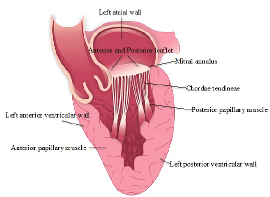
Figure 1 Mitral valve apparatus. Valve leaflets, mitral annulus, chordae tendineae, papillary muscles, left ventricular and atrial walls form the mitral valve appa-ratus. The mitral annulus is a discontinuous fibrous ring that has elastin, dense collagen fibers, and sparse cartilage. It reinforces the myocardium internally, an-chors the valve leaflets, prevents excessive dilation of valvular orifices; serves as an insertion point for atrial and ventricular myocyte bundles, and delays conduc-tion of electrical impulses between atria and ventricles. It is a dynamic structure, whose size and shape change during the cardiac cycle.
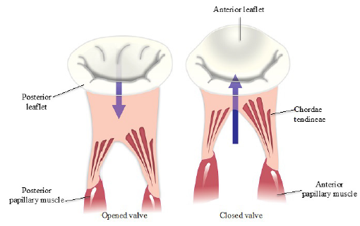
Figure 2 Mitral annulus, chordae tendineae and papillary muscle during the cardiac cycle. The posterior leaflet is smaller, thinner, but more compliant, with more chordae tendineae attachments providing more mechanical support compensating its delicate structure, but permitting it tolerate a higher compressive load. The anterior leaflet has a higher modulus of elasticity due to a thicker fibrosa layer. Leaflets have a curvature that reduces peak leaflet stress. This mechanical advantage is important during left atrial and ventricular contractility and atrial ventricular pressure diferencial, whict generate leaflet stress. Papillary muscle dynamics may play a role to facilitate leaflet apposition. Shortening of the papillary muscle throughout left ventricular isovolumic relaxation may contribute to mitral valve opening, while elongation of the papillary muscle during late diastole permits closure of the mitral valve leaflet.
Effective closure of the mitral valve requires complex, temporal. and geometric coordination of each component. The valve leaflets are adjoined to the anterior or posterior papillary muscles or occasionally, directly to the ventricular wall by CT. Together these structures work in a coordinated manner to prevent mitral valve prolapse and regur-gitation (Figure 2) 6,15.
Biomechanics of the histological layers
Leaflets are heterogeneous, laminated, structures composed of four distinct layers. From the atrial to ventricular aspect, the order is atrialis located on the inflow side, spongiosa, fibrosa and ventricularis (Figure 3) 15,36.
The spongiosa is thought to manage the mechanical compressive forces of the closed leaflets 11, allowing the other 3 layers to move towards each other during valve flexure and provides nutritional support by promoting fluid movement during flexure 36. The fibrosa withstands tensile forces of the closed valves and contributes to the flexibility during opening and closing of the leaflet 15,36. The ventricularis is similar to the atrialis but without smooth muscle cells, it is the thinnest of the layers 11.
Physiologically the mitral valve undergoes constant cyclic bending, hemodynamic shear stress, and heterogeneous strain 20,42. Extracellular matrix (ECM) of the mitral valve reflect the mechanical forces each layer sustains during the cardiac cycle: elasticity of the atrialis, stability of the fibrosa and flexibility of the spongiosa 3. Recent study has focused on the biomechanics of the mitral valve, and the possible implications of myxomatous degeneration 20,42.
Myxomatous mitral valve degeneration
Myxomatous mitral valve degeneration (MMVD) is the most common heart disease in dogs 5,29,41accounting for approximately two thirds of the cardiac caseload seen in small animal practice 1,2,38. MMVD is the most accepted term because it describes the degenerative and pathologic features of this disease 1,2,15.
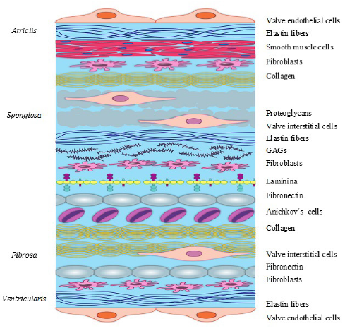
Figure 3 Histological layers of the mitral valve. The atrialis comprises a thin layer of endothelial cells supported by radially-oriented elastin fibers that minimize the shear forces on the flow side of the leaflet, allowing them to undergo significant stretch and recoil back; there are also fibroblasts, smooth muscle and collagen I, III, and VI fibers. The spongiosa is rich in proteoglycans and glycosaminoglycan (GAGs), elastin fibers, fibroblasts, Anichkov´s cells, laminin, fibronectin and collagen I, III, and VI fibers. In the anterior leaflet there are adipose cells. The fibrosa is a dense and thick layer of compact collagen I, III, VI fibers, and fibronectin, with scattered amount of fibroblasts. The ventricularis has elastin fibers thru out the valve and circumferentially oriented and small quantities collagen fibers.
MMVD has been reported as early as 1817, when Delabere Blaine noticed that some dogs had a palpable thrill on the chest, different from the beating of the heart of a healthy dog 6 . In 1935, a study in 100 dogs confirmed a chronic valve disease, mainly affecting the mitral valve, often in combination with the tricuspid valve 4,5,6. The incidence and progression of MMVD is strongly associated with age, breed, body size 6,34,36 and gender 15,30,41. It is particularly evident in small size breeds 3,29,37, frequent in medium size dogs 3,4,30but could also occur in large breeds dogs 15,17,30 where it could develop concurrently with dilated cardiomyopathy, but often overlooked 15. It is frequent in ol-der dogs, and males develop MMVD earlier than females 3,15,29. Cavalier King Charles spaniel (CKCS) 4,11,29 and bull terrier breeds can have MMVD from an early age 15. In some breeds MMVD incidence can approach 90-100% 6,30,37.
The mitral valve is most affected, but all cardiac valves could be involved. The proportion of myxomatous changes in the mitral valve is highest 1,21,38, tricuspid and mitral valve in second place and sole tricuspid involvement is least frequent 1,11,15. In older dogs mild aortic insufficiency associated whit mxyomatous changes can be detected by echocardiography 15. Valve degeneration is the principal feature of this disease 4,5,17, however myocardial intramural arteriosclerosis and fibrosis have been recognized 44,48,39.
MMVD progress over the years and the morbidity is directly related with the degree of valvular insufficiency and volumen overload 5,24,34. It could be relatively benign in mildly affected dogs, whith a 1-6 grade murmur intensity which helps predict diseasu status 4,24. A variety of clinical signs can be present 38,39,41, in advanced cases congestive heart failure (CHF) can develop due to valvular insufficiency and volume overload, which confers a poor long-term prognosis 1,38,39.
Severe MMVD can generate high velocity jets of regurgitation that could reach the atrium causing injury to the endocardium, which results in focal, thickened, fibrotic, endocardial lesions or tears (Figure 4) 15,44. When the left atrial ruptures, endocardial and endomyocardial splits could perforate the atrial septum, generating an acquired atrial septal defect, leading to right heart failure. Left atrial rupture is a major com-plication of MMVD in dogs (Figure 4) leading to hemopericarduim 6,44.
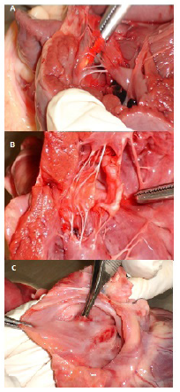
Figure 4 Normal mitral valve, macroscopic MMVD changes and atrial rupture. A. Normal mitral valve, from the atrial aspect appears smooth and transparent. From the ventricular aspect it is fasciculated and irregular, associated with chordae tendi-neae attachments. B. White nodules of variable sizes often starting at the margin of the leaflet. Thickening of the mitral leaflets, large grayish-white nodules and diffuse opacity and the chordae tendineae elongated decreasing extensibility. C. Focal, thickened, fibrotic, left atrial endocardial lesion caused by high velocity jet of regurgitation that caused a tear.
A consensus in 2009 established guidelines and a new classification for the diagnosis and treatment of MMVD based on the disease stage and clinical status. The classi-fication is as continue: Stage A, dogs at high risk of developing CHF but without any structural abnormality. Stage B are patients with a structural irregularity of MMVD but without clinical signs of CHF, generally having a heart murmur. This stage is divided into B1 and B2. Stage B1 are dogs that have hemodynamically insignificant mitral murmur without cardiac remodeling, and B2 have a hemodynamically signi-ficant murmur and cardiac remodeling is present. C1 are patients with structural heart changes that have had or have currently clinical signs of CHF with require acute hospital-based therapy. C2 patients can have a home-based treatment. D1 are patients with clinical signs of CHF refractory to standard therapy that require acute hospital based therapy, D2 are patients that can be managed with home based the-rapy 1,2,21.
MMVD has a long asymptomatic period (stages B1 and B2) 1,7,10. A study by Borgarelli et al., (2012) revealed a median survival time of 27.6 months in 256 dogs with Stages B1 y B2 7. Recent studies revealed that most cardiac enlargement occurs in the year preceding the onset of CHF 5,6,7.
In the last couple of decades a lot of pharmacological studies have undergone, with big advances in this area 9,10,17 the guidelines were updated in 2017 (unpublished) based on the results of the multicenter clinical trial with pimobendan 9,17. Pimoben-dan is a benzimidazole-pyridazinone derivative that functions as an arteriodilator, venodilator, and positive inotrope, and is therefore classified as an inodilator 9, 17, 21. The main focus is to treat the symptoms and clinical signs of this disease, by blocking the effects of the neurohumoral 9,10,21 and prolonging the asymptomatic period previous to CHF 1,2,32.
Surgical options for dogs are different from those in humans 46, the procedures mostly used in persons are mitral valve repair, valve replacement with a mecha-nical valve or bioprosthetic valve 33,35,45. There are very few comparative studies of veterinary patients that have undergone valve surgery, so there is not a clear criteria for which techniques have the best outcome 45,46,47. Recent reports reveal that the surgical option of mitral valve repair has a better outcome in dogs, using the mitral chordal replacement, with mitral annuloplasty 33,45,46, valvuloplasty 47 or resection of the prolapsed segment 8. Current pilot studies are focused on devices that repair mitral valve percutaneously or transapicaly 33,35,47 without the need of extracorporal circulation and cardioplegic cardiac arrest 8. Costs and availability are still an issue, but hopefully these devices promise a new therapeutic approach 8,35,45.
Etiology
Etiology hasn’t been completely understood, a heritable basis suggests a polygenic mode of inheritance 3,4,16. In the CKCS two loci have been recently associated with MMVD 4,32,37. This might indicate that morphologic changes associated with diminu-tive size could alter the heart valves 34 or that miniaturization genes carry the MMVD mutation 37. Breeds are in general closed populations, so a genetical disease per-petuates easily in lineage 16, 37. Interestingly MMVD affects a variety of small breeds inclusive crossbreeds 29, 30, 37. It is possible that small dogs with MMVD have inherited the same mutations from a common ancestor, but this is not the sole reason of this disease 16,32,37. Recent data, could not identify a genetic cause in the genes associated with the human form of MMVD in the genes of CKCS and Dachshund studied 32.
Insulin-like growth factor 1 gene (IGF1) is a major determinant of small size in dogs, and it is also involved in cardiac development. In a IGF1 negative mice experiment, their body size was reduced to a third but heart size reduced by less than half. It is possible that this gene regulated improperly could lead to valve malformations. There are other genes linked to body size that possibly could be associated with ca-nine heart development: high mobility group AT-hook 2 (HMGA2), insulin-like growth factor 2 mRNA binding protein 2 (IGF2BP2), Obscurin Like 1 (OBSL1), Stanniocalcin-2 (STC2), Suppressor Of Cytokine Signalling 2 (SOCS2) and Mothers Against Decanta-plegic homolog (SMAD2) (TGFb) 37, but this still is not clear.
Pathophysiological changes
Affected dog valves with MMVD have greyish-white nodules of variable sises often starting at the margin of leaflet 11,15,28. As MMVD advances the leaflets thickens and roll inward, protruding towards the atrial aspect, the nodules coalesce involving a larger portion of the leaflet 15,28 possible valve lengthening 18.
Pathological changes associated with MMVD involve progressive expansion of the spongiosa layer and disruption of the fibrosa layer 15,18. The spongiosa becomes thic-kened with increased extracellular matrix composed of glycosaminoglycan (GAGs) and PG, and proliferation of the extracellular matrix (ECM). In the fibrosa collagen be-comes disrupted and attenuated 15,28,34. In advance stages, it is difficult to recognize distinct spongiosa and fibrosa layers 15,28.
Moderate MMVD is characterized by a moderate increase of GAGs and proteoglycan deposition in the distal half of the spongiosa, mild degeneration of collagen bundles in the fibrosa, but normal structure in the proximal half of the valve 3, 15. Severe MMVD is characterized by severe increase of GAGs and proteoglycan deposition resulting in complete displacement of the fibrosa25, disrupted collagen bundles in the valve, and fibroblastic proliferation in the atrialis and fibrosa3, 15.
Whitney in 1965 classified MMVD into 4 types based upon the degree of valve no-dularity, thickening, and deformity 15. Types 1 have few small nodules with areas of diffuse opacity in the proximal valve. Types 2 have thickening of the mitral leaflets, larger grayisg-white nodules and difduse opacity but the CT are unchanged. In Type 3, the nodular thickening is severe which tend to coalesce and extend to the CT, valvular incompetence could be present (Figure 4) 15. Type 4 the valves have a ballooning disposition, the CT are thickened and occasionally ruptured, valvular incompetence is present 6,11,15. A mitral valve can have different stages at once 15.
Pathologic changes that alter the mitral apparatus restrain valve mechanics, affect fluid dynamics, and promote valvular insufficiency. Tethering and coapting forces act on the leaflets, as well as forces that affect annular size, papillary muscle position, and transvalvular pressure 15. In some dogs CT is elongated 3, 15, 18, which is not clear if it is a morphologic variation or a initial pathologic change of MMVD 15. Data reported previously showed that an increase in stiffnes can increase stress in the CT and leaflets, reducing the probability of valve coaptation 42. When a CT is ruptured left ventricular geometry can change altering the pressure volume relation 15.
The mechanical strength of the leaflets and CT depends generally on collagen, which is deranged losing their ability to maintain mechanical stability and flexibility. Overall collagen content is not reduced significantly, but there is a reduction in organized collagen fibers. The form of collagen organization defines the distribution and magnitude of strain of the valve during the cardiac cycle 42. Mechanical studies of MMVD indicate tensile weakening of the valve apparatus, being a consequence of increased matrix catabolic enzymes 20,28.
Mitral valve innervation is altered in aged dogs and in dogs with MMVD 11,15. Significant loss of innervation was detected in association with reduction in myocytes with an increase in adipocytes within mitral valvular leaflets as a senile feature. MMVD may develop before reduced innervation with advanced age. It is uncertain whether muscle loss occurs secondary to loss of innervation, or whether reduced innervation density was caused by muscle loss due to MMVD 11.
While much is known about the structural and cellular changes in canine MMVD, less is known about the molecular mechanisms and biochemical changes 28,42,43. It is necessary to understand the signalling pathways and discern the mechanisms involved, which in the future could possibly lead to reduce the presentation of the most frequent cardiac disease in dogs.
Signalling pathways for MMVD
In heart valves the extracellular matrix (ECM) is a complex dynamic physical and che-mical network consisting of collagen, elastin, fibronectin, laminin, but mainly of GAGs and PG 3,11,36. ECM is constantly being remodeled, keeping a balance of synthesis and degradation to ensure a normal valve 3,11,22. ECM is produced by valve endothelial cells (VECs) and valve interstitial cells (VICs), although they have different phenotype post-natal, they share a common embryonic origin 11,36.
A single layer of VECs cover the surface of the valves continuing to the endocardium 11 and perform similar functions as the endothelial vascular cells 3,19,34. Damage to this layer can induce synthesis of endothelin, a potent vasoconstrictor, which stimulates proliferation of fibroblasts and collagen 11,19.
VICs are fibroblastic mesenchymal cells that help neutralize biomechanical forces of the valve: tension (prevents reversal of blood flow), shear (blood passing), compression and flexure (opening and closing) 11,20,34. VICs are present in all the valve layers 3. Because of their function, VICs are proposed to be the mechanical stimuli of valve degeneration 26. Increased numbers have been reported in MMVD, mainly in the atrial layer. VICs can undergo phenotypic transformation turning into alpha smooth muscle actin (αSMA), which are thought to mediate degeneration producing catabolic enzymes 3,22,28 which generates an inappropriate remodeling of the valve 20,22,25. It seems that the mecha-nisms are the activation of the endothelial or damage which activates Transforming growth factor β signalling (TGFβ)/Serotonin (5-hydroxytryptamine, 5HT) signalling pathway, enabling transformation to a VICs phenotype (Figure 5) 3,26,28.
TGFβ action is mediated through two serinethreonine kinase receptors, TGFβ receptor I (RI) and receptor II (RII), increased levels of both receptors has been reported in dogs with MMVD 23,34. In porcine cultured aortic valve, serotonin induced TGFβ 1 mRNA expression. This data suggest a strong relation between serotonin signalling, TGFβ signallig, and expression of myxomatous effector genes and proteins 23,34. It is hypothesized that local serotonin synthesis plays a role in arresting osteogenesis and encouraging chondrogenesis (Figure 5) 20,23,34.
Endothelial to mesenchymal transition (EndoMT) and an increase expression of the genes actin alpha 2 smooth muscle aorta (ACTA2), CTNNB1 (β-cateninin), Snail Family Transcriptional Repressor 1 (SNAI1) and hyaluronan synthase (HAS2) has been deter-mined in canine MMVD valves. It also seems that EndoMT is an alternative pathway, justified on that not all of the αSMA positive cells, are derived from VICs 25,28,36.
Han et al., (2013) documented reduction in total VECs numbers and morphology al-teration, and increased number of VICs in MMVD valves 19. It is still not clear the role of VECs in the pathogenesis of MMVD, VICs have been documented to some degree, or even if these changes are due to myxomatous degeneration or reflect long-term valve remodeling 19,28.
Serotonin (5-hydroxytryptamine, 5HT) is involved in the embryogenesis, valvular and myocardial function. The majority of 5HT is synthesized in the enterochromaffin cells of the intestine 24,27. Almost all circulating 5HT is contained within platelets and re-leased into serum during platelet aggregation and activation. Serotonin has a rapid metabolism by platelet uptake and liver and lung clearance 13,24.
In humans high levels of 5HT induced by serotonergic drugs can cause carcinoide valvulopathy 13,24,34. This valvulopathy has gross and histologic similarities with MMVD 23,24,27. It is also possible that local 5HT is an initiating chemical stimuli, which correlates with an increase in tryptophan hydroxylase (TPH1) expression, enzyme involved in serotonin synthesis in dogs with MMVD 12,20,34 making tensile strain a plausible initiating mechanism 20,34, which is a known risk factor for MMVD in humans 20. MMVD alters the mechanical ability of the affected valve, and the change on the mechanics have an important impact on the degeneration 22,28,42. 5HT is a possible initiating chemical stimuli for MMVD 13,20,34 it is associated with PG/GAGs deposition 34.
Cremer et al., (2015) reported an increased presence of alpha smooth muscle actin (αSMA) positive cells in the marginal zone of myxomatous nodules and in papillary muscle, demonstrating a positive association between gene expression of 5HT2BR (Receptor 2B, 2BR) 12. This finding associated with increased concentrations of 5HT in the mitral valve and myocardium with MMVD, may indicate 5HT signalling in cli-nically affected dog with MMVD 13,12 . Dogs with MMVD and unaffected CKCS have been identified to have high serum levels of platelet-5HT, also mitral valve leaflets and left ventricular myocardium in dogs with MMVD 13. Ljungvall et al., (2014) found higher serum 5HT levels in CKCS compared to other breeds 24, serum 5HT concen-trations related to severity of MMVD 12. While Mangklabrunks and Disatian (2014), found no difference in platelet 5HT, plasma 5HT in healthy predisposed small breeds and MMVD dogs 27.
Whit age the valve increases its stiffness in the radial and circumferential direction, and extensibility decreases and stress relaxation increases. Basically this is explai-ned by the thickening of the fibrosa layer and an increase in collagen production 11. Changes of ECM composition in MMVD diminish the ability of the cells to communi-cate contributing to the dysregulation of the cellular synthesis and alter the strain distribution in the valve 42. Progressive deterioration of ECM age related could be the relationship between age and MMVD 18,34.
Lacerda et al., (2012) described that in cultured canine mitral valves subjected to static or tensile strain displayed increased expression of αSMA, Embryonic smooth muscle myosin heavy chain (SMemb), matrix metalloproteinases (MMP)-1, MMP-13, cathepsin K, xylosyl transferase, and decorin (PG core) in comparison to control mitral valves 20,34. An in vivo sheep experiment concluded that a turbulent flow proximal to a mitral leaflet can cause myxomatous pathology, being possible that abnormal shear forces may have an important role in initiating and propagating MMVD 34.
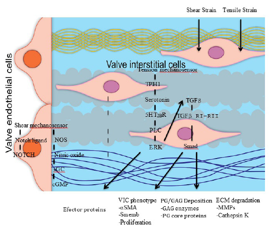
Figure 5 Signalling pathways of MMVD. Abbreviations. Neurogenic locus notch homolog protein (NOTCH), nitric oxide synthetase (NOS), soluble guanylate cyclase (sGC), Cyclic guanosine monophosphate (cGMP), tryptophan hydroxylase 1 (TPH 1), serotonin type 2B receptor (5HT2BR), phospholipase C (PLC), extracellular-signal regu-lated kinase (ERK), transforming growth factor β (TGFβ), Receptor I (RI), Receptor II (RII), mother against decantaplegic homolog (Smad), smooth muscle actin (αSMA), non-muscle embryonic myosin (Smemb), glycosaminoglycan (GAG), proteoglycan (PG), matrix metalloproteinases (MMPs).
Endothelial cells in addition to the immune system also express cytokines 31,40. In a research of dogs with heart failure due to MMVD, elevated circulating concentrations of monocyte chemoattractant protein 1 (MCP-1) (proinflammatory cytokine and key attractant for mononuclear cells) was found in comparison to healthy control dogs, correlating to the finding of increased transcriptional expression in MMVD tissue 40,48. Myocardial fibrosis and arterial luminal narrowing is speculated to be caused by locally produced MCP-1. Also decreases of interleukin (IL)-2 and IL-7 as MMVD increased in severity, and IL-8 decreased with increasing severity of left ventricular function, could indicate de-creased immunological activation in this disease 31,40,48. Mavropoulou et al., (2016) found IL-8 increased with severity of MMVD, and βTGF was higher in asymptomatic MMVD dogs, suggesting involvement of cytokines at different stages of the disease 31.
Nitric oxide (NO) is involved in a variety of biological functions 14. Increased NO activity is associated with MMVD 14,28,34 and with increased endothelin (ET) receptor expression, with a strong correlation in the areas of greatest pathologic change 14,43. It is uncertain the role of NO and ET in MMVD, as a mediator of pathologic gene expression or denotes a ECM response 28,34. Also increased Galectin 3 in plasma concentrations and in cardiac muscle, a biomarker for cardiac fibrosis, was found in dogs with MMVD when compared to normal dogs 43.
Gene ontology and network analysis identified relevant biological functional clusters related to MMVD: cell movement, ECM organization and EndoMT, cardiovascular de-velopment, and inflammation. The gene families that have been detected to be re-levant to MMVD are collagens, ADAMTS, PGs, MMPs, TIMPs, cathepsin S, basement membrane, integrins, tight junction cell adhesion proteins, cadherins, and members of the serotonin (5-HT)/transforming growth factor β signalling pathway. Three canonical pathways have been recognized: caveolar mediated endocytosis, which controls endo-thelial cell growth and migration; remodeling of epithelial adherens junctions, which can affect cell proiferation and migration; endothelin signlling which can cause ECM remodeling 26,28.
It is yet not clear the specific initial stimuli for the pathologic protein expression in MMVD, it could be chemical and mechanical stimuli that triggers signal pathways which through transcription factor control gene expression 20,34. Advances in specific canine molecular tools have been made, making possible research in the signalling pathways of canine MMVD. These efforts are currently being made. It is necessary to understand the pathobiology of this disease and reveal the changes at the levels of gene and protein expression, to possibly develop ways to block these mechanisms.
Final thoughts
In 2012, the Journal of Veterinary Cardiology dedicated an entire issue to this di-sease that determined more research in this field. It is evident that investigators dedicated to this area have not agreed on the same term for this disease. Terms like “chronic mitral regurgitation”, “degenerative mitral valve disease” and “mitral valve disease” are used as synonyms. This alone makes it harder to look up the latest studies on this disease. It is necessary to unify a term for MMVD. Based on the definitions the authors agree and recommend the term MMVD because it describes the degenerative and pathologic features of this disease. Most of the research has been centered on the mitral valve, and some on the tricuspid valve, but few is known about the myxomatous pulmonary and aortic valve disease.
It is important that veterinarians acknowledge the ACVIM Consensus, which provides a guideline to diagnosis the stage and stage-specific recommended treatment of MMVD. The statements of the Consensus stand on the foundation of evidence-based medicine; some non-consensus treatments are stated due to the lack of clinical drug efficacy and safety trials in this patient population. The concensus classification of the clinical stages of MMVD is more adequate then the adapted New York Heart Association (NYHA) and International Small Animal Cardiac Health Council1 (ISACHC), which are based on functional classification. The newer classification aims to categorize patients objectively in the course of their heart disease and to link the severity of signs to adequate treatments at each stage.
The focus on differente factors that could alter the biomechanics of the valve, as possible concurrent initiation mechanisms of valve degeneration, discloses processes that could have an important impact on the degeneration. Also initiating chemical stimuli, as serotonin, cytokines, nitric oxide and endothelin still have a strong basis for contributing to this disease.
The main characteristics of canine MMVD are increased cellularity, disorganization of the valve structure and phenotype transformation of VIC and VEC cells 3. It is uncer-tain the role of biomechanical overload and aging in the pathogenesis of MMVD 20,34. It is unclear that the same pathologic process is equal in the different breeds of dogs, or if the predisposed breed like CKCS have specific features 15. It is relevant to identify the molecular biology, signalling pathways, and gene activation sequences to comprehend the pathobiology and possible aim to developed effective diagnostic and therapeutic strategies for MMVD.
Current therapies are aimed to slow down the preclinical period and to minimize clinical signs and symptoms, but not to its underlying pathology. The couple of last decades the focus has been on developing effective treatments and improving diagnostics. But the last five years have been dedicated to pursue canine molecular biology, so new advances hopefully will be made in MMVD.

