


 English (pdf)
 Article in xml format
 Article references
 Send this article by e-mail
 Cited by SciELO
  Cited by Google
  Similars in
SciELO
Similars in
SciELO  Similars in Google
 Permalink
Introduction
Some optic neuropathies cursing with the degeneration of ganglion cells and their respective axons in the retina lead to changes in the optic disc and in some cases; even loss of vision knows glaucoma. After diabetic retinopathy, glaucoma is the second cause of blindness in humans and the third in canines 10,19,31. Glaucoma is classified depending on the characteristics of the iridocorneal angle as an open angle or closed angle or, depending on the origin, as primary or secondary. One of the most common types of this disease in humans is primary open-angle glaucoma, however in canines we find angle-closure glaucoma more frequently 10,31. Intraocular pressure (IOP) represents the primary risk factor for developing glaucoma (6,13,19. IOP depends on the flow rate of aqueous humor and its resistance to outflow; thus, the result of the relationship between intraocular fluid, elasticity and stiffness of the cornea and sclera would be called intraocular pressure 6,10,29.
Aqueous humor is a hydatoid fluid, which is formed by a passive ultrafiltration process and by an active secretion of the epithelium that covers the ciliary body. The constant flow of aqueous humor supplies nutrients to the avascular cornea and lens, also removes metabolic waste. The level of production of the aqueous humor should be equal to its level of output. The formation of aqueous humor depends on ciliary arterial blood pressure, which is equal to the intraocular pressure 29. Aqueous humor circulation begins when the fluid produced in the posterior chamber passes through the pupil and enters the ciliary opening, which contains the trabecular meshwork. The mesh filters this liquid to reach the vessels of the scleral venous plexus and then from there to the venous system 6,10,25.
The procedure to measure intraocular pressure is called tonometry, which can be indentation, applanation or rebound 20,24,28. With tonometry, more precise IOP data can be obtained, with indentation tonometry being the most imprecise of all. Elements such as the thickness and biomechanics of the cornea; as well as edematous, ulcerative and pigmentary alterations can affect the determination of IOP by introducing noise in the tonometry 3,4,7.
Systemic blood pressure has been documented to influence intraocular pressure, becoming a risk factor for the development and progression of glaucoma. On the one hand, the microvasculature of the retina, the choroid and the optic nerve can present a series of pathophysiological changes that can manifest in conditions such as retinopathy, choroidopathy and hypertensive optic neuropathy 9,10,31. Alternatively, it has been shown that the nocturnal arterial hypotension that some patients present with favors glaucomatous optic neuropathy progressively due to a reduction in the perfusion of the optic nerve head, leading to a greater deterioration of the visual field 6,13,17.
Calcium antagonist drugs are effective for treating cardiovascular diseases, including high blood pressure. Calcium enters excitable cells through voltage-gated calcium channels 8. Amlodipine is a third-generation drug, dihydropyridine calcium antagonist, selective for vascular smooth muscle, with little effect on cardiac contractility; it exerts its action through the inhibition of calcium entry into vascular smooth muscle and myocardial cells, decreasing peripheral vascular resistance. It has an elimination half-life of 40 h (h) to 60 h, which confers several pharmacokinetic characteristics that are not seen with other calcium antagonist drugs. The bioavailability of the orally administered drug is between 60% and 80% and accumulates to a steady state with administration once daily for a period of approximately 1 to 1 and a half week 8,26.
Due to the devastating impact that glaucoma has on the quality of life of animals, more efforts are required to understand this pathology and develop new therapeutic and prevention options. Rabbits have played an important role as an animal model in ophthalmological research processes, allowing the development of surgical techniques, as well as in the evaluation and monitoring of new therapeutic options 8,16,33. However, in the environment there has been a deficiency of sensitive equipment in the measurement of intraocular pressure for this species. Therefore, we set ourselves the objective of comparing rebound and applanation tonometry in healthy New Zealand rabbits treated with Amlodipine.
Materials and methods
Animals
Following the arrive guidelines, twelve healthy male New Zealand white rabbits, 5 months old and an average weight of 3.1 kg were used in this experimental study. Which were raised under convectional bioterium conditions and handled according to the institutional guidelines of the Central Animal Facility, individually housed in a cage with controlled temperature and humidity conditions, rabbit´s commercial food and water ad libitum.
Two groups treated (T) and control (C) were randomized, each group with six individuals (12 eyes per group for each measurement). The treated group received amlodipine at a dose of 5 mg/kg 15 diluted in 2-mL water administered orally, once a day for a month. The control group received only 2-mL water,
Blood pressure
A Contec® veterinary blood pressure monitor with a neonatal cuff was used to blood measure in the left forelimb (brachial/radial artery) before treatment and 6 h after treatment on days 1 - 4 - 8 - 12 - 16 - 20 - 24 - 28 and 32. Systolic and diastolic blood pressure was recorded in both groups.
Tonometry
For measuring intraocular pressure, the rabbits were gently manipulated without applying pressure to the neck or eyelids. A rebound tonometer (TONOVET Plus®, Nasdaq, Helsinki) and an applanation tonometer (TONO-PEN VET®, Buffalo, New York) were used following the indications of each manufacturer. It was started using a rebound tonometer and then the applanation one for IOP measurement (after 1 min of topical application of a drop of proparacaine hydrochloride ophthalmic solution, Alcaíne®), maintaining a methodical order starting with the left eye and then the right. The intraocular pressure and blood pressure were measured on days 1 - 4 - 8 - 12 - 16 - 20 - 24 - 28 and 32 before and 6 h after treatment. At the end of the study, data from 216 eyes for each group were analyzed.
Data analysis
In order to contrast normality, Shapiro Wilk was performed. Two-way ANOVA was performed and for the multiple comparisons of the results, Tukey and Sidak tests were performed using the Graphpad Prism® Version 6. Values of P<0,05 were considered statistically significant. Data were expressed as the median and interquartile range.
Results
Clinical signs
Before the start of the study, all the animals underwent general clinical and ophthalmological assessments, ruling out health problems in all individuals. All received placebo (C) and treatment (T) according to the experimental group to which they belonged, without any manifestation of clinical alteration or side effects, and they completed the study according to the schedule. Once the study was finished and it was verified that they were healthy, the rabbits were given up for adoption.
Blood pressure
Both the control group (C) and the treated group (T) presented average normal values in both systolic (a) and diastolic (b) pressure without showing statistically significant differences between treatment days (Figure 1). However, a trend is noted where the group of animals treated with amlodipine® (T) presents lower values than the control group, both in systolic and diastolic pressure.
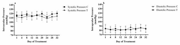
Figure 1 Average systolic and diastolic blood pressure. The figure shows the global average values of Systolic pressure (a) and Diastolic pressure (b) measured in millimeters of mercury (mmHg). Each point represents the mean value ± S.E.M for each experimental group (n = 12) of control (C) group and treatment (T) group.
The systolic pressure was 116.5 ± 7.9 mmHg and 117.4 ± 8.9 mmHg, for experimental and control groups, respectively. The blood pressure before and after treatment in the treated was 113.6 ± 7.9 mmHg and 111.8 ± 10.0 mmHg (pretreatment and posttreatment respectively). No statistically significant differences were observed; nevertheless, when comparing the blood pressure before and after treatment between the control group and treated group differences were observed with p = 0.0314 (pretreatment C vs T) and p = 0.0042 (posttreatment C vs T). The control group showed a diastolic pressure of 83.0 ± 7.3 mmHg (pretreatment) and 83.9 ± 7.4 mmHg (posttreatment) while, the treated group had a diastolic pressure of 84.9 ± 8.0 mmHg (pretreatment) and 82.9 ± 10.3 mmHg (posttreatment). No statistically significant differences were observed between control and treated groups neither pre and posttreatment (Figure 2).
Tonometry
In the first instance, the TONOVET Plus® was used, which is a rebound tonometer, starting with the left eye and then the right, then the measurement was taken with the TONO-PEN VET®, which is of flattening, in the same way starting with the left eye and then the right eye (after instillation of topical anesthetic) (Figure 3).
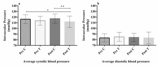
Figure 2 Pretreatment and posttreatment global average systolic and diastolic blood pressure. The figure shows the global average systolic (a) and diastolic (b) blood pressure measured before treatment (Pre) and six hours after treatment (Post) in control (C) and treatment (T) group. Each point represents the mean value ± S.E.M for each experimental group (n = 12) of the Pretreatment C group. * Pretreatment C vs Posttreatment T, (p-value=0.0314). ** Posttreatment C vs Posttreatment T, (p-value =0.0042).
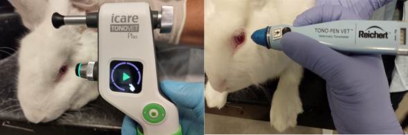
Figure 3 Rebound (TONOVET Plus®) and applanation (TONO-PEN VET®) tonometers. The image on the left shows the TONOVET Plus® tonometer and the image on the right shows the TONO-PEN VET® tonometer, both of them about to perform measurement intraocular pressure in New Zealand rabbits.
In the control group, no significant differences were observed between measurements before treatment and 6 h after treatment for each tonometer. The TONOVET Plus® did not present statistically significant differences between days of follow-up. The TONO-PEN VET® for its part presented statistically significant differences between day 1 (19 ± 3 mmHg) and days 4 (19 ± 3 mmHg vs 15 ± 4 mmHg), 8 (19 ± 3 mmHg vs 16 ± 4 mmHg), 16 (19 ± 3 mmHg vs 16 ± 2 mmHg) and 32 (19 ± 3 mmHg vs 16 ± 3 mmHg); similarly, there were differences between days 4 (15 ± 4 mmHg) and days 12 (15 ± 4 mmHg vs 18 ± 2 mmHg), 20 (15 ± 4 mmHg vs 18 ± 3 mmHg) and 24 (15 ± 3 mmHg) and 24 (15 ± 4 mmHg vs 19 ± 2 mmHg). Likewise, between days 8 and 24 (16 ± 4 mmHg vs 19 ± 2 mmHg), 16 and 24 (16 ± 2 mmHg vs 19 ± 2 mmHg) and days 24 and 32 (19 ± 2 mmHg vs 16 ± 3 mmHg). Alternatively, statistically significant differences were found between TONOVET Plus® and TONO-PEN VET® on days 4 (19 ± 3 mmHg vs 15 ± 4 mmHg), 8 (19 ± 4 mmHg vs 16 ± 4 mmHg), 16 (19 ± 2 mmHg vs 16 ± 2 mmHg), 28 (20 ± 3 mmHg vs 17 ± 4 mmHg) and 32 (20 ± 3 mmHg vs 16 ± 3 mmHg). When comparing the results obtained in the measurements of the control group, we can observe in a general way that the values obtained using the TONOVET Plus® (19 ± 3 mmHg, n = 216 eyes) are on average higher than the values obtained using the TONO-PEN VET® (17 ± 3 mmHg, n = 216 eyes) (Figure 4).
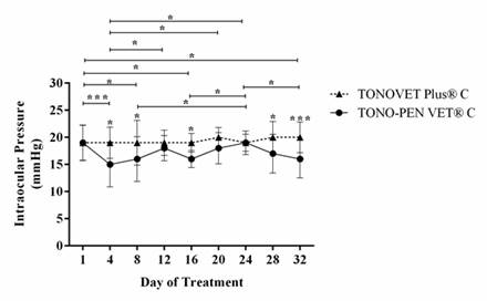
Figure 4 Control group tonometry measured using a rebound (TONOVET Plus®) and applanation (TONO-PEN VET®) tonometers. The figure shows the measures in millimeters of mercury (mmHg) of the intraocular pressure in the control group obtained using two tonometers. Each point represents the mean value ± S.E.M for the control group h(n = 12) measured using the TON.
When comparing the control group with the treatment group for both TONOVET Plus® and TONO-PEN VET® (Figure 5), we can see that TONOVET Plus® (a) could identify statistically significant differences on day 20 of follow-up (20 ± 2 mmHg vs 17 ± 2 mmHg) between the control group and the treatment group, respectively. Alternatively, between day 8 of the treatment group and days 20 (17 ± 2 mmHg vs 20 ± 2 mmHg), 28 (17 ± 2 mmHg vs 20 ± 3 mmHg) and 32 (17 ± 2 mmHg vs 20 ± 3 mmHg) from the control group. Similarly, between day 20 of the treatment group and days 28 (17 ± 2 mmHg vs 20 ± 3 mmHg) and 32 (17 ± 2 mmHg vs 20 ± 3 mmHg) of the control group; For its part, the TONO-PEN VET® (b) indicated statistically significant differences between day 1 of the control group and days 4 (19 ± 3 mmHg vs 15 ± 2 mmHg), 8 (19 ± 3 mmHg vs 15 ± 3 mmHg), 16 (19 ± 3 mmHg vs 16 ± 1 mmHg) and 20 (19 ± 3 mmHg vs 16 ± 2 mmHg) of the treatment group. Likewise, between day 4 of the treatment group and days 20 (15 ± 2 mmHg vs 18 ± 3 mmHg), 24 (15 ± 2 mmHg vs 19 ± 2 mmHg) of the control group. In both tonometers, it was possible to show the trend to lower intraocular pressure values in the group treated with amlodipine® than those of the control group (TONOVET Plus® control (19 ± 3 mmHg, n = 216) vs TONOVET Plus® treatment (18 ± 2 mmHg, n = 216); TONO-PEN VET® control (17 ± 3 mmHg, n = 216) vs TONO-PEN VET® treatment (16 ± 2 mmHg, n = 216).
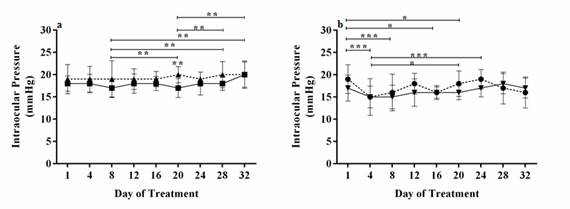
Figure 5 Intraocular pressure comparation between control and treatment groups measured using a rebound (TONOVET Plus®) and applanation (TONO-PEN VET®) tonometers. The figure shows the average of intraocular pressure comparing the control and treatment groups measured using TONOVET Plus® (a) and TONO-PEN VET® (b). Each point represents the mean value ± S.E.M for each experimental group (n = 12) of TONOVET Plus® a control group, TONOVET Plus® treatment group, TONO-PEN VET® control group, TONO-PEN VET® treatment group. (*p-value=0.0238), (**p-value=0.0036) (***p-value=0.0001).
When comparing the measurements obtained in the group of individuals treated with amlodipine® with both tonometers (Figure 6). TONOVET Plus® and TONO-PEN VET®, statistically significant differences between both tonometers can be observed on day 4 (18 ± 2 mmHg vs 15 ± 2 mmHg) and 32 (20 ± 3 mmHg vs 17 ± 2 mmHg). El TONO-PEN VET® showed statistically significant differences between days 4 and 28 (15 ± 2 mmHg vs 18 ± 2 mmHg) and days 8 and 28 (15 ± 3 mmHg vs 18 ± 2 mmHg). TONOVET Plus® indicated differences between days 8 and 32 (17 ± 2 mmHg vs 20 ± 3 mmHg) and days 20 and 32 (17 ± 2 mmHg vs 20 ± 3 mmHg).
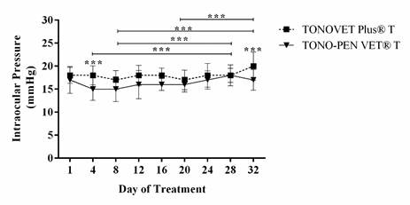
Figure 6 Intraocular pressure comparation between the treatment group measured with a rebound (TONOVET Plus®) and applanation (TONO-PEN VET®) tonometers. The figure shows the average of intraocular pressure between the treatment groups measured with TONOVET Plus® and TONO-PEN VET®. Each point represents the mean value ± S.E.M for each experimental group (n = 12) of the TONOVET Plus® treatment group. TONO-PEN VET® treatment group. (***p-value=0.0007).
Discussion
Animal models have allowed great advances in different aspects of both human and veterinary medicine. Rabbits, specifically, have played an important role in research processes, both for developing surgical techniques, and in the evaluation and monitoring of new therapeutic options. At the ophthalmology level, they have also made it possible to understand the pathophysiology of such dire diseases as glaucoma 8,16,32.
Systemic blood pressure can affect the structure and function of the eye as has been reported in different studies indicating that it has an influence on intraocular pressure, becoming a risk factor for the development and progression of glaucoma 2,6,17. In these studies it has been indicated that not only hypertension, but also hypotension are risk factors for the development of glaucoma, since it can generate a reduction in the perfusion pressure of the central retinal artery and the posterior portion of the optic nerve (produced by cardiovascular changes that increase resistance to blood flow and / or reduce perfusion) 5,17. On the other hand, it has been reported that the systemic arterial pressure fluctuates in relation to the circadian rhythm and in the hours of the night it can appear in normotensive and hypertensive patients, a reduction between 10% and 20% of the systemic arterial pressure while they sleep; This is due to a reduction in the activity of the sympathetic nervous system, to then return to its basal values early in the morning 6,31. A study in healthy rabbits where intraocular pressure was monitored at different times of the day indicated that it is higher at 6 am than in the following hours of the day 30. For the above reason, in our study, the arterial and intraocular pressure measurements were performed after 7 am, so as not to interfere with the circadian rhythm of the rabbits.
Alternatively, amlodipine® is effective for treating many cardiovascular diseases, including high blood pressure. Due to the above, we wanted to evaluate its effect on systemic arterial pressure in healthy rabbits; being able to show a trend where the group of animals treated with amlodipine® presents normal values but lower than the control group, both in systolic and diastolic pressure. Statistical differences were evidenced between the control group and the post-treatment group with amlodipine®, and there were no statistically significant differences between pretreatment and 6 h post-treatment within each group. The latter could be explained by the pharmacodynamics of amlodipine®; after oral administration, the onset of the effect is gradual, reaching a peak plasma concentration at 6 or 8 h, gradually decreasing blood pressure, which tends to slowly return to its initial pressure after a dose, at 24 to 72 h, without severe effects on heart rate. In chronic oral treatments, where amlodipine® is administered once a day, it has been noted that blood pressure decreases from the start of treatment showing little fluctuation during the dose interval. If the treatment was suspended after several days of supply, the blood pressure would gradually return to its initial value 7 to 10 days later, without the rebound effect. Given these characteristics, this drug has been used as a complement in the therapy of ocular pathologies such as glaucoma 1,11,22.
Alternatively, the intraocular pressure (IOP) represents the primary risk factor for developing glaucoma; if a reduction in intraocular pressure is achieved, the incidence and progression of glaucoma can be controlled; in contrast, an increase in IOP can cause microangiopathies and a reduction in ocular blood flow 13,19,31. Therefore, within the ophthalmological evaluation, tonometry is a highly valuable diagnostic aid, allowing the identification of intraocular pressure alterations. Rebound tonometry (TONOVET Plus®) measures intraocular pressure by quantifying the de-acceleration of the probe after impact with the corneal surface while the applanation tonometer (TONO-PEN VET®) quantifies the force required to flatten a certain area of the cornea. Both types of tonometry can be affected by corneal thickness, as well as ulcerative, pigmented or edematous affectations 7,12,21, therefore, this study was aimed to compare TONOVET Plus® and TONO-PEN VET® in healthy animals and to demonstrate their ability to detect changes in intraocular pressure under the effect of amlodipine®, a drug that regulates systemic arterial pressure. Intraocular pressure was measured for each rabbit with both tonometers, however, measurements were routinely started with the TONOVET Plus® and then with the TONO-PEN VET® because reports indicate that the first one may be affected by the application of topical anesthetics, which must perform applanation tonometry with the TONO-PEN VET®. Some of these reports even indicate a reduction in intraocular pressure immediately after the application of topical anesthetics with a possible duration of effect of up to 20 minutes 27. There have been several possible explanations given to explain this phenomenon; within them are the relaxation of the pressure exerted by the eyelids on the eyeball, corneal relaxation, the eyeball and ciliary muscles; understanding that corneal-scleral stiffness and extra-ocular muscle tone can affect intraocular pressure 4,14,18.
We observed that the control group, who received only water and the rabbits were adapted to the same management protocol as the animals treated with amlodipine®, did not present significant differences in intraocular pressure measurements before treatment and 6 h posttreatment with neither of the two tonometers. Alternatively, the TONE-PEN VET® of the control group showed greater variability between days of follow-up (significant statistical differences between day 1 and days 8, 16 and 32; on days 4 and days 12, 20 and 24; likewise, between days 8 and 24, 16 and 24, 24 and 32); while the TONOVET Plus® of the control group did not present statistically significant differences between days of follow-up. When comparing both tonometers, significant differences were found on days 4, 8, 16, 28 and 32, possibly due to the greater variability of the data obtained using the TONO-PEN VET®, we were able to observe that when measuring intraocular pressure in the control group, the values obtained in the same individuals with TONOVET Plus® (19 mmHg ± 3, n = 216 eyes) are on average higher than those obtained using TONO- PEN VET® (17 mmHg ± 3, n = 216 eyes). A similar trend was observed in the group treated with amlodipine® where the TONOVET Plus® indicated an average intraocular pressure of 18 mmHg ± 2 (n = 216 eyes) and the PEN-TONE VET® of 16 mmHg ± 2 (n = 216 eyes).
This phenomenon has been documented in other studies where a difference has been found in both tonometers between approximately 2 and 3 mmHg 18,30. As we mentioned previously, it cannot be ruled out that this difference is partly caused by the effects of topical anesthetic that must conduct the measurement using the applanation tonometer; In some sensitive patients, it has been possible to identify reductions in intraocular pressure after the application of the topical anesthetic of up to 6-8 mmHg 23, suggesting that the need for the use of topical anesthetic induces an underestimation of intraocular pressure.
Both TONOVET Plus® and TONO-PEN Vet® showed a lower intraocular pressure tendency in rabbits treated with amlodipine® (who also showed a lower systemic arterial pressure tendency than the control group), validating the fact that intraocular pressure is influenced by systemic arterial pressure. Additionally, the results also validate that dihydropyridine calcium antagonists, L-type selective for vascular smooth muscle such as amlodipine®, have great potential as supportive therapy for patients with glaucoma secondary to systemic hypertension.
Conclusions
It was possible to validate the relationship between systemic arterial pressure and intraocular pressure in white New Zealand rabbits, reaffirming the use of rabbits as an animal model for the study of some ophthalmologic alterations. It was also possible to demonstrate the effect of amlodipine on systemic arterial pressure and its subsequent influence on intraocular pressure. Both rebound tonometry and applanation tonometry allowed measuring normal values of intraocular pressure in healthy rabbits; in addition, they were able to detect sensitive changes in intraocular pressure after amlodipine administration; however, rebound tonometry showed less variability in the results than applanation tonometry.

