


Serviços Personalizados
Journal
Artigo
 Inglês (pdf)
 Artigo em XML
 Referências do artigo
 Enviar este artigo por email
Indicadores
-
 Citado por SciELO
 -
 Acessos
Links relacionados
-
 Citado por Google
 -
 Similares em
SciELO
Similares em
SciELO -
 Similares em Google
Compartilhar

 Permalink
Tecciencia
versão impressa ISSN 1909-3667
Tecciencia vol.10 no.19 Bogotá jul./dez. 2015
https://doi.org/10.18180/tecciencia.2015.19.6
DOI: http://dx.doi.org/10.18180/tecciencia.2015.19.6
Images compression process in the radiological service unit as an infrastructure component in the Colombian health model
Proceso de comprensión de imágenes en las unidades del servicio radiológico como un componente de la infraestructura en el modelo colombiano de salud
Lilia Edith Aparicio Pico1, Julián Luciano Cárdena2, Alexandra López Sevillano3
1 Universidad Distrital Francisco José de Caldas, Bogotá, Colombia, medicina@udistrital.edu.co
2 Universidad Central Marta Abreu de las Villas, Santa Clara, Cuba, julian@uclv.edu.co
3 Universidad Distrital Francisco José de Caldas, Bogotá, Colombia, alelose@gmail.com
* Corresponding Author. E-mail: medicina@udistritaLedu.co
How to cite: Aparicio Pico, L.; Cardena, J.; Lopez Sevillano, A.; Images compression process in the radiological service unit as an infrastructure component in the Colombian health model, TECCIENCIA, Vol. 10 No. 19., 33-42, 2015, DOI: http://dx.doi.org/10.18180/tecciencia.2015.19.6
Received: March 30 2015 Accepted: July 30 2015 Available Online: August 6 2015
Abstract
This article depicts the process of images compression for the Discrete Cosine Transform (DCT). By being one of the most significant transformations in the area of digital image compression the DCT transforms a block of data into a new set of values.
This work presents a review of images compression applied to the field of health specifically for the radiology service, where the Discrete Cosine Transform (DCT) is used as the Inverted Discrete Cosine Transform (ICDT). In this particular case, a rapid algorithm is used for DCT, which is figured out by applying the parallel arithmetic allowing the designed architecture to reach a better performance in software implementations.
Firstly the different imaging modalities that are introduced into the compression system to obtain results through simulations in Matlab are included here. Subsequently, based on the results the application of radiology service is seen within the infrastructure component in the health sector and finally an analysis of production from 2011 to 2014 of hospitals is observed.
Keywords: Compression process development, health informatics, Discrete Cosine Transform (DCT), image processing, 33 image compression, infrastructure in the causal model, infrastructure component, radiology.
Resumen
Este artículo presenta el proceso de compresión de imágenes de la Transformada Discreta de Coseno (DCT, por sus siglas en inglés). Al ser una de las transformadas más significativas en el área de la compresión digital de imágenes, la DTC transforma un bloque de datos en un nuevo conjunto de valores fijos.
Este trabajo presenta una revisión de la compresión de imágenes aplicada al campo de la salud, específicamente en los servicios de radiología, donde la Transformada Discreta de Coseno (DCT) es utilizada como la Transformada Discreta del Coseno Inverso (IDCT, por sus siglas en ingles). En este caso particular, un algoritmo rápido es usado para la DTC, que es resuelto mediante la aplicación del paralelo aritmético, permitiendo que el diseño arquitectónico alcance un mejor desempeño en la implementación del software. Inicialmente se incluyen las diferentes modalidades de imágenes, que son introducidas en el sistema de comprensión para obtener resultados a través de simulaciones en Matlab. Posteriormente, los resultados se basan en la aplicación del sistema en los servicios de radiología dentro de los componentes de infraestructura en el sector de salud y finalmente se realiza un análisis de la producción del 2011 al 2014 en los hospitales observados.
Palabras clave: Desarrollo del proceso de comprensión, salud informática, Transformada Discreta de Coseno (DCT), procesamiento de imágenes, comprensión de imágenes, modelo causal de infraestructura, componentes de infraestructura, radiología.
1. Introduction
In Latin America and the Caribbean, there are notorious inequities in health (WHO, 2013). A set of components limits the access to a timely, and good quality health care. The lack of infrastructure resources, do part of a series of studies in the regional program of social policy in Latin America [1], equipping (technology), drugs, human talent and geographical distance, Health and TIC [2], between the public or private supply and the demanding population for health care.
Despite the progress achieved in the different iterations in each of the above-listed components, there are still difficulties in terms of integrating. One of the possible solutions is the approach proposed by Joseph, where the essential components such as: data information (information architecture), Hardware-Software-Network (Architecture of Technology), processes-tasks-applications (domain architecture) and the users' management (Architecture Control) is the general structure of information systems for health management [3]. Leaving aside the component of quality of service supply and the management of technological and social issues. Another similar proposal is the strategy and IT system architecture of health care of Bourke 1994, which does not incorporate the item of information management.
It is, therefore, important to develop simulation scenarios of the scalable health model under a healthcare service integration and levels approach.
In this regard in many places of Colombia due to the lack of resources and/or geographic isolation, there is a lack of specialists in different pathologies affecting the patients to be treated. It is for this reason that one single image is not sufficient to make a diagnosis by the specialist. In some cases, a diagnosis could require between four to one hundred images, which makes it complicated either transmitting or storing them. If a remote diagnosis or medical consultation is required, this situation will be a challenge. A study of images compression is conducted to solve these problems. The different modalities commonly related in the healthcare field are used. The objective of such tasks is to reconstruct the images without an apparent loss of quality for achieving a better utilization of the available channel capacity in some places. It occurs in the case of storage in medical offices or in a database of images to reduce the sizes of imaging studies that are carried out.
To compress the images, the Discrete Cosine Transform (DCT) is used, allowing to obtain a picture matrix with a unique feature that facilitates compression. With the results, an evaluation of final images is made by specialists determining a maximum level of allowed compression for each of the modalities and thus giving to it an application in the Colombian health model.
2. Modalities of medical imaging in the component of infrastructure
The term medical imaging modalities means the different imaging achieving techniques, being the type of energy used the essential element defining such modalities. [4]. The elaboration of images involves irradiating the sample on the patient under treatment with some energy; its electromagnetic and ultrasonic character will determine or define the contrast of the picture. The basic modalities in medicine are: Radiology (electromagnetic radiation: X-rays), Echography (ultrasonic energy), Nuclear Medicine (electromagnetic radiation: gamma radiation) and Magnetic Resonance (electromagnetic radiation: radio waves).
A large number of images produced for diagnostic in clinics or hospitals, especially when they should be printed and filed, has made their administration too complicated. Hence, an alternative are the efficient digital images through networked devices, which together offer a series of products to support the operation of radiology services. Hence, for having an excellent clinical and healthcare acceptance, we should consider the easiness, speed, security in images access and quality in the supply of healthcare services. Donabedian (2001) was the first to argue that the methods for assessing the quality of health care can be applied to three essential elements of the system: structure, process, and outcomes. This approach is kept to monitor the quality of health and hospital care. Additionally, the benefits of current technology can be utilized to offer additional features such as: display of multiple images on one screen, image processing to correct them, voice recording concerning the diagnosis, and the computer-assisted diagnosis. [5].
2.1 Classification of modalities
Modalities can be classified into morphological or structural and functional. Figure 1 defines the non-linear compression form that allows the reconstruction of the image, same as the original image. The algorithm examines and transforms the image compression provided that it complies with the following two conditions. 1) The transformed image should be represented by the same number of coefficients as they are present in the original image. 2) The filtering must be performed on pixels that are neighbors one to the other in the original form. The merging of a lossless algorithm (coding by bit planes) and a loss algorithm (vectors quantization) is based on the idea of the stratification of the image coding. In the first phase, the encoding with losses is performed to yield reconstructed images featuring some distortion elements. For the next step, the extraction on the encoder side of the image is required, with the difference or error image that appears between the original and the reconstructed version of the input image. This image differs from the image undergoing the successive stages of encoding with no losses. A series of symbols is generated, which are tuned into the frames of bits constituted to the encoded image. In that frame, the different stages of coding are unified.
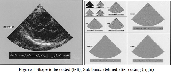
Figure 2 of digital X-ray facing the viewer has not suffered any visual loss with respect to the original image. The transformation of the image represents a correlation of the signal, a reduction in its dynamic range which removes redundant information. The transformed coefficients must be statistically independent, and the transformed image energy must be compacted into coefficients minimum number. [6]
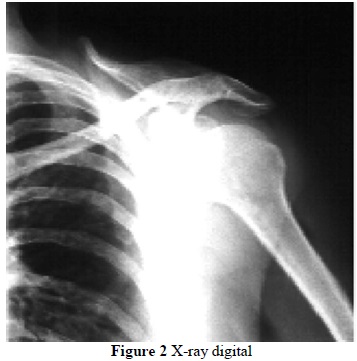
Figure 3 includes the Original image (a), the detail original region (b-e) Analysis of the region of interest (ROI) using different DCT methods. (d) The principal component of analysis (PCA) (e) Vector Quantization (f) Estimated Coding. Figure 3 presents transform encoders where the distribution of values representing the intensity level is observed where many of them can be eliminated or at least quantified with very few bits.
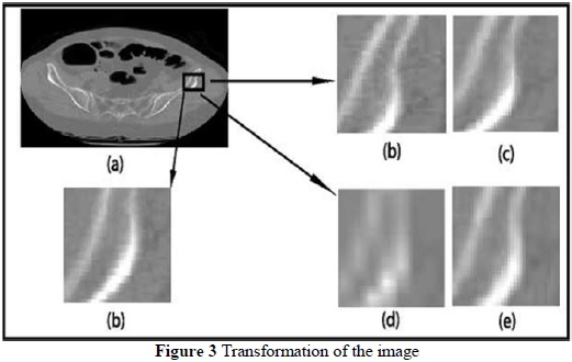
Thus, a decrease of the statistical dependence of pixels before moving to the quantization stage is achieved. The transformation is not performed on the entire image, but the image is divided into blocks of size (typically 8 x 8 or 16 x 16 pixels). Afterward, the transformation is made reducing computers cost considerably.
The former images feature excellent resolution with very fine details of the patient's anatomy. The latter are characterized by providing information on the functioning of the different organs or systems.
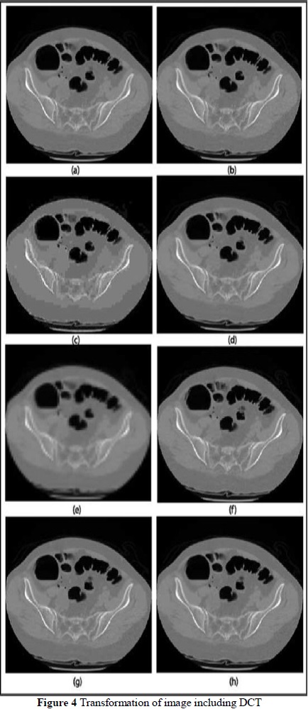
Figure 4 exhibits the original image (a), the DCT with 128 size quantization (b), the DCT step with 1024 size quantization (c), the PCA (d), the quantization vector with 7 x 7 blocks (e), the estimated coding (f), the ROI with 8 x 8 blocks (g), the ROI with 16 x 16 blocks (h). [7]
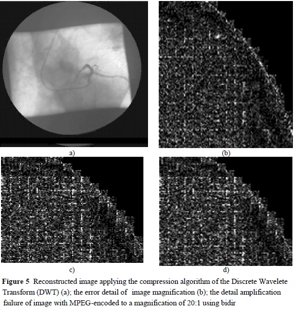
The compression process of Figure 5 was carried out with the discrete compression irreversible transform. The figure includes the reconstructed image applying the compression algorithm of the Discrete Wavelete Transform (DWT) (a); the error detail of image magnification (b); the detail amplification failure of image with MPEG-encoded to a magnification of 20:1 using bidirectional prediction (c); and the detail of amplification error with MPEG-encoded image for an enlargement of 20:1 (d). The result of the prediction is identical to the amplification processed for Figure 5 (b), (c) and (d). The image was performed irreversible to compressive at an enlargement scale of 20: 1, to compare the approach developed on the basis of the Discrete Wavelet Transform (DWT) block. [4]. For the MPEG (Moving Picture Experts Group) standard video compression method based on Discrete Cosine Transform (DTC) block and currently used for many medical applications.
3. Digital Cosine Transform (DCT)
There are many mathematical transformations that convert a data set of a measurement system into another. [8]. Choosing the Digital Cosine Transform (DCT), where the representation of data in the system (DCT), has properties that facilitate the compression. The Discrete Cosine Transform (DCT) is one of the most significant transformations in the area of digital image compression because it transforms a block of data into a new set of values.
Moreover the inverse DCT reverses this process, recovering the original data values. In theory, during the process of transformation and anti-transformation no information is lost, however, in practice, there is a little loss of information due to the following two factors:
-
Cosine values can not be accurately estimated.
-
The final results of calculations are affected by the rounding off errors due to the limited accuracy of the data representation.
Differences between recovered data and original data are nil, but they depend on the method used to calculate the DCT. The DCT can be applied to blocks of data of any size, but the most common practice is to use 8 x 8 blocks. This is because 8 x 8 blocks does not demand large memory requisite. Additionally, the algorithmic complexity of such blocks is compatible on most platforms. On the other hand, from the point of view of the compression factor, if we use large blocks we will not get large enhancements.
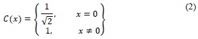
The definition of DCT is given by the equation 1
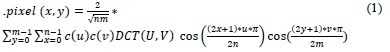
Where for both cases, u=0,.. ..,n and v=0,......, m.
DCT can be implemented according to its definition, but there are faster incremental methods, where the method starts from some pixels to be increased until all the input space is covered. If any item is not covered by the characteristics of the existing pixels, one more would be added. The advantage of this approach is high speed.
It may not be intuitive for its definition, but each calculated value in the transformation process involves a transformation matrix (base function) different from n x m components. For the most common case of 8 x 8 blocks, there are 64 transformation matrices, each of 8 x 8.
4. Modes of image compression
At present, there is a vast amount of image formats. Each one has its respective strong and weak points depending on the application for which they were designed such as quality, compression ratio, efficiency, costs, patents, licenses, standards, among others.
In mid-1980, members of the International Telecommunication Union (ITU) and the International Standards Organization (ISO) started a partnership to conduct a standard for images compression in grayscale and color. This standard was known as Joint Photographic Experts Group Associates. This JPEG standard was called. [7]
The JPEG committee decided to develop another standard for image compression called JPEG2000. It was a response to the growing demands of multimedia, the internet and a great variety of digital imaging. However, in terms of the compression methodology, these two standards are very different while the original JPEG is supported on DCT, the JPEG2000 is grounded on the DWT. [8].
JPEG images can be of any resolution and color space, both with algorithms with/out losses. The JPEG standard is a general purpose item and it has many features and possibilities. For example, by adjusting the parameters, a decision can be made to compromise between the image quality and its compression factor. The compression ratio is very broad: from about 100: 1 with significant visual degradation to about 3: 1 indistinguishable from the original image. The compression factor threshold for perceiving differences between the original image and the reconstructed image is within the range from 10: 1 to 20: 1 depending on the original image.
While the JPEG format was developed with the intention of compressing images, it has proven satisfactory for video compression. Especially when it comes to video in real time as it is more efficient computationally than other solutions, and as MPEG format, it is also useful for editing. However, the attained compression factor is not very high since it does not take advantage of the real redundancy between frames. Once the editing process has been completed, the video can be converted to a more appropriate format for a higher compression. [9]
The JPEG format defines four different operating modes: with no loss, sequential, progressive and hierarchical. The no-loss coding is based on a spatial algorithm on the pixels domain. A prediction of the value of a sample is carried out, considering up to three neighboring samples. Then, the prediction value is deducted from the actual value, and the difference is encoded with no loss using the Huffman coding or the arithmetic coding. The no-loss compression achieves a compression factor of approximately 2 to 1 as shown in equation 1.
The other three compression modes are based on DCT. The sequential scheme can use either Huffman encoding, the JPEG default option (Sequential baseline scheme) as arithmetic coding. When the image is reconstructed each row (8 x 8-pixel blocks), it is decoded from left to right, from top to the bottom, and it is sequentially presented with all its accuracy and resolution.
The progressive scheme presents the image in multiple steps. When the image is immediately decoded, an approximation of the complete picture is obtained. Quality is progressively improving until its complete accuracy is attained. This is ideal for the applications of searching into images databases or websites exploration. Spectral selection can be used, as well as successive approximation or both. The spectral selection encodes the low frequency coefficients of DCT at the beginning in order to quickly getting the image. [10] followed by the high frequency coefficients to add the details.
Successive approximation encodes the most significant bits of the DCT coefficients, followed by the least significant bits.
Finally, the hierarchical mode represents an image in multiple resolutions, for example, if there are three versions of an image: 512 x 521, 1024 x 1024 and 2048 x 2048, the images featuring the best resolution are coded as differences from the smaller image below, requiring fewer bits than it would need if individually stored.
Of course, the total number of bits is greater than that required to store the higher resolution image. Note that individual images in a hierarchical sequence can be gradually encoded. Figure 6 shows a schematic diagram of a sequential compressor.
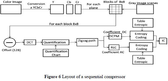
If the image to be compressed is a color image, the first step is to convert that color (YCbCr) image because the JPEG standard does not define a specific color space for the input image. Thus, it is possible to make some sub-sampling of Cb and Cr channels. The JPEG standard allows any of the sampling formats of YCbCr [11]. For the proper decompression, the header stores information of the vertical and horizontal sampling factor. Then each color space plane is discretized into 8 x 8-pixel blocks. In case of an image in grayscale, the image is discretized without any preprocessing.
The next step is to perform a shift of the values of components in each block since DCT produces better values for compression if original data are focused on zero (0). Since the original range is from 0 to 255, then by subtracting 128 the new range will be from 128 to 127.
Next, each block is subjected to DCT. Then each block is quantified considering a quantification table of the attained quality. Once quantified, the block is traversed in zigzag, to accumulate the maximum number of zeros at the end of the running. Afterward, the DC coefficients of blocks are encoded by DPCM and AC coefficients are coded by RLC. Right after this first encoding, each set is independently coded for the second time. In this case, encoding is performed by an entropy coder of the Huffman coding type, as it is the only possibility in the baseline sequential scheme to perform such a process. Once these processes are completed all the compressed information will be available, and ready to be stored or transmitted.
To decompress the image, the whole process is simply reversed. Entropy decoders, RLC decoder, and a DPCM decoder are used. Blocks are reconstructed, un-quantified, and anti-transformed and finally the recovered image is obtained.
5. Application of radiology services and diagnostic imaging
One of the applications for the results attained of compressed images is the decreasing of transmission times according to the available channel capacity. Without compressing images, the time ranges from 630 milliseconds to 113 seconds. It is important to point out that in some places the switched network connection is still used.
In the process of diagnostic of medical imaging the professional observes a set of images to grant an assertive diagnosis. It is a mistake to perform these studies or consultations with just one image; therefore, some studies consist of a vast number of images that give the advantage to specialists.
The objective is to minimize the format, which is the study performed for diagnostic and remote consultation. A large number of images produced for diagnosis has made its handling be complicated, especially when they should be printed and filed. An alternative is the efficient management of digital images through networked devices that offer a range of services that support the operation of an area of Radiology; [12]. Also, the current technology offers additional features such as: displaying of multiple images on one screen, image processing to correct or improve them, voice recording for the computer-aided diagnosis, among others.
6. DX Diagnosis support
There is a summary of the behavior of production in diagnostic support services tracers of the 22 State Social Enterprises (ESE) assigned to the District Department of Health (SDS) between 2011 and 2014. It is presented, according to information reported by them in compliance with Decree 2193 of 2004. The data are described considering the affiliation type of the cared for the population to the General System of Social Security in Health (SGSSS), levels of care and total general of the public network.
It is remarkable to highlight that the information corresponding to the Non-POSS activities is reported by hospitals in an aggregate form to the Production of Poor
Uninsured Population (PPNA). The Financial District Health Fund (FFDs) economically supports this activity. Fundamental issues include the analysis of the production of general and specific services of the following activities: outpatient, emergencies and hospitalization for radiology and diagnostic images.
Dx diagnostic supports services that are offered in the 22 state owned social enterprises of the SDS network, specifically nuclear medicine, polysomnography, audiology tests and radiology and diagnostic images. During 2011, 952.938 activities in all regimes of affiliation were observed, from them, 373.645 were related to outpatients; 367.033 to emergency activities and 212.260 for hospitalization activities. This represents about 39.21%, 38.52% and 22.27% respectively (Tabla 1).
In 2012, the production in all affiliate schemes was seen for diagnostic laparoscopy support, nuclear medicine, radiology and polysomnography and diagnostic images including abdominal ultrasound, gynecological ultrasound, obstetrical ultrasound, electroencephalography, electromyography, mammography, fetal monitoring, other ultrasound, special x-rays and x-rays simple. As a result of that, 879.525 of total activities were registered, from which 330.769 were related to outpatients, 339.877 to emergency issues, and 209.279 regarding hospitalization activities. This corresponds to 37.59%, 38.63%, and 23.78% respectively.
Likewise, the production in all affiliate schemes for diagnostic radiology and diagnostic imaging support (abdominal ultrasound, gynecological ultrasound, obstetrical ultrasound, electroencephalography, electromyography, mammography, fetal monitoring, other ultrasound, X-ray special and X-ray simple were observed in 2013. A total of 898.387 were registered. From these 339.684 activities were related to outpatients, 321.488 to emergency activities and 23.215 to hospitalization activities. Finally, the registers of 2014 were analyzed. For a total of 951.126 activities, 391.876 were related to outpatients, 315.106 to emergency activities and 244.140 to hospitalization activities (Table 2).
7. Conclusions
The decision making process related to relocation, optimization, enlargement or reduction of physical infrastructure, human talent and the existing biomedical technology, to achieve the maximum possible efficiency in the use of available resources use, allowing to define the lines of investment of public resources in physical infrastructure and equipment, aimed at improving quality of the supply of health services and technically assess the needs of health institutions was analyzed.
Moreover, the process of image compression for the Discrete Cosine Transform (DCT) provides the information required to guide the application of technical criteria. Meanwhile, this enhances the definition of scenarios allowing to model the behavior of supply and demand, according to the redistribution of services and resources among the different institutions making up each network.
The behavior of reconstructed images of the compression system both objectively and subjectively was observed, to get bigger rates than those supported by the uncompressed methods. This is due to the features of results obtained through the discrete cosine transform (DCT) which generates coefficients correlated to concentrate the important image information in some coefficients. This advantage permitted to increase the elimination threshold, and the compression ratio started to be varied.
References
[1] Programa Regional de Políticas Sociales en América Latina (SOPLA), «Aplicaciones automatizadas en tiempo real basada en componentes software,» 2012. [ Links ]
[2] Economical Commission for latin America (CEPAL), «Equipping and technology,» CEPAL, 2012. [ Links ]
[3] J. K. H. Tan, Healt management Information Systems: Methods and practical applications, Aspen Publication, 2001. [ Links ]
[4] L. Alfaro Ferreres, M. García Rojo y A. Puras Gil, Manual de telpatología, Pamplona: Club de informatica aplicada de la sociedad española de anatomí patológica, 2001. [ Links ]
[5] M. A. Martínez, A. J. R. Jiménez, B. V. Medina y L. J. Azpiroz, «Los sistemas PACS,» Manual Matrox Corona II. Universidad Autónoma Metropolitana Iztapalapa, Iztapalapa, 2003. [ Links ]
[6] M. Martín- Fernández, «Image Compression,» Laboratory of Mathematics in imaging, 2003. [ Links ]
[7] S. B. Gokturki, C. Tomasi, B. Girod y C. beaulieu, «Medical Image compression based on region of interest (ROI) with application to colon ct images,» Radiology departament. Stanford University, 2004. [ Links ]
[8] J. D. Bronzino y D. R. Peterson, The Biomedical Engineering Handbook. Fourth Edition, CRC Press, 2000. [ Links ]
[9] R. C. González y R. Woods, Digital Image Processing. Second Edition, Pearson Education, 2003. [ Links ]
[10] W. Kou, Digital Image Compression: Algorithms and standards, Springer Science, 1995. [ Links ]
[11] Unión internacional de telecomunicación UIT, «Digital compression and coding of continuous-tone still images. T.84,» UTI, 1997. [ Links ]
[12] G. E. 0ien, «L2-optimal Attractor Image Coding with Fast Deoder Convergence,» PhD thesis, the norwegian institute of technology, Norway, 1993. [ Links ]
