


 English (pdf)
 Article in xml format
 Article references
 Send this article by e-mail
 Cited by SciELO
  Cited by Google
  Similars in
SciELO
Similars in
SciELO  Similars in Google
 Permalink
INTRODUCTION
In the Nariño Department, potato production ranks third in Colombia, and 24,906 ha were planted in 2019, with a yield average of approximately 578,695 t (FEDEPAPA, 2019). The many phytosanitary problems that affect potato cultivation include the black scurf and stem canker caused by the fungus Rhizoctonia solani Kühn (Tsror, 2010; ICA, 2011; Álvarez-Sánchez et al., 2018). Rhizoctonia solani causes damage in all stages of potato development crops, including emerging shoots, young neck tissues of the seedlings, roots and stolons, where necrotic brown lesions can strangle them. In adult plants, necrotic lesions interfere with the normal movement of nutrients, inducing the formation of aerial tubers in the axils of the leaves. Developing tubers remain small or deformed, and the surface of tubers have black crusts or sclerotia, detracting from quality (Tsror, 2010; ICA, 2011). Worldwide, this disease causes important decreases in yield and quality and is responsible for losses of up to US$75million/year in several countries (Das et al., 2014).
The fungus R. solani (anamorph of Thanatephorus cucumeris (Frank.) Donk) belongs to Class Basidiomycetes. The hyphae are dark brown, septate and multinucleated, where cells have 2-18 nuclei (Misawa and Kurose, 2018, 2019). The hyphae fusion, known as anastomosis, is a particular characteristic of this species (Carling, 1996). This condition has been considered for classification in anastomosis groups (AGs), which differ from each other in phylogenetic, morphology, physiology and pathogenicity (Fiers et al., 2011; Ferrucho et al., 2012; Muzhinji et al., 2015). To date, 13 AGs has been identified worldwide, and the more important are AG3 and AG4. AG3 is the most pathogenic and is characterized mainly by the formation of sclerotia on tubers (Ferrucho et al., 2012; Das et al., 2014; Muzhinji et al., 2015). In Venezuela, Ecuador and Colombia, AG3 has been determined as the most pathogenic in potato cultivars (Escalona et al., 2011; Alban, 2015; Ferrucho et al., 2012, 2013).
The study of population genetics of plant pathogens is useful for understanding epidemiology, ecology and evolutionary trajectory to effectively estimate the pathogen potential evolution and dispersion under natural ecosystems. This knowledge evaluates the sensitivity of pathogen isolates for agrochemicals and development of resistant cultivars (Zhan, 2016; Chañag et al., 2018). Because of the importance of R. solani in potato production in the Nariño Department, the objective of this research was to obtain a preliminary identification and characterization of R. solani populations in the region. The following studies expand knowledge of this pathogen and evaluate management strategies.
MATERIALS AND METHODS
Sampling and isolation of R. solani
The sample collection was done from September to November, 2019, on commercial potato farms in the municipalities of Pasto, Ipiales, Tuquerres and Ospina, south of Colombia (1º12’ N and 77º16 W to 0º49 N and 77º38’ W), at altitudes of 2,915 to 3,179 m a.s.l. The laboratory work was carried out in the phytopathology laboratory of the Faculty of Agricultural Sciences of the Universidad de Nariño (Pasto, Colombia), with an altitude of 2,527 m a.s.l. In each municipality, potato crops in the harvest stage were randomly selected, collecting 10 tubers with sclerotia/place. The sclerotia were removed using a scalpel, disinfested with sodium hypochlorite (3%), placed on potato dextrose agar (PDA) containing streptomycin sulphate (20 mg L-1), and incubated for 3 d at 25ºC, according Das et al.(2014) and Misawa et al. (2018). Rhizoctonia-like fungi colonies were identified according to Sneh et al. (1991), and the purification was done with the hyphal tipping technique on PDA.
Morphological characterization of isolates
The strains were grouped according cultural appearance, such as pigmentation, growth pattern (GP), and pattern of sclerotia formation (PSF), according Escalona et al. (2011) and Dubey et al. (2014). Of these groups, 30 strains were selected to continue the morphological study on PDA and incubation in dark for 15 d at 25°C. Four repetitions were made for each isolate, placing 3 mm diameter plugs taken from the colony margins in the center of the plates. Strains were monitored daily for the following characteristics, according Dubey et al. (2014): - Mycelial growth, measuring the colony diameter (mm/ day), mean of two measurements; pigmentation, with evaluation at 8 and 15 d, according to a color chart (Kramer, 2004); - mycelium texture: cottony (aerial mycelium), plush attached to the medium or grainy; - mycelial growth pattern (MGP) at 15 d, classified according to Dubey et al. (2014) as: central, simple radial, complex radial, dispersed and star-shaped; - Sclerotia characteristics: PSF, as peripheral, central, scattered, star-shaped or absent; size (using the software of imageJ program), categorized as microsclerotia (<1 mm) and macrosclerotia (>1 mm); shape (regular, irregular, globular and powdery); coloration and amount visually estimated. According to the sclerotia abundance, the isolates were classified as: 1-100 (scarce), 100-200 (moderate) and > 200 (abundant), as reported by Dubey et al. (2014).
The mycelial growth rate (MGR) was obtained in mm day-1 using the method of Dubey et al. (2014), and the data were subjected to ANAVA and Tukey's test (P = 0.05%). According to the MGR, the isolates were classified as: slow (10 mm day-1), medium (> 10-12 mm day-1) and fast (> 12 mm day-1). With qualitative variables (pigmentation, mycelium texture, MGP and PSF), the averages of the proportion (%) of isolates were obtained for each one.
Microscopic characteristics were made using method of Gondal et al. (2019); slides with mycelium printed with adhesive tape and stained with lactophenol blue (0.05%) were used to observe the hyphal morphology of R. solani, (four plates/repetition) in 8-d-old cultures. The morphology of each culture was compared with previous descriptions, including width and length of the hyphal (Misawa et al., 2018).Number of nuclei per cell were counted by staining the hyphaewith Safranin O + a drop of 3% KOH on a portion of mycelium 48 h after incubation (Bandoni, 1979). Five observations of each characteristic/repetition were examined microscopically at 100X. The data were subjected to ANOVA and Tukey’s test (P=0.05).
Pathogenicity tests
The inoculum was prepared by colonizing isolates of R. solani on rice grains for 18 d (Castro et al., 2013). Potato seedlings (S. tuberosum Group Phureja) sown in plastic cups (one tuber/cup) were used. The seed-tubers were checked to be free of sclerotia and disinfested in water with 1% sodium hypochlorite. Each cup contained 350 g of a soil-rice husk mixture (3:1), sterilized with hot water. Eighteen-day-old seedlings were inoculated with 6.0 g of rice colonized with each isolate; the inoculum was mixed in the upper 4 cm layer of the soil. Five plants were inoculated by strain, and a control free of pathogen was included. The plants were placed on tables under a mesh house, at 11-20oC. Thirty-five days after inoculation, plant infection was evaluated, observing secondary symptoms (wilting and yellowing): lesions on roots, shoots, stolons or stem canker (primary symptoms). The results were expressed as a percentage of plants infected by isolate. Re-isolations of the pathogen were done to confirm the pathogenic nature of the isolates.
RESULTS AND DISCUSSION
Sampling and isolation of R. solani
The pathogen was 100% prevalent in all locations, where tubers with sclerotia were found, indicating the presence of this pathogen in the potato production area of the department. Out of the 50 samples, 17 were found in crops in Pasto, 12 in Ipiales, 9 in Tuquerres and 12 in Ospina. The commercial potato varieties with R. solani included the species S. tuberosum and S. tuberosum Group Phureja(Fig. 1). The sclerotia observed on the tubers had dark brown and black colors, with varied shape and size, from 1 to 4 mm in diameter, as reported by Misawa and Kurose (2018). From the sclerotia, 494 pure isolates were obtained with typical features of R. solani: hyphae with right-angle branches at the distal septae of cells, dolipore septum, and constriction at the branch, similar characteristics described by Sneh et al. (1991). No conidia or conidiophores were observed.
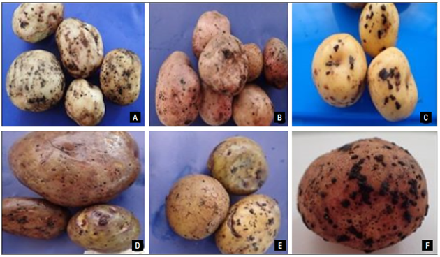
Figure 1. Potato tubers with R. solani sclerotia. A) Solanum tuberosum var. Unique, B) S. tuberosum var. Parda Suprema, C) S. tuberosum Group Phureja, D) S. tuberosum var. Diacol Capiro, E) S. tuberosum var. Superior and F) S. tuberosum var. Red Huila.
In this study, the pathogen sampling focused exclusively on the collection of tubers with sclerotia (Atkinson et al., 2010; Dubey et al., 2014; Muzhinji et al., 2015), or even from soil (Alban, 2015). The R. solani isolation from the sclerotia of commercial varieties of S. tuberosum, as reported by Escalona et al. (2011) for Venezuela, by Ferrucho et al. (2012) in Colombia, and, in Ecuador, by Alban (2015), obtained isolates from soil.
Morphological characterization of isolates
The 494 isolates showed diversity in pigmentation, GP and PSF at 15 d after incubation, as reported by Misawa and Kurose (2018). Predominantly white colonies and cream, light brown and salmon colors, with simple radial, complex radial and star-shapes were noticed. The sclerotia were arranged in the center, periphery, scattered or absent. These diversities coincide with several authors (Escalona et al., 2011; Oliveira et al., 2014; Abdel-Sattar et al., 2017), who suggested that such variability may be related to the culture medium, temperature, colony age, AG group or host plant species.
Based on the cultural features expressed by the R. solani isolates, 15 groups were classified, as shown in table 1. A variable number of isolates of each group were selected, with 30 isolates for the morphology and pathogenicity (Tab. 2).
Table 1. Isolates of R. solani grouped according cultural features in the PDA medium.
Group 1 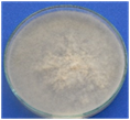 Plush and white mycelium attached to the medium. No sclerotia Plush and white mycelium attached to the medium. No sclerotia |
Group 2  Plush and white mycelium attached to the medium. Late and white sclerotia formation Plush and white mycelium attached to the medium. Late and white sclerotia formation |
Group 3  Plush and cream mycelium attached to the medium, with radial complex circles. Central and brown sclerotia Plush and cream mycelium attached to the medium, with radial complex circles. Central and brown sclerotia |
Group 4 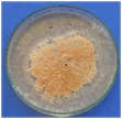 Plush and salmon mycelium, attached to the medium, with simple radial growth central and salmon sclerotia Plush and salmon mycelium, attached to the medium, with simple radial growth central and salmon sclerotia |
Group 5 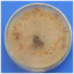 Abundant cottony mycelium, aerial, brown, with complex radial growth. Brown sclerotia formation on the lid Abundant cottony mycelium, aerial, brown, with complex radial growth. Brown sclerotia formation on the lid |
Group 6 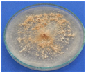 Pale salmon colony, mycelium attached to the medium. Scattered salmon sclerotia Pale salmon colony, mycelium attached to the medium. Scattered salmon sclerotia |
Group 7 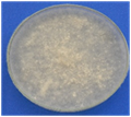 Pale cream colony. Abundant aerial mycelium. Scattered white sclerotia Pale cream colony. Abundant aerial mycelium. Scattered white sclerotia |
Group 8 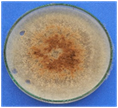 Mycelium and sclerotia brown, mostly central or scattered Mycelium and sclerotia brown, mostly central or scattered |
Group 9 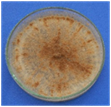 White colony. Brown sclerotia in radial growth, towards the edge White colony. Brown sclerotia in radial growth, towards the edge |
Group 10 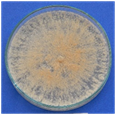 Pale salmon colony, star-shaped growth. Scattered and abundant salmon sclerotia Pale salmon colony, star-shaped growth. Scattered and abundant salmon sclerotia |
| Group 11 Dark salmon colony, sparse mycelium. Sclerotia scattered, dark brown or light brown |
Group 12 Pale cream colony, with radial growth and wavy edges. Cream sclerotia scattered with gum exudates |
Group 13 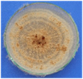 Cream and fluffy mycelium, with wavy edges. Beige, scattered and central sclerotia Cream and fluffy mycelium, with wavy edges. Beige, scattered and central sclerotia |
Group 14 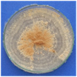 Sparse and cream mycelium, granular (pasty) texture and wavy edges. Central brown sclerotia Sparse and cream mycelium, granular (pasty) texture and wavy edges. Central brown sclerotia |
Group 15 Sparse dark cream and plush mycelium. Scattered brown sclerotia |
Table 2. Rizoctonia solani isolatesandcultural characteristics identified in this study.
| Isolate* | Location of collection | Potato variety | Group** | Mycelial growth rate (mm/day)*** | Nuclei number | Hyphal width-length (μm) |
|---|---|---|---|---|---|---|
| RsPN-400 | Pasto | S. tub. G. Phureja (Criolla amarilla) | 3 | 18.13±0.09 a | 7.3±0.3 a | 10.1±1.0 a 108.9±13.9 a |
| RsPN-232 | Pasto | S. tub. G.Phureja (Puerreña) | 13 | 16.87±0.16 abcde | 6.3±0.4 a | 8.5±0.7 a 99.9±13.6 a |
| RsPN-135 | Pasto | S. tuberosum (Parda Suprema) | 9 | 16.84±0.27 abcde | 5.7±0.3 a | 8.8±0.5 a 82.9±6.3 a |
| RsPN-183 | Pasto | S. tuberosum (Diacol Capiro) | 13 | 16.80±0.27 abcde | 5.3±0.6 a | 9.9±0.3 a 93.4±5.0 a |
| RsPN-109 | Pasto | S. tuberosum (Única) | 1 | 16.65±0.20 abcde | 6.7±0.8 a | 8.8±0.5 a 105.8±9.4 a |
| RsPN-195 | Pasto | S. tuberosum (Diacol Capiro) | 1 | 16.60±0.09abcde | 6.6±0.4 a | 9.5±0.9 a 90.4±12.0 a |
| RsPN-325 | Pasto | S. tuberosum (Superior) | 2 | 16.58±0.11 abcde | 5.8±0.1 a | 8.1±0.7 a 72.7±2.5 a |
| RsPN-158 | Pasto | S. tuberosum (Diacol Capiro) | 11 | 15.99±0.35 bcde | 6.5±0.4 a | 10.5±1.3 a 102.9±3.2 a |
| RsPN-175 | Pasto | S. tuberosum (Diacol Capiro) | 8 | 14.98±0.96 e | 6.4±0.5 a | 8.1±0.1 a 118.4±18.9 a |
| RsPN-24 | Ipiales | S. tuberosum (Diacol Capiro) | 5 | 16.94±0.22 abcde | 7.2±1.0 a | 7.9±0.2 a 74.1±1.7 a |
| RsPN-7 | Ipiales | S. tub. G. Phureja (SuaPa) | 11 | 16.88±0.13 abcde | 8.2±0.8 a | 8.3±0.6 a 94.3±8.9 a |
| RsPN-346 | Ipiales | S. tub. G Phureja (Ocarina) | 15 | 16.76±0.15 abcde | 6.3±0.6 a | 8.1±0.6 a 106.3±11.5 a |
| RsPN-345 | Ipiales | S. tuberosum (Diacol Capiro) | 15 | 16.53±0.42 abcde | 7.0±0.4 a | 8.2±0.2 a 96.4±1.4 a |
| RsPN-374 | Ipiales | S. tuberosum (Diacol Capiro) | 3 | 16.46±0.26 abcde | 8.0±1.3 a | 7.4±0.2 a 104.9±14.1 a |
| RsPN-3 | Ipiales | S. tuberosum (Superior) | 10 | 16.28±0.25abcde | 6.1±0.2 a | 11.2±0.8a 88.7±12.9 a |
| RsPN-36 | Tuquerres | S. tuberosum (Roja Huila) | 6 | 17.66±0.20 abc | 6.4±0.3 a | 7.4±0.1 a 114.2±12.1 a |
| RsPN-274 | Tuquerres | S. tuberosum (Diacol Capiro) | 6 | 18.09±0.16 a | 5.8±0.4 a | 7.8±0.4 a 75.0±3.7 a |
| RsPN-269 | Tuquerres | S. tuberosum (Diacol Capiro) | 8 | 17.07±0.15 abcd | 6.3±0.7 a | 9.2±0.8 a 92.6±6.3 a |
| RsPN-262 | Tuquerres | S. tuberosum (Diacol Capiro) | 5 | 16.98±0.11 abcd | 4.9±0.2 a | 7.9±0.3 a 101.6±2.3 a |
| RsPN-290 | Tuquerres | S. tuberosum (Diacol Capiro) | 4 | 16.94±0.02 abcde | 6.2±0.8 a | 12.9±4.9 a 91.0±19.6 a |
| RsPN-99 | Tuquerres | S. tuberosum (Única) | 12 | 15.79±0.23 cde | 5.9±0.3 a | 7.8±0.5 a 84.1±5.7 a |
| RsPN-289 | Tuquerres | S. tuberosum (Diacol Capiro) | 12 | 12.43±1.34f | 5.2±0.1 a | 8.7±0.5 a 99.2±5.2 a |
| RsPN-487 | Ospina | S. tuberosum (Única) | 4 | 17.85±0.09ab | 6.3±0.5 a | 8.6±0.1 a 95.9±5.0 a |
| RsPN-469 | Ospina | S. tuberosum (Diacol Capiro) | 9 | 16.87±0.14 abcde | 5.8±1.0 a | 8.0±0.6 a 102.5±6.8 a |
| RsPN-434 | Ospina | S. tuberosum (Diacol Capiro) | 14 | 17.31±0.14 abc | 7.3±0.3 a | 8.1±0.5 a 77.6±3.4 a |
| RsPN-484 | Ospina | S. tuberosum (Diacol Capiro) | 14 | 17.30±0.17abc | 5.8±0.5 a | 8.4±0.3 a 90.5±3.8 a |
| RsPN-432 | Ospina | S. tuberosum (Diacol Capiro) | 10 | 17.21±0.17abcd | 6.9±0.5 a | 8.6±0.3 a 86.0±4.3 a |
| RsPN-482 | Ospina | S. tuberosum (Diacol Capiro) | 2 | 16.45±0.14 abcde | 6.9±0.8 a | 6.6±0.1 a 103.2±3.5 a |
| RsPN-486 | Ospina | S. tuberosum (Única) | 7 | 16.34±0.09 abcde | 6.7±0.6 a | 10.0±0.8 a 89.2±2.4a |
| RsPN-442 | Ospina | S. tuberosum (Diacol Capiro) | 7 | 15.28±0.15de | 6.5±0.7 a | 8.4±0.8 a 106.5±8.1 a |
*Isolates encoded: R. solani (Rs), potato (P), Nariño (N).
** Groups according cultural preliminary features on PDA (related to Tab. 1).
Means are mean±standard error. Means followed by the same letter in a columns do not differ according to Tukey’s test (P≤0.05).
Strains pigmentation. During the first 2 d, the colonies formed hyaline mycelium that adhered to the medium, characteristic of R. solani populations according to Dubey et al. (2014). In the following days, the mycelium had different pigmentations: white, cream to pale beige. According to the color table (Kramer, 2004), at 8 d-old, pale brown predominated in 50% of the isolates, with cream in 30%, and the rest had the two colors. At 15 d, pale brown was the predominant color in 26% of the isolates, with cream in 10%, beige in 10% and brown in 6.6% of the isolates. The rest of colonies had similar colors, two were salmon. Several authors (Muzhinji et al., 2015; Abdel-Sattar et al., 2017) have reported that the pigmentation of R. solani colonies is a variable characteristic exhibited by isolates from all species of plants affected by this pathogen. It appears to be related to the culture medium, temperature, age, and possibly the genetics of the fungus. In potato isolates, Misawa and Kurose (2018) found white pale brown, slightly gray and brown colors; while Muzhinji et al. (2015) reported R. solani isolates in different AG groups, with white to brown mycelium. Oliveira et al. (2014) found colonies with colors from beige to brown. In the present study, this characteristic was probably associated with strain age in the PDA medium.
Strain texture. At 8 d-old, the plush mycelium adhered to the medium in 63.3% of isolates, as reported by Carling and Leiner (1990). Cottony and raised mycelium was observed in 25% of the isolates, and the rest had lower proportions of both types. Four isolates had granular texture. After 15 d, 94% of the isolates had plush mycelium adhered to the medium, and the rest of colonies exhibited cottony, sparse, aerial mycelium adhered to the lid of the Petri dish. This last characteristic was not highly variable although several authors have reported colonies with abundant aerial mycelium (Abdel-Sattar et al., 2017; Misawa and Kurose, 2019).
Strain growth pattern.Thevaried GP initially exhibitedcolonies, and it changed over time, as reported by Abdel-Sattar et al. (2017). A simple radial pattern had 30% of isolates at 8 d-old, followed by 13.5% with a complex radial pattern (several circles). The rest of the isolates had varied forms, such as scattered and star-shaped. At 15 d, 26.6% of the isolates had complex radial forms, followed by 23.3% with a simple radial, and the rest exhibited scattered and star forms (Fig. 2). This variability, as reported by Abdel-Sattar et al. (2017) and Misawa and Kurose (2019), coincided in some cases with the formation of simple and multiple concentric circles, changing over time.
Sclerotia characteristics. Sclerotia formation began at 6 d-old in 86% of the strains, similar to that described by Abdel-Sattar et al. (2017) and Gondal et al. (2019), with varying colorations between white, cream and brown, either rough or smooth. Fifteen days later, 35% of the isolates showed brown sclerotia, followed by beige (25%) and cream (15%). The rest were white, cream and orange. Three isolates did not form sclerotia (Fig. 2A). The 27 strains showed varied PSF; 50% were scattered, 40% were peripheral, and the rest were central, with a star shape, or combined both star/peripheral (Fig. 2A), similar to the results of Misawa and Kurose (2019). Forty-six percent of the strains were classified with a low sclerotia/plate (1-100), 42% were moderate (between > 100- 200), and 11.5% had abundant sclerotia (> 200) (Fig. 2B). The size and shape of the sclerotia was variable; macro (>1 mm) and microsclerotia (<1 mm), jointly in 90% of the isolates, with irregular or undefined shapes, and the rest presented microsclerotia with regular borders (Fig. 2C). These results coincide with those recorded by Dubey et al. (2014), who reported sclerotia diameters between 0.10-5.4 mm and with a peripheral, central and dispersed arrangement. Abdel-Sattar et al. (2017) reported the formation of brown sclerotia, 5.0 mm in diameter, while Escalona et al. (2011) found abundant dark brown sclerotia located only in the center of the colonies, similar to Misawa and Kurose (2019).
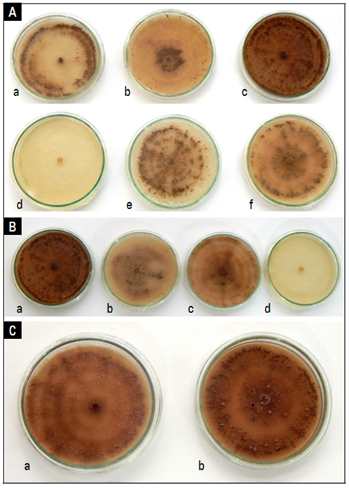
Figure 2. Mycelial growth pattern and sclerotia of R. solani on PDA. A) Arrangement, a: peripheral, b: central, c: scattered, d: absent, e: starry and f: starry/peripheral. B) Quantity, a: abundant, b: moderate, c: scarce, d: absent. C) Size, a: microsclerotia, b: macrosclerotia and microsclerotia.
Mycelial growth rate. This variable was evaluated during the first 6 d, when all isolates filled the plate. All strains had rapid growth, as observed by Dubey et al. (2014), with a MGR higher of 12 mm d-1. Although the ANOVA showed statistical differences (P<0.0001) for the rate of mycelial growth between the isolates, no relationship was observed with the 15 initially determined morphological groups, neither with the geographic location of the cultures nor with the potato variety. According to Tukey’s test (P=0.05), only one isolate (RsPN-289) had the lowest MGR with 12.43 mm d-1, and the maximum was 18.13 mm d-1 (RsPN-400) (Tab. 2). Escalona et al. (2011) and Dubey et al. (2014) indicated that the MGR of R. solani depends on the culture medium and the incubation temperature. At 10ºC, the full plate can occur between 7-11 d, while, at 20ºC, it occurs in 4 d. At 30ºC, it occurs in 16 d. In our study, the full of plate was in 6 d at 25ºC. However, the MGR may be related to AG groups or pathogenicity level, aspects that will be addressed to the next phase of the research.
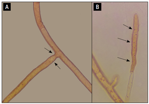
Microscopic characterization. All isolates showed the typical characteristics of R. solani: hyphae with right-angle branches, branches at the distal septae of cells, dolipore septa, constriction at the junction of these hyphae, and multinucleate cells (Fig. 3 A and B), as described by Dubey et al. (2014) and Abdel-Sattar et al. (2017). No statistical differences were found in hyphae dimensions; the width ranged between 6.6 and 12.9 μm (average 8.7 μm), and the hyphal distance between two septae varied from 72.7 to 118.4 μm (average 95.0 μm) (Tab. 2). These values are similar to those reported by Dubey et al. (2014) ‒4.1-10.3 μm‒, who found some isolates with values of > 8 in the groups AG1 to AG5. No differences were found in nuclei number, with a minimum of 4.9 and maximum of 8.2, within the ranges reported by Tsror (2010) and Misawa and Kurose (2019) with (4.4-10.9 nuclei).
Pathogenicity tests
Thirty-five days after inoculation, some plants showed secondary symptoms, such as yellowing, wilting and reduction of plant development, as compared to the control (Fig 4A). Reduction of root area, necrotic lesions in root tips and sections, in stolons and in some cases in shoot tissues were observed (Fig. 4 B and C). Out of 30 isolates, 24 caused root lesions in 100% of the plants, as well as reduction of more than 50% of roots, as compared with the control, coinciding with symptoms mentioned by several authors (Abdel-Sattar et al., 2017). These 24 isolates produced some small sclerotia on developing tubers (12.5-100% of plants). Six isolates caused both secondary and primary symptoms (50- 87.5% of plants). No symptoms of stem canker were observed. External symptoms, mainly plant reduction of development, were related with primary symptoms at the variable level. Colonies of the pathogen were recovered from root lesions. The results showed the susceptibility of the potato variety S. tuberosum Group Phurejain this first stage of plant development. According to Dubey et al. (2014), R. solani is more aggressive in young plants because, in older plants, tissues resist attack. However, new stolons and roots would be re-infected as well as the development of sclerotia in tubers, as observed in the potato crops.
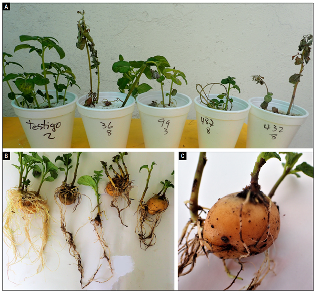
Figure 4. Symptoms caused by different strains of R. solani in potato seedlings (S. tuberosum Group Phureja). A-B) Secondary symptoms and reduction of roots, as compared to the control (far left), and C) Necrotic lesions on roots.
On the other hand, several authors (Dubey et al., 2014; Alban, 2015; Abdel-Sattar et al., 2017) suggested differences in pathogenicity or no infection with different isolates within a population, which is apparently related to the state of the potato crops the isolates come from. Genetic aspects, differences in virulence, and AGs present in populations under natural conditions are also factors mentioned by several authors (Carling, 1996; Das et al., 2014), including differences in pathogenicity level related with AGs on different organs of the plant (roots, stems and tubers). This may explain the lower infection observed with six isolates in this research. It is important to mention the efficacy of the R. solani inoculation method, using an enriched natural substrate to multiply this type of soil pathogens, such as parboiled rice (Castro et al., 2013). Several authors (Castro and Rivillas, 2012; Abdel-Sattar et al., 2017; Misawa and Kurose, 2018) suggested other substrates such as wheat, sorghum, carrot, or stems of Typha latifolia, since long-term preservation of the inoculum from mycelium in culture medium would affect fungus infectivity.
Finally, the evaluation of symptoms is a topic for future research because of the ability of R. solani to cause multi-symptoms. Preliminary pathogenicity verification of isolates was one of the objectives of the present research. However, studies on genetic diversity and AG groups suggest a greater demand in the inoculation standardization, evaluation methods, and experiment design to determine the incidence and severity of isolates on different stages of plant development (Fiers et al., 2011). This is also true in the case of evaluations under controlled conditions throughout the crop cycle to find the presence of symptoms and signs (mycelium and sclerotia) in roots, stems and tubers, as suggested by Alban (2015) and Misawa and Kurose (2018). This will allow greater precision in results that determine the presence of pathogenic populations under field conditions.
CONCLUSION
The presence of R. solani was verified in all potato producing areas in the Department of Nariño.
Rhizoctonia solani could be isolated from different potato varieties of S. tuberosum and S. tuberosum Group Phureja.
The R. solani isolates showed morphological variability in terms of color, texture and colony growth pattern.
Variability was also noticed in the production and characteristics of the sclerotia and mycelial growth rate.
For the variability of R. solani, all isolates were pathogenic, indicating their ability to cause damage in crops.

