


 Inglés (pdf)
 Articulo en XML
 Referencias del artículo
 Enviar articulo por email
 Citado por SciELO
  Citado por Google
  Similares en
SciELO
Similares en
SciELO  Similares en Google
 Permalink
Introduction
Anomia, nominal aphasia, or amnesic aphasia, has been described in the classic aphasia syndromes as an alteration in the production of words, this deficit is more prominent to nouns than for verbs, characterized by a preservation of understanding and repetition (Webb, 2017; Caplan, 2003; Fernández-Blázquez et. al., 2012). During naming process difficulties are in lexical access and lexical retrieval of word (Fernández-Turrado et al., 2006). The neurologic conditions causing aphasia and anomia are: stroke, dementia, epilepsy and traumatic brain injury (TBI) (Fridriksson, 2011; Laganaro, Di Pietro, & Schnider, 2006; Hoffmann & Chen, 2013; Lorenz and Ziegler, 2009). Additionally, anomia is one of the most common symptoms of patients with post-stroke aphasia (Van Hees, McMahon, Angwin, de Zubicaray, & Copland, 2014; Fridriksson et al., 2007). The neuroanatomical correlates are inferior parietal lobe, plus connections between parietal and temporal lobes (Caplan, 2003).
It is estimated that one million people in the USA has an aphasia (The National Institute on Deafness and Other Communication Disorders -NIDCD-, 2015), and the prevalence of these is between 20% and 40% after a cerebrovascular disease around the world (Martínez, Reyes-Saborit, Turtós-Carbonell and Dusu -Contreras, 2014). One of the most common characteristics is the difficulty to find words, this is present in approximately 70% of aphasias, it even manages to configure an independent disorder (anomic aphasia).
Dell, Chang and Griffin (1999) propose that the Parallel Distributed Processing models (PDP) are the best models to explain production of words and they integrate linguistic and no-linguistic features. In this models, structure and order are entwined in the sequential schemata that develop from the superimposed weight changes associated with the training set.
Structural Priming Model and Phonological Error Model are both PDP Models. The first is about the grammatical encoding feature while the second is about the phonological component (See Figure 1).
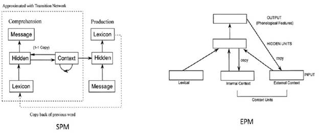
Figure 1 Structural Priming Model (SPM) and Phonological Error Model (PEM) (each rectangle indicates a group of units). Source: Dell et al. 1999.
In its treatment, there have been evidence of changes in the neural activity in both hemispheres (Fridriksson et al., 2007). Fridriksson (2011) suggests that rehabilitation depends of the anomia subtype and the best results are seen in disorders adjusted to the specific language of each individual therapy.
Treatment for anomia is an objective of the therapies of language and cognitive disorders. The more common difficulties in this kind of patients have trouble retrieving names of places, people, monuments, this impairment has an impact in everyday life such as in social, working and other contexts.
Knowing more precisely the neurological components associated with anomia has two important implications, the first is to establish if the brain areas are more related to the phonological component (PEM) or the grammatical component (SPM); the second one would be to know areas or related brain networks that would allow to give indications for possible intervention strategies, both pharmacological and cognitive. This led to ask, what are the neuroanatomical correlates of anomia?
Metodology
Design
This study is developed through the methodology of scoping review (Levac, Colquhoun, & O'Brien, 2010; Pham et al., 2014). This review synthetized the literature on a particular topic and provide evidence on that field.
Selection criteria
The papers should be in English or Spanish, should be published between the years 2009-2016; and as the main criterion, the paper should establish neurological findings associated with anomia. Those items showing evaluation results of other language disorders, such as dyslexia or other aphasia were excluded.
The data bases used were: EBSCOhost and Science Direct, using the keyword for the search terms "anomia" and "neurological".
The procedure was to search items in the database containing the previously named certain keywords, for which the criteria of inclusion and exclusion are considered. Was important that the articles were about research, not reviews or considerations about the topic.
Analysis plan
We considered the next components for the article analysis:
- Author(s), year of publication and study location
- Sample size
- The analysis focus on the anatomic structure and cognitive functionality in accordance with the results of the studies reviewed.
Ethical Standards
The authors assert that all procedures contributing to this work comply with the ethical standards of the relevant national and institutional committees on human experimentation and with the Helsinki Declaration of 1 975, as revised in 2008.
Results
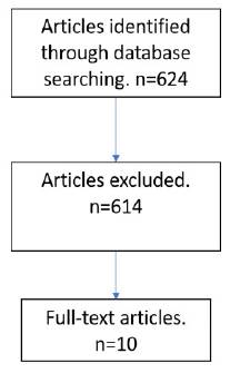
After the bibliographic search, only 10 articles remained (See Figure 2).
When we reviewed all the articles, we found that many of them appeared based on the fact that the topic is common in journals and texts of neurology and neuropsycho-logy, without an analysis of brain activity, focusing on the psychological characteristic and on the results of applications of neuropsychological tests and their relationship with other psychological or linguistic features. Of these few found texts, almost all are methodologically focused on magnetic resonance studies (Magnetic resonance imaging -MRI- and functional MRI -fMRI-) and only one of Evoked Related Potential (ERP) (See Table 1).
About the last one, Neumann, Obler, Gomez and Schafer (2009) studied the brain activity to a phonological response. Thirty-two people divided into two age groups (young and old) were evaluated with a naming exercise and response times to different stimuli were obtained. They found that there are no substantial differences between the two groups, people got less time between 300ms and 450ms, seniors got time between 351ms and 500ms. The role of the phonological component is discussed in the response naming.
The second group of articles found were about magnetic resonance imaging (MRI). In 2012, Kraft et. al. defined the anatomic correlates of neuropsychological dysfunctions using mapping symptoms and voxel-based lesion in 61 patients with cerebral stroke; patients underwent a neuro-ophthalmological, neurological and neuropsy-chological evaluation test and a brain MRI. They highlight the importance of additional neuropsychological deficits to visual present in lesions of the aforementioned areas, also showing that anomia and some memory impairments are related to lesions in the lower occipital rotation in the lingual gyrus in hippocampus and injuries of some major white matter tracts.
In 2014 were published two papers, in the first one, Van Hees et al. (2014) established the relationship of anomia with neural activation in the context of a series of interventions to improve such neurolinguistic condition. Evaluated eight people diagnosed with speech pathology after stroke (Cerebrovascular Accident -CVA-); assessed language with Boston-Naming-Test (BNT) and battery name based on international project database naming photographs and for neuronal activation images were used a MRI. For the study, had fourteen healthy control patients with similar demographic characteristics. Before the intervention found that areas related to anomia were the superior frontal gyrus, the angular gyrus and the superior temporal gyrus and a half I left mainly
They highlight the importance of additional neuropsycholo-gical deficits to visual present in lesions of the aforementioned areas, also showing that anomia and some memory impairments are related to lesions in the lower occipital rotation in the lingual gyrus in hippocampus and injuries of some major white matter tracts.
They found activation in the left superior temporal gyrus before the intervention; after the intervention activity was seen in the upper, middle and inferior temporal gyrus, plus activations were found in the fronto-medial turn and right turn supramarginal.
hemisphere. While intervention shows changes in brain activation for the eight different patients, it was found that an improvement in the treatment is generally associated with an activation on the left supramarginal gyrus and the right precuneus.
In the second one, Toumiranta et al. (2014) conducted a study with patients with extensive brain damage in the left temporal lobe and phonological impairment, in order to test the claim that the ability to learn and use new words depends on the left temporal lobe. Patients and controls results were compared. In MRI was observed in the left hemisphere a complete shutdown of the white matter of the arcuate fasciculus, which was confirmed by tractography.
The remaining articles were about functional magnetic resonance imaging (fMRI), that uses a technology based on oxygen rich-blood. This articles were clasified in two, treatment and no treatment articles.
In the first group found studies about brain activity after therapy. Dressel et al. (2010) examined changes in brain activity associated with a treatment for anomia. With a unique case of a patient with semantic dementia they used different neuropsychologi-cal and neurolinguistic evidence, including the Naming Test and the BNT, in addition to conducting fMRI. They found activation in the left superior temporal gyrus before the intervention; after the intervention activity was seen in the upper, middle and inferior temporal gyrus, plus activations were found in the fronto-medial turn and right turn supramarginal. In the left hemisphery the middle frontal and cingulate gyrus were activated, also the putamen, caudate and thalamus. However, the authors emphasize that right temporal region, the superior temporal and inferior gyrus were more representative.
In a similar study, Rochon et al. (2010) studied the characteristics of neural processing skills associated with access to the words after treatment for anomia. The research was made with two patients and ten controls were applied to different neu-ropsychological instruments, including the Boston-Naming-Test and the Philadel-phia-Naming-Test. After the speech, the inferior frontal and middle temporal gyrus in the left hemisphere and the left cuneus the right precuneus and supramarginal turn in both hemispheres were activated. The authors discuss about the importance of phonological route in the recovery of anomia.
Fridriksson, Richardson, Fillmore and Cai (2012) studied the treatment of anomia and the left hemisphere in 2010. In 2012 added more patients to the sample and improved methods of study. The study aimed to characterize precisely the changes in the left hemisphere taking into account a larger patient sample; plus: 1. Compare the role of perilesional cortex, and 2. Understand of how brain circulation and brain activation evaluated before starting treatment is associated with improvement in the naming after treatment. Thirty patients were administered the Western Aphasia Battery (WAB). Patients received treatment of anomia for three hours a day, half received phonological treatment using keys in the first week and then treated using semantic keys. Twenty-nine patients underwent fMRI and participated in naming tasks. Subsequently they conducted a multivariate statistical analysis and concluded that treatment increases brain activation in areas surrounding the injured cortical area while improving the naming, through modulation of the left frontal lobe, independent of the type of aphasia.
In the second group we found two studies, Bonelli et al (2011) researched the correlation of hippocampal activation with visual confrontation naming in patients with temporal lobe epilepsy; made a retrospective investigation of the relationship between the name and integrity of the networks of language using fMRI with simple tasks of verbal fluency. Included 22 healthy subjects and 66 patients with temporal lobe epilepsy candidates for surgery, all with preoperative evaluation and pharmacological treatment; the test were McKenna naming, verbal fluency test, and underwent structural and fMRI. They demonstrated the importance of the hippocampus in the denomination key to patients and controls.
In 2009, Mesulam et al. studied the brain areas related to anomia. They studied seven patients (four women) with a part of the Peabody Picture Vocabulary Test (PPVT-IV) and functional MRI. Founded an atrophy in the left inferior temporal orders, upper half, the fusiform gyrus and the anterior parahippocampal gyrus.
Finally, Hoffmann and Chen (2013) studied the spectrum of aphasia, subtypes and etiology after subacute stroke. Of the 625 patients who met the selection criteria, 165 patients had anomic aphasia, this corresponds to 26.40%, making it the second most common after Broca's aphasia (27.20%). They found that anomia occurred most commonly in small vessel disease (subcortical level), specifically in the thalamus, corona radiata and the globus pallidus. They concluded that aphasia subtypes are heterogeneous in etiology and could be useful to adopt a new taxonomy of aphasia based on independent component analysis, which may be based on studies with fMRI.
Related to the time when the damage that generates anomia occurred, an activation of temporary gyrus and frontal gyrus in the left hemisphere was found. This evidence suggests that anomia is not dependent on specific structures, since these areas are also related to memory tasks.
The main findings were that prior language intervention the brain areas implicated are the temporal and frontal gyrus of the left hemisphere. After intervention, the activation is in the temporal gyrus, precuneus and the supramarginal gyrus of the right hemisphere (See Figure 3).

Discussion
According to the findings published in recent years, there is evidence for differential naming tasks: before and after non-pharmacological intervention.
Related to the time when the damage that generates anomia occurred, an activation of temporary gyrus and frontal gyrus in the left hemisphere was found. This evidence suggests that anomia is not dependent on specific structures, since these areas are also related to memory tasks. These findings are like those reported in the literature, but expand understanding of related areas since reportedly are the areas 39 and 40 of Brodmann those involved in the process of anomia (Montañés & de Brigard, 2001).
These findings show the importance of the connections in the left hemisphere in temporal and frontal areas in anomia.
It is also evidence related to the neurological processes in naming tasks after a non-pharmacological intervention. Then it found that after surgery the temporal gyrus, that the right precuneus, cuneus and supramarginal gyrus are active. This would show that connections between areas that link the sensory, memory and executive functions of the right hemisphere occur in the naming recovery.
This evidence implies that naming is a complex process that involves primary, secondary structures and its connections in a multimodal manner. This categorization shows the similarity in recognition and motor interaction. Probably in language development in childhood same areas are activated due to sensory multimodality, then other areas leading to the consolidation and specialization of the areas in the left hemisphere. This evidence supports the PDP models (Dell et al., 1999). This would be an indication of processes compensation, restitution and replacement of the right hemisphere when intervention.
Such evidence would give us two lines:
First, the naming and its alteration could be a linguistic marker for the presence of Alzheimer's Disease. In turn, detection of abnormalities in these brain areas could help the remediation of functional alterations in Alzheimer's Disease (Cuetos-Vega, Arango-Lasprilla, Uribe-Pérez, Valencia-Marín, & Lopera-Restrepo, 2007).
Second, pharmacological and non-pharmacological stimulation centered interventions in temporal gyrus, precuneus, cuneus and supramarginal gyrus. This could end up slowing the progression of impairment associated with Alzheimer's disease. Other dementias such as fronto-temporal could be affected positively to the patient, interventions in this regard.
Limitations
The information about the anomia is subject to the presence of aphasia, and in some studies it is not specified about the characteristics of the aphasias, much less the anomia. Another problem lies in the methods used, since in some studies the MRI is used and in others the fMRI, we suggest that in future studies more robust methods such as fMRI should be considered. Finally, there are several strategies for measuring the denomination and the lexicon, such as the BNT or the PPVT, this could cause problems in the conclusions of the different anomia studies.
The number of articles included in the study draws attention, this shows a low production in these issues in recent years, possibly because the rise of research in this subject in previous years.

