


 English (pdf)
 Article in xml format
 Article references
 Send this article by e-mail
 Cited by SciELO
  Cited by Google
  Similars in
SciELO
Similars in
SciELO  Similars in Google
 Permalink
INTRODUCTION
Extranodal natural killer/T-cell lymphoma, nasal type (NKTL), was the term endorsed by the Revised European American Lymphoma Classification (REAL) in 2008 1 to refer to what was previously known as malign granuloma or polymorphic malignant reticulosis. This lymphoma is one of the most lethal mid-line granulomas, which are characterized for the extensive destructive lesions in the mucosa of the superior airway and middle facial third 1,2.
It is a rare disease that accounts for 7-10% of non-Hodgkin lymphomas in Asia and Latin America 3 and affects mostly adult men aged between 30 to 40 years. Most of the patients affected by this disease present symptoms for six months prior to the first consultation 2-4, with 70% to 90% of cases with a recent or latent infection with Epstein - Barr virus (EBV) 4. The survival rate ranges between 35% and 85% depending on the severity of symptoms and the local extension. Different case series show a high local recurrence rate of 50% 5 and metastasis in only 13% of patients in skin, lungs and gastrointestinal tract, which represents the order of frequency and worst prognosis 6,7. A correct diagnosis is important to define the type of treatment, which may include surgical procedures, chemotherapy or radiotherapy 8.
CASE PRESENTATION
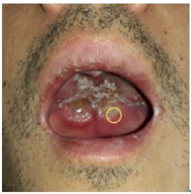
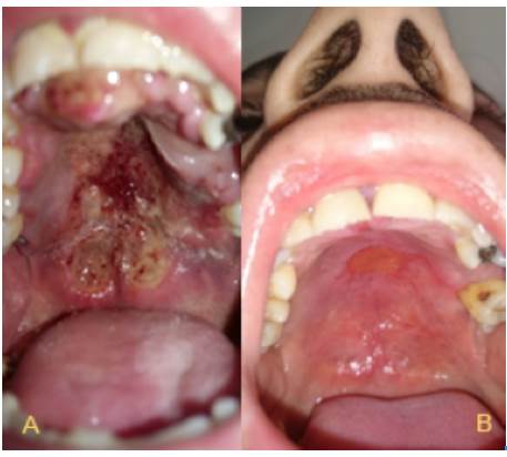
This case presents a 31-year old mestizo male patient, born in Fusagasugá, Colombia, with a history of inhaled cocaine and tobacco abuse. He works as a painter, with constant exposure to inhaled chemicals. The month prior to consultation, he was treated for acute sinusitis with amoxicillin for 10 days due to bilateral nasal obstruction and occasional epistaxis. Three weeks before consultation, he noted a painful ulcer in the hard palate, although, no systemic symptoms were reported. On general examination he was cachectic but stable, and presented with hyponasal speech. Rhinoscopy showed a bilateral mass in the nasal cavity with septal perforation in Cottle zones 2 - 3. Hard and soft palate showed a necrotic lesion with irregular borders, mucosa swelling without active bleeding. He had no palpable adenopathy on head and neck (Figure 1).
Source: Own elaboration based on the data obtained in the study.
Figure 1 Ulcerated lesion in hard palate with necrotic borders. Circle: Delimitation of the site of the biopsy.
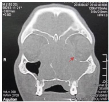
Blood tests were negative for human immunodeficiency virus (HIV), venereal disease research laboratory (VDRL) and hepatitis B and C, and C-reactive protein test (CRP) was less than 10 mg/L. Computed tomography scan in face and neck showed a mass occupying the nasal cavity, extended to the nasopharynx. The paranasal sinuses were occupied by homogeneous soft tissue density material; left lamina papyracea was eroded and the periorbital fat had inflammatory changes (Figure 2). Lymph nodes of the neck were compromised at zones IIa y IIb. The upper airway was patent. No lesions were identified in CT scans of thorax and abdomen. The patient was hospitalized and multiple punch biopsies of the soft palate were taken.
Source: Own elaboration based on the data obtained in the study.
Figure 2 CT scan of nose and paranasal sinuses. Coronal view: complete occupation of left maxillary sinus, frontal and bilateral ethmoidal sinuses. Arrow: Eroded left lamina papyracea.
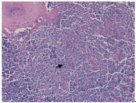
Broad spectrum antibiotic and pain control treatment were administrated, but during the first week of hospitalization the patient presented left proptosis and gradual loss of vision. He was taken to the operation room for a left lateral canthotomy, but three days later, the patient developed irreversible left amaurosis. The histopathology test for the soft palate biopsy reported polymorphous lymphoid infiltrate with angiocentric distribution and extensive necrosis (Figure 3). Immunohistochemistry confirmed lymphoid phenotype with positive T/NK CD3, CD4, BCL2 and CD56; lactate dehydrogenase (LDH) was negative, and IgG for EBV was positive.
Source: Own elaboration based on the data obtained in the study.
Figure 3 Soft palate biopsy. NK/T extranodal nasal type lymphoma. Arrow. Polymorphous lymphoid infiltrate with angiocentric distribution.
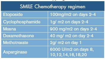
The final staging was T4, N1, M0, ECOG 1, IPI of low/intermediate-risk in a patient with poor health condition. The patient underwent chemotherapy with SMILE protocol (Table 1). Only 2 courses of SMILE were administered with adequate tolerance and some side effects, which were easily managed, such as queasiness, vomiting, weight loss and alopecia. Figure 4 shows the improvement of the lesion seven months after initiation of treatment; finally, after ten months of clinical and radiological surveillance, there was a complete remission of the lesions in oral and nasal cavity. In addition, the patient reported improvement of nasal obstruction and pain in the palate by 90%, but left amaurosis was persistent.
DISCUSSION
Most of the lethal midline granulomas correspond to NK-cell lymphomas, in which an angiocentric and angiodestructive lymphocytic proliferation occurs along the midline tissue with a fast growing rate. Despite being a low-prevalence pathology in our continent, the NK-cell lymphoma is being more frequently diagnosed and patient's survival rate has increased. The initial symptoms are non-specific and include nasal obstruction and rhinorrhea with recurrent bacterial sinusitis 5,6. As the disease progresses, a unilateral collapse of the nasal cavity and oronasal fistula may appear due to edema, necrosis and major destruction of the tissue in the facial midline 6. Cutaneous manifestations have the highest prevalence among systemic symptoms.
The most accurate test for diagnosis in order to find atypical lymphocytic infiltrates with angiocentric distribution is biopsy, and immunohistochemistry is always positive for tumoral markers CD3, CD4, CD56, CD40, CD40 RO. Infection with EBV and high levels of LDH can be found and constitute findings of poor prognosis 8. Medical imaging with computed tomography and magnetic resonance are useful for determining the size of the lesion, the presence of osteolysis, as well as extension to adjacent structures 8,9. In Latin America, discarding infectious diseases such as fungal infections, tuberculosis or tertiary syphilis, and granulomatous diseases like Wegener's granulomatosis, other non-Hodgkin's lymphomas, aggressive NK cell leukemia and malignant epithelial midline tumors is highly important 1,4.
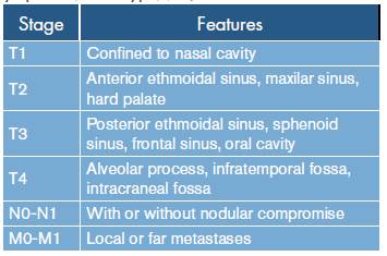
Early diagnosis is of great relevance for better prognosis in order to achieve a higher survival rate. Bad prognosis markers at the time of diagnosis are extensive local invasion, lymph nodes compromise, metastases, high levels of LDH, history of EVB infection and systemic inflammatory response syndrome 6. Likewise, classifying the disease based on the TNM system is essential to define the treatment and the prognosis 10 (Table 2). Numerous scales of functionality for oncologic patients have been described; therefore, ECOG performance status and NCCN-IPI are recommended since they are the most effective and practical ways to define the stage and prognosis of a lymphoma 11,12.
The treatment depends on the staging of the disease. NKTL staged as low risk, stage I or II, is treated with chemotherapy and radiotherapy with a 5 year survival rate of 91%, compared to 54% for only chemotherapy and 76% for only radiotherapy 13. Better results have been accomplished with doses higher than 50 Gy 8-15. Advanced stages III or IV, or with NCCN-IPI greater than 4, with or without extra nasal compromise, show better response to only chemotherapy.
Multiple protocols of chemotherapy are described with asparaginase, cyclophosphamide, etoposide, among others, which can be used in advanced stages, but survival rates have a clear decrease 8-14. Stem cell transplant in advanced stages and relapses is currently being considered as an alternative therapy with good results for improving quality of life and higher survival rates 14,15. Multidisciplinary approaches of this disease are fundamental for the treatment of the patients; new studies are required to evaluate possible alternatives for mid-face reconstruction in patients without relapse.
CONCLUSIONS
Emphasizing on the early diagnosis of one of the most lethal midline pathologies can improve prognosis and quality of life. Although, diagnosis is made based on the first biopsy, usually, more than two or three biopsies are necessary. For advanced stages, only chemotherapy is mandatory in order to reduce mortality probabilities.

