


 English (pdf)
 Article in xml format
 Article references
 Send this article by e-mail
 Cited by SciELO
  Cited by Google
  Similars in
SciELO
Similars in
SciELO  Similars in Google
 Permalink
INTRODUCTION
Toxocariasis is a neglected parasitic disease that affects mostly poor and isolated communities in low-income countries. For this reason, little attention has been paid to this condition in terms of surveillance, prevention and control. 1-3 Toxocariasis is distributed worldwide and the seroprevalence of Toxocara infection varies from 2.4% to 76.6%. 4-5 This zoonosis is caused by Toxocara nematodes, particularly Toxocara canis, a dog parasite and main etiological agent, and Toxocara cati, found in the intestine of cats. 1,3,6
In general, humans are infected through ingestion of embryonated eggs in contaminated soil and, therefore, contaminated hands. In consequence, children of preschool and school age are the most affected, although adults may also develop the disease. 1,6 Ocular toxocariasis occurs when larvae migrate to the eye and cause inflammation and scarring that can lead to vision loss. 1
The purpose of this work is to report a case of ocular toxocariasis in an adult patient, highlighting the limitations found when establishing the correct diagnosis in adults, even though this is one of the most common zoonotic infections in the world. The relevance of this clinical case is that it provides specific clinical signs of ocular toxocariasis that help to achieve a diagnosis using complementary serological methods that provide evidence on Toxocara infection to minimize anatomical and functional sequelae.
CASE PRESENTATION
22-year-old female university student, living in Bogotá D.C.-Colombia, without a significant pathological history, who referred vision loss in the left eye of 3 months of evolution, accentuated in the last 2 weeks, accompanied by eye pain, diplopia, photophobia and bilateral red eye. The patient presented retinal detachment in the left eye diagnosed by ultrasound. She consulted with ophthalmology, where a differential diagnosis of retinoblastoma and pars planitis was made. Symptomatic management was initiated with oral and topical corticosteroids. She was referred to the Instituto Nacional de Salud (National Health Institute) due to suspicion of ocular toxocariasis.
Upon reviewing her socioeconomic background, coexistence with dogs and cats during childhood in rural Caquetá was observed. At the time of consultation, she had been living for 6 months with a dog and two kittens that had not been vaccinated nor dewormed. The woman said that she occasionally took her pets to the park, where they were in contact with soil and feces and reported feeling better with the drugs prescribed by ophthalmology, although her visual acuity continued to be affected.
Upon physical examination, her vital signs were within the normal range and without fever. Ophthalmological examination showed visual acuity in the right eye sc: 1.00 and in the left eye sc: 0.5. Hyperemic tarsal papillae were also observed in both eyes. Ophthalmoscopy in the right eye was normal, while a peripheral granuloma and a fibrous band pulling the macula were observed in the left eye. The rest of the physical examination did not show any alterations.
When analyzing the complete blood count made on March 9, 2016, slight leukocytosis and lymphocytosis without eosinophilia were observed. However, the values returned to normal after the symptomatic treatment ended on June 14, 2016 (Table 1). The results of the coprological and partial urine tests were normal. In addition, anti-Toxoplasma gondii and anti-Taenia solium cysticerco antibodies test for T. canis was positive, with IgG titres of 1:64 (positive ≥1: 32, specificity >90%).
Table 1 Results of blood counts at two different times
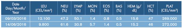
LEU: leukocytes; NEU: neutrophils; LYMP: lymphocytes; MON: monocytes; EOS: eosinophils; BAS: basophiles; HEM: hemoglobin; HCT: hematocrit; PLAT: platelets; CEL: cells.
Source: Own elaboration based on the data obtained in the study.
Management was initiated with two doses of oral prednisone 2 mg/kg/day, topical prednisolone 1 drop/2hr and albendazole 400 mg/day for 5 days. The patient required surgical management with vitrectomy in the left eye to release the vitreous band, making the diplopia disappear. However, residual peripheral granuloma persisted and will be operated by ophthalmology. Hygiene recommendations were provided to the patient and her relatives, the consumption of well-cooked meats was suggested and control was scheduled at 1 month.
DISCUSSION
Knowing about this clinical case is relevant to the medical and scientific community for three reasons: first, because this is a case of ocular toxocariasis, a parasitic disease neglected worldwide that is of special interest for Latin American countries such as Colombia, where it is considered endemic 7; second, because current medical literature mentions that toxocariasis is predominant in children, but some cases are associated with the adult population 8-11, and third, because it is related to the importance of differentiating this ocular pathology from others that require differential diagnosis such as retinoblastoma, toxoplasmosis and syphilis. 8
Toxocariasis has been described more frequently in children of preschool and school age due to the permanent presence of risk factors in this particular age group. 1 Nevertheless, this disease should not be ignored in the adult population, in which cases have also been reported, since a less common way of acquiring the infection is through the intake of raw or undercooked foods contaminated with the larvae of the parasite. 3,7,12 In addition, it is worth mentioning that the type of syndrome that appears due to infection with Toxocara seems to be related to age, as visceral larva migrans appears mostly during childhood, whereas ocular toxocariasis is seen in advanced ages, but still, there is controversy around this particular issue, since many authors report that it is predominant during childhood. 7,13
Definitive diagnosis of ocular toxocariasis is obtained by demonstrating the presence of migrating larvae in the biopsies of the compromised tissues, which is rather exceptional since it is an invasive procedure. Coproparasitological examination is not useful because the parasite is unable to mature inside a human host. 3,13,14 Therefore, current diagnosis is made based on typical ophthalmological signs, which, in general, are unilateral and supported by immunological tests. 3
The indirect ELISA test, used as an aid to obtain a diagnosis, uses excretion/secretion antigens of the T. canis larvae to detect anti-Toxocara spp. in serum or other body fluids such as vitreous humor, which is used especially in ophthalmic cases. The sensitivity of this test is 80-100%, with specificity of 9095%, but these figures may vary according to the geographical region where it is applied and the quality of the antigen obtained. 13 According to other sources, an ELISA test with serum titers >1:32 has a sensitivity of 73% and a specificity >90% 2, although a titre of 1:8 in serum is sufficient to support the diagnosis if the patient has clinical manifestations compatible with this zoonosis. 3 Due to potential cross-reactivity with the ELISA test, some authors suggest confirming the result with a Western Blot test. 13
On the other hand, although eosinophilia is an important marker of systemic toxocariasis, it is not usually observed in ocular form 1,3, but its presence may indicate the coexistence of both forms of toxocariasis in the same patient. 3 Needless to say, for accurate diagnosis, it is necessary to know the complete clinical history of the patients, as well as their signs and symptoms and socioeconomic situation, to identify predisposing factors such as cohabitation with dogs or cats and geophagy. 14
There are four clinical presentations of ocular toxocariasis, but only two are more common: posterior pole granuloma and peripheral granuloma. 3 They usually present with unilateral vision loss, sometimes with strabismus and sometimes with leukocoria, although granulomatous inflammation can cause diverse manifestations such as keratitis, iridocyclitis, chronic endophthalmitis, retinal detachment and optic neuritis. 7 For this reason, differential diagnosis should be made with other ocular granulomatous diseases such as ocular toxoplasmosis, sarcoidosis, tuberculosis and fungal infections. 3,15 Since leukocoria and strabismus are the two most common signs of retinoblastoma, it is also necessary to perform a differential diagnosis with this neoplasm. 7,13,16 The patient described in this case did not present any of those ocular alterations.
CONCLUSIONS
Diagnosis of ocular toxocariasis is based on the identification of particular signs and symptoms; it should be supported by a complete clinical history that provides a detailed report of socioeconomic background and should be complemented by an indirect ELISA test performed on a serum sample or vitreous humor. This process is carried out in order to avoid erroneous diagnoses such as retinoblastoma and other granulomatous diseases of the eye.
Since ocular toxocariasis is a neglected and global disease, it is important to implement prevention and control measures to reduce the prevalence of this parasitic disease in the population. For this purpose, awareness must be generated in the community about this type of preventable diseases through the promotion of good hygienic practices such as hand washing, identification of transmission sources and reduction of exposure to etiological agents. Furthermore, it is necessary to develop programs for deworming pets, both dogs and cats, in order to control the transmission of this disease.

