SciELO - Scientific Electronic Library Online

 
vol.5 issue1GIANT CELL TUMOR OF THE DORSAL VERTEBRAE. CASE REPORTPEDIATRIC DIABETIC KETOACIDOSIS IN A PATIENT WITH DOWN SYNDROME. CASE REPORT author indexsubject indexarticles search
Home Pagealphabetic serial listing  

Services on Demand

Journal

Article

Indicators

Related links

  • On index processCited by Google
  • Have no similar articlesSimilars in SciELO
  • On index processSimilars in Google

Share


Case reports

Print version ISSN 2462-8522

Case reports vol.5 no.1 Bogotá Jan./June 2019  Epub June 13, 2019

https://doi.org/10.15446/cr.v5nl.75350 

CASE REPORT

INTUSSUSCEPTION SECONDARY TO INFLAMMATORY MYOFIBROBLASTS TUMOR OF THE SMALL INTESTINE. CASE REPORT

David Yépez-Yépeza  * 

Betsy Macías-Jarab 

a Lúver Macías-Jara Hospital Especializado Mariana de Jesús - Department of General Surgery - Guayaquil - Ecuador.

b Hospital General de Chone Dr. Napoleón Dávila Córdova - Department of General Surgery - Chone - Ecuador.


ABSTRACT

Introduction:

Intussusception occurs when part of the intestine slides into an adjacent intestinal segment. Inflammatory myofibroblast tumor is a rare cause of this condition, and is observed in 5% -16% cases in adults.

Case presentation:

A 41-year-old woman presented with abdominal pain and distension. A exploratory laparoscopy was performed, finding ileocolic intussusception into the transverse colon. Due to uncontrollable bleeding, the procedure was converted to laparotomy; resection and latero-lateral ileocolic anastomosis were performed. Histopathology reported inflammatory myofibroblastic tumor, with a favorable postoperative evolution. The patient was discharged on the sixth postoperative day.

Discussion:

When located in the small intestine, 57% of the tumors that cause intussusception are benign, including the myofibroblastic tumor in this patient. The symptoms and signs associated with this neoplasm are cramp-like abdominal pain, nausea and vomiting. Although imaging studies may lead to suspect this diagnosis, in most cases it is made intraoperatively. Surgical resection of the affected intestinal segment is curative, with favorable prognosis.

Conclusions:

This case is considered as a rare cause of intussusception. It had a benign course and is still under study since its patho-physiology has not been fully understood.

Keywords: Intussusception; Intestinal Obstruction; Intestinal Neoplasm

RESUMEN

Introducción.

Se denomina intususcepción a la introducción de un segmento intestinal a otro distal, siendo esta la causa posterior del tumor miofibroblástico inflamatorio en el 5-16% de los adultos.

Presentación del caso.

Paciente femenino de 41 años con presencia de dolor y distensión abdominal. Se practica exploración quirúrgica laparoscópica, observando intususcepción ileocólica hasta colon transverso. Por sangrado no controlable se realiza conversión a laparotomía, se reseca y se realiza anastomosis ileocólica latero-lateral. La histopatología reporta tumor miofibroblástico inflamatorio, con evolución postquirúrgica favorable. Se da de alta al sexto día postquirúrgico.

Discusión.

En el intestino delgado, 57% de los tumores que originan intususcepción son benignos, como el tumor miofibroblástico que presentó la paciente reportada. Los síntomas y signos de esta neoplasia son dolor abdominal tipo cólico, náusea y vómito. Aunque los estudios de imágenes pueden dar una sospecha del diagnóstico, en la mayoría de los casos se hace intraoperatorio. La resección quirúrgica del segmento intestinal afectado es curativa, con pronóstico favorable.

Conclusiones.

El presente caso representa una causa poco frecuente de intususcepción intestinal, de curso benigno, la cual continúa en estudio ya que no se ha logrado entender por completo su fisiopatología.

Palabras clave: Intususcepción; Obstrucción intestinal; Neoplasia intestinal

INTRODUCTION

Intussusception occurs when a part of the intestine and its mesentery slide into an adjacent intestinal segment. 1 It is most frequently observed in children, and only 5% to 16% of the cases occur in adults. 2 Currently, this neoplasm represents 1% of all causes of intestinal obstruction. 3,4

Inflammatory myofibroblastic tumor (IMT) is a rare mesenchymal neoplasm caused by the multiplication of spindle cells in variable inflammatory patterns. 5-8

Although the etiology of intussusception is unknown, previous surgeries, infections, trauma, immune reactions, radiation therapy and steroids have been suggested as probable causes. 9-11 The onset of this condition in 52% of cases is associated with malignant tumors and exacerbation of acute abdominal pain; in addition, data on pathological involvement have been obtained in the small intestine and, less frequently, in the colon and the gastroduodenal portion. Imaging studies help to make the diagnosis; however, exploratory laparotomy supports the diagnosis in 68% of cases. 12,13 It is worth mentioning that it can be confused with cancer due to similar clinical and imaging characteristics, causing difficulties in its subsequent treatment. 5,12

IMTs that extend through the gastric wall and into neighboring organs such as the esophagus, duodenum, pancreas, peritoneal cavity, and liver are known as inflammatory infiltrates and simulate malignancy in endoscopy and imaging. 7,13 Regarding management, despite pathological findings, it has been proven that complete resection is possible; this is the treatment of choice and is considered as a cure as symptoms and even recurrences tent to decrease. 6,14-16

CASE PRESENTATION

Female patient of 41 years, of mixed race, from the rural area of Babahoyo (Ecuador), housewife and of a low-income household who consulted for pain and abdominal distension associated with vomiting, constipation and diarrhea of three months of evolution prior to admission.

No relevant medical history was reported. Initial care provided found normal vital signs, and physical examination revealed general abdominal pain with response to deep palpation in mesogastrium and hypogastrium, abdominal distention and increased hydro-aeric sounds, accompanied by nausea that led to vomiting and diarrheic stools. Finally, laboratory results showed anemia and leukocytosis without additional blood tests.

Anteroposterior abdominal x-ray, in standing position and supine decubitus position, showed accentuated distension and dilatation of thin intestinal loops and hydro-aerial levels (Figure 1).

Source: Document obtained during the study.

Figure 1 X-ray of abdomen in standing position showing thin bowel loops with hydro-air levels. 

Likewise, abdominal ultrasound reported concentric thickening of the wall of the ascending and transverse colon with preservation of the visualization of its layers (Figure 2).

Source: Document obtained during the study.

Figure 2 Concentric thickening of the colonic wall compatible with endoluminal tumor or intussusception. 

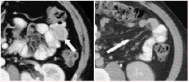
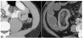
Diffuse wall edema of the transverse ascending colon and splenic angle, image of invaginated appearance and space-occupying lesion at this level can be observed in abdomen and pelvis tomography (Figures 3 and 4).
Source: Document obtained during the study.

Figure 3 Crescent in doughnut sign. Imaging of oral phase showing enteroenteric intussusception. The arrow points the external muscularis of intussusception. 
Source: Document obtained during the study.

Figura 4 A. Sausage pattern with alternate areas of low and high attenuation. B. Intraluminal lesion as the lead point of intussusception. 

Based on the result of the admission examination, a diagnostic exploratory laparoscopy was performed to identify the location, causality and correct procedures of the treatment. During surgery, dilation of the small intestine was observed and the diagnosis of ascending intestinal intussusception was confirmed; due to uncontrollable bleeding, the procedure was converted to laparotomy (Figures 5 and 6).

Source: Document obtained during the study.

Figure 5 Ileocolonic intussusception. 

Source: Document obtained during the study.

Figure 6 Intussusception of ascending bowel to transverse colon. 

Resection of the segment of terminal ileum affected by the tumor mass and bowel transit reconstruction were performed by means of termino-terminal ileocolic anastomosis (Figure 7).

Source: Document obtained during the study.

Figure 7 Affected bowel segment. 

When analyzing the sample collected during surgery, a polypoid formation of 6cm of diameter, on a pedicle and ulcerated that occupied of 85% the lumen, with smooth and whitish external surface was observed. A homogeneous and elastic hard consistency was evident after cutting.

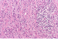
Then, histological cutting was performed (Figure 8) and taken to a laboratory where proliferation of spindle cells (which surround the vessels) and abundant inflammatory infiltrate were identified.
Source: Document obtained during the study.

Figure 8 Microscopy of the tumor segment in sectioned terminal ileum. 

The definitive treatment in this case was surgical, that is to say, resecting the segment of intestine that had the tumor and restoring intestinal transit with termino-terminal ileocolic anastomosis. The patient had a good post-operative evolution and no adverse reaction; she was discharged six days after surgery, with favorable prognosis and without complications.

DISCUSSION

Intussusception in adults occurs after an aggression in the intestinal wall causes alteration of peristalsis, in turn leading to a proximal segment sliding into a distal segment. If the mesentery is involved, it causes vascular compression, wall edema and necrosis of the intestinal loop. Its etiology in 70-90% of cases is an organic lesion of malignant origin, frequently in the colon and small intestine. 17 57% of tumors that cause intussusception are benign, including myofibroblastic tumor, also known as granuloma; 30% of cases are malignant. The most common malignant tumor is melanoma and its metastases 18. Other less frequent causes are Meckel's diverticulum, adhesions or hematoma of the wall. 19

Nevertheless, adhesions are the most common extraluminal lesions; they originate after being pulled into an intestinal segment, which causes a fold and invagination into the segment that produces the pulsation due to the intestinal movement. 20 According to the location of the adhesions, and of intussusception in general, the lesions are classified into four categories: 1) enteroenteric (75% and with greater recurrence), 2) colocolic (14% recurrence), 3) ileocecal (8% recurrence) and 4) ileocolic (5% recurrence). 17,21

If the alteration is caused by the colon, the most common cause of intussusception is adenocarcinoma, followed by leiomyosarcoma, liposarcomas, reticular cell sarcoma and metastases. 22

It is important to assess symptoms and signs; those related to intestinal obstruction, according to frequency, are: colicky abdominal pain (75%-85%), nausea (50%), vomiting (30%) and constipation. Other less common symptoms include diarrhea, weight loss, melena, fever, and palpable abdominal mass. 17 The case reported here presented with abdominal pain and bloating. Because of these symptoms, diagnosis is made preoperatively only in a third of the cases, of which 50% are diagnosed as intestinal obstruction, 11% as abdominal tumor and 2-5% as bleeding in the digestive tract. 17

This correlates well with the ultrasound studies of the abdomen performed worldwide; target sign imaging have shown that it is possible to identify multiple thin, parallel, hypo-echoic and ecogenic layers in longitudinal planes. 23

Pathognomonic findings in tomography include bowel thinning, space-occupying lesion and an area of fat and vessel hyperdensity. The computed tomography (CT) performed on the reported patient showed diffuse edema of the transverse ascending colon wall and splenic angle, image of invaginated aspect and space-occupying lesion. 24 Nuclear Magnetic Resonance Morphology is similar to CT and consistent with other studies. 25

It is worth noting that surgical exploration is the main means of diagnosis and that, despite other simpler methods, it is the only one that allows providing a definitive treatment by intestinal resection and primary anastomosis with restoration of the intestinal transit. This is mostly curative and to some extent coincides with the literature and the surgical treatment provided to this patient, which was resection of the affected ileal segment and termino-terminal ileocolic anastomosis. 26

CONCLUSIONS

This case corresponds to a very rare cause of intussusception due to the specificity of the clinical presentation, the diagnosis and the immediate need for resection. It should be noted that the definitive diagnosis is made through a histopathological study. The pathophysiology of this disease is not correctly established yet, so this neoplasm is still under study.

REFERENCES

1. Coran AG. Intussusception in adults. Am J Surg. 1969;117(5):735-8. http://doi.org/bz3p66. [ Links ]

2. Eisen LK, Cunningham JD, Ausfses AH. Intussusception in adults: institutional review. J Am Coll Surg. 1999;188(4):390-5. http://doi.org/dwzzbn. [ Links ]

3. Laws HL, Aldrete JS. Small-bowel obstruction: A review of 465 cases. South Med J. 1976;69(6):733-4. [ Links ]

4. Stewardson RH, Bombeck CT, Nyhus LM. Critical operative management of small bowel obstruction. Ann Surg. 1978;87(2):189-93. [ Links ]

5. Company-Campys MM, García-Bonafé MM, Salva F, Terrasa F, Ibarra J, Ramos B. Tumor miofibroblástico inflamatorio ileal: Presentación de un caso. In: VIII Congreso Virtual Hispanoamericano de Anatomía Patológica; 2006. [ Links ]

6. Lazure T, Ferlicor S, Gauthier F, Doz F, Courtrier J, Fabre M, et al. Gastric Inflammatory Myofibroblastic Tumors in Children: An Unpredictable Course. JPediatr GastroenterolNutr. 2002;34(3):319-22. [ Links ]

7. Kim KA, Park CM, Lee JH, Cha SH, Park SW, Hong SJ, et al. Inflammatory myofibroblastic tumor of the stomach with peritoneal dissemination in a young adult: imaging findings. Abdom Imaging. 2004;29(1):9-11. http://doi.org/fjfz6p. [ Links ]

8. Pettinato G, Manivel JC, De Rosa N, Dehner LP. Inflammatory myofibroblastic tumor (plasma cell granuloma). Cliniopathologic study of 20 cases immunohistochemical and ultrastructural observations. Am J Clin Pathol. 1990;94(5):538-46 [ Links ]

9. López-Ibarra S, Calderón-Elvir C, Carrasco-Daza D, Medina-Vega A, Méndez-Martínez D. Tumor Miofibroblástico Inflamatorio Gástrico. Informe de un caso y revisión de la literatura. Revista Mexicana de Cirugía Peditrica. 2007;14(2):77-82. [ Links ]

10. Hamidah A, Khu SY, Thambidorai CR, Muhaizan WM, Zarina AL, Jamal R. Recurrent gastric inflammatory myofibroblastic tumor in a 13-year-old male. Pediatr Surg Int. 2007;23(6):601-3. http://doi.org/bjtn5w. [ Links ]

11. Curtis JL, Burns RC, Wang L, Mahour GH, Ford HR. Primary gastric tumors of infancy and childhood: 54-year experience at a single institution. J Pediatr Surg. 2008;43(8):1487-93. http://doi.org/cdmf2g. [ Links ]

12. Dehner LP. Inflammatory Myofibroblastic Tumor: the continued definicion of one type of so-called inflammatory Pseudotumor. Am J Surg Pathol. 2004;28(12):1652-4. [ Links ]

13. Karnac I, Senocak ME, Ciftci AO, Caglar M, Bingöl-Kologlu M, Tanyel FC, et al. Inflammatory Myofibroblastic Tumor in children: diagnosis and treatment. J Pediatr Surg. 2001;36(6):908-12. http://doi.org/d2w439. [ Links ]

14. Coffin CM, Dehner LP, Meis-Kindblom JM. Inflammatory myofibroblastic tumor, inflammatory fibrosarcoma, and related lesions: an historical review with differential diagnostic considerations. Semin Diagn Pathol. 1998;15(2):102-10. [ Links ]

15. Cho MY, Min YK, Kim NR, Cho SJ, Kim HK, Lee KC, et al. Fever of unknown origin as a presentation of gastric inflammatory myofibroblastic tumor in a two-year old boy. J Korean Med Sci. 2002;17(5):699-703. http://doi.org/czpx. [ Links ]

16. Narla LD, Newman B, Spottswood SS, Narla S, Kolli R. Inflammatory pseudotumor. Radiographics. 2003;23(3):719-29. http://doi.org/fsjgdc. [ Links ]

17. Azar T, Berger DL. Adult intussusception. Ann Surg. 1997;226(2):134-8. [ Links ]

18. López A, Droguett ME. Paciente con obstrucción intestinal. Rev Chil Radiol. 2003;9(1):36-7. http://doi.org/b678vr. [ Links ]

19. Marinis A, Yiallourou A, Samanides L, Dafnios N, Anastasopoulos G, Vassiliou I, et al. Intussusception of the bowel in adults: A review. World J Gastroenterol. 2009;15(4):407-11. http://doi.org/c89pwf. [ Links ]

20. Huang BY, Warshauer DM. Adult intussusception: diagnosisand clinical relevance. Radiol Clin North Am. 2003;41(6):1137-51. http://doi.org/b94wtw. [ Links ]

21. Amr MA, Polites SF, Alzghari M, Onkendi EO, Grotz TE, Zielinski MD. Intussusception in adults and the role of evolving computed tomography technology. Am J Surg. 2015;209(3):580-3. http://doi.org/f65zgj. [ Links ]

22. Warshauer DM, Lee JK. Adult intussusception detected at CT or MR imaging: Clinical-imaging correlation. Radiology. 1999;212(3):853-60. http://doi.org/czpz. [ Links ]

23. Anderson D. The pseudokidney sign. Radiology. 1999;211(2):395-7. http://doi.org/czp2. [ Links ]

24. Visvanathan R, Nichols TT, Reznek RH. Acquired immune deficiency syndrome-related intussusception in adults. Br J Surg. 1997;84:1539-40. http://doi.org/cbq8tv. [ Links ]

25. Gourtsoyiannis NC, Papakonstantinou O, Bays D, Malamas M. Adult enteric intussus-cepction: Additional observations on enteroclysis. Abdom Imaging 1994;19(1):11-7. [ Links ]

26. Zumarán-Cuéllar O, Robles-Vidal C, Vi-llarreal-Colín P, Olivares-Mendoza H, Sa-rue-Saed N. Intususcepción en el adulto. An Méd (Mex). 2006;51(4):188-92. [ Links ]

CONFLICT OF INTEREST None stated by the authors.

FUNDING None stated by the authors.

Received: October 03, 2018; Accepted: December 17, 2018

* Corresponding author David Yépez-Yépez. Departamento de Cirugía General, Hospital Especializado Mariana de Jesús. Guayaquil. Ecuador. Email: dayeye1988@gmail.com.

Creative Commons License This is an open-access article distributed under the terms of the Creative Commons Attribution License