


 Inglês (pdf)
 Artigo em XML
 Referências do artigo
 Enviar este artigo por email
 Citado por SciELO
  Citado por Google
  Similares em
SciELO
Similares em
SciELO  Similares em Google
 Permalink
INTRODUCTION
Systemic lupus erythematosus (SLE) is a multisystem chronic autoimmune disease with an unpredictable relapsing-remitting course 1, occasionally limited to certain organs, with extremely varied clinical manifestations, and a complex pathogenesis, that is diagnosed based on clinical grounds and serological abnormalities. Multidisciplinary care is required and depends on joint patient-physician decision 1. The hematologic manifestations of the disease include the presence of autoantibodies (especially anti-dsDNA and anti-Sm), hypocomplementemia, and antiphospholipid antibodies (which is associated with a worse prognosis). Patients usually experience certain damage induced by the disease and the implemented therapy 1,2.
SLE has several signs and symptoms that involve constitutional, musculoskeletal and dermatological manifestations, although these are not the only ones 2. Other manifestations used to classify it are non-scarring alopecia, oral ulcers, subacute cutaneous or discoid lupus, acute cutaneous lupus, antiphospholipid antibodies, low C3 and/or C4, and anti-dsDNA or anti-Sm antibodies 3. Recently, a study highlighted the role of C1q in suppressing the activation and expansion of CD8+ in a SLE model, supporting a strong association between clinical condition and complement C1q deficiency 4. Moreover, the production of autoantibodies is associated with deregulation of human T helper cell factors 5.
Due to this wide display of manifestations, classification of lupus can be a challenging and complex endeavor. There are different forms of SLE that may be mistaken for other diseases, especially because they have similar names. Three of such diseases are lupus pernio (a type of sarcoidosis) 6, chilblain lupus (also known as lupus perniosis, a manifestation seen in some forms of SLE) 7, and lupus pernio (a rare inflammatory condition) 8. Treatment of SLE involves hydroxychloroquine, which is recommended for all patients, and glucocorticoids in maintenance doses (prednisone equivalent). Cyclophosphamide can be used for the initial treatment of organ-threatening disease; if the disease is refractory, biologic therapies, such as rituximab, can be used and should be considered 9.
This case report describes a complex case of SLE in a female patient who presented with the characteristics of lupus perniosis, which was classified as lupus pernio, a type of sarcoidosis.
CASE PRESENTATION
A 29-year-old Colombian mestizo woman, from a middle-income household, housewife, from Tenjo, Cundinamarca, Colombia, a cold region of the country, went to the emergency room of a tertiary care center in Bogotá, due to symptoms of asthenia, adynamia, sleepiness, and articular pain. During examination, she reported having regular menstrual cycles, using male condoms as contraception method, no children, no previous pregnancy, and no previous surgeries. She also stated that she occasionally consumed alcoholic drinks and that she smoked 1 or 2 cigarettes per day. Family health history was negative for autoimmune, inflammatory, or cutaneous diseases.
The following is the timeline of her symptoms:
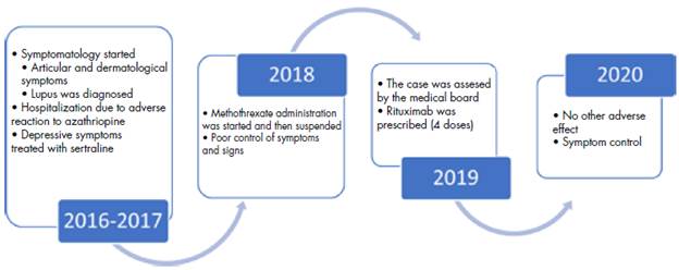
2016 - 2017: The patient experienced asthenia, adynamia, sleepiness, and articular pain, consequently, she consulted the emergency room. Thyroid evaluation was performed by a general physician, obtaining normal thyroid function results, so she was prescribed paracetamol 500mg per os every 6 hours for 5 days and physical activity. Symptoms did not recede and, in the following weeks, multiple reddish punctuate pruritic lesions appeared, at first in the right and then in the left hand, and later ulcerated. In June and July, she had pain in knees and ankles, for which she was prescribed paracetamol 500mg per os every 6 hours for 5 days, as well as topical medications (betamethasone 0.1% every 12 hours for two weeks), which she used for six months without success. At the end of this six months, hip pain, malar rash and alopecia appeared, so she sought consultation with the dermatology service.
Due to the onset of these new symptoms and the persistence and aggravation of the previous ones (especially articular pain), the dermatologist suspected an immune disease, therefore, lab tests and biopsy were requested in the first two weeks of 2017. Biopsies (Table 2) were performed in the lateral aspect of the nose and in the second finger of the left hand. Immunological studies were started, but treatment was not implemented until receiving the results.
After that, follow-up testing was performed, revealing hypocomplementemia and elevated transaminases levels, with a slight elevation at baseline with subsequent normal successive measurements. Antimitochondrial antibodies (AMA) and anti-smooth muscle antibodies (ASMA) were negative. Hepatitis C, HIV, and non-reactive hepatitis B surface antigen were negative as well. Blood cells were normal at most moments of the follow-up, but lymphocytes, monocytes and eosinophiles presented low values on two occasions. Urinalysis was normal, with no hematuria, proteinuria, or active inflammatory sediment.
After considering multiple differential diagnosis (dermatological, immunological, and infectious), the dermatology reached the lupus pernio diagnosis.
As a result, the patient attended an appointment with rheumatology , which agreed with these findings, so chloroquine 250 mg per day per os and prednisolone 15mg per day per os were started. This therapy was administered for 1 month until a new follow-up appointment was scheduled. During this appointment, which took place in February 2017, the rheumatology service confirmed the diagnosis, and more tests were requested to assess renal function. The prescription was continued.
Two weeks later, due to severe hypocomplementemia, azathioprine was started 2 times per week. After 15 days of treatment, a rash appeared in the left thigh, so a biopsy was taken based on the rheumatologist advice. The patient continued taking the medication, but the rash expanded to the right thigh and lower back; it was highly pruritic. No other medication was added, but due to the poor improvement of the lesions and the intense pruritus that disrupted her sleep, the patient consumed over-the-counter antihistamines and the lesions progressed.
She attended once again the emergency department of the same institution, where she received initial therapy with chloroquine and prednisolone (60 mg/day); the latter was suspended to initiate methylprednisolone (80mg per day for three days IV). At this point, the rheumatology service diagnosed Stevens-Johnson's syndrome, and at the end of February and beginning of March 2017 she was hospitalized for 10 days.
During the hospitalization, renal and pulmonary function tests were carried out, as well as X-rays of the chest and hands, which were all normal. Azathioprine 1mg/kg per os (only taken for two weeks) was suspended and switched to hydroxychloroquine (200 mg every 12 hours per os) and chlorpheniramine (4mg every 12 hours per os). Unfortunately, on the third day, another allergic reaction occurred, so hydroxychloroquine was suspended, and chlorpheniramine was complemented with methotrexate (7.5mg per week per os).
Due to the allergic reaction, laboratory tests were performed once again, revealing neutrophilia and a dermatological reaction. Methylprednisolone was suspended, and prednisolone (5mg per day per os) was reinstated. Dermatological symptoms improved, but residual hypochromia and desquamation remained. She was discharged after 10 days with a prescription of prednisolone 30mg per day per os, methotrexate 7.5mg weekly per os -which she is still using-, hydroxyzine 20mg per every 8 hours on demand, and folic acid 1mg per day per os alongside methotrexate.
2017 and 2018: Articular symptoms improved, but cutaneous signs and symptoms did not. Follow-up appointments with the rheumatology service were scheduled every three months. She was showing signs of depression, so the psychiatry service was consulted, and sertraline 25mg/day per os started with increments until an effective dose was achieved (50mg/day).
2019: Rituximab was prescribed, and symptoms and active lesions improved significantly. It was administered at a dose of 1g IV in weeks 0 and 2, and the dosage was repeated annually. However, the patient had an allergic reaction, so a corticoid was prescribed for a short period of time (hydrocortisone 100mg IV single dose). Protein levels (measured on 05/08/2019) were normal, except for a slight increase in the gamma fraction (1.6g/dL, normal: 0.6-1.5).
21/08/2020: Two biopsies were taken, one from the right nasal sinus and one from the right thigh. Immunofluorescence was required to confirm the diagnosis, but the patient did not return due to clinical improvement with the established therapy.
Late 2020: Dermatological signs and symptoms, as well as pain control, improved following rituximab therapy (20/09/2020), although periods of alopecia persisted. The dosage of the other medications was reduced. Ocular and visual function testing was normal throughout the follow-up. The parieto-occipital region of the scalp was infiltrated with injectable and topical corticoid and antihistamine.
Table 1 below presents the results of the immune panel, while Table 2 describes the results of the biopsies taken from the finger lesions (diagnosis of lupus pernio) and from the facial lesions (diagnosis of lupus erythematous).
Table 1 Findings in clinical tests: antigens, blood chemistry, and antibodies (only abnormal results are shown).
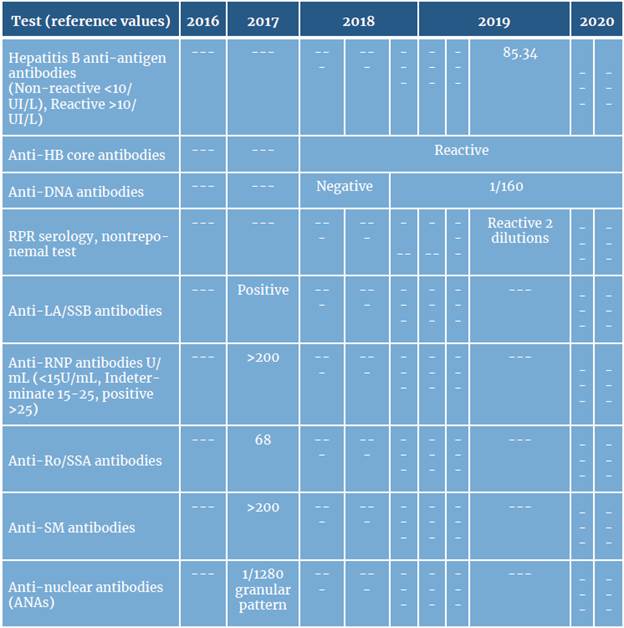
Source: Own elaboration.
Table 2 Biopsy results.
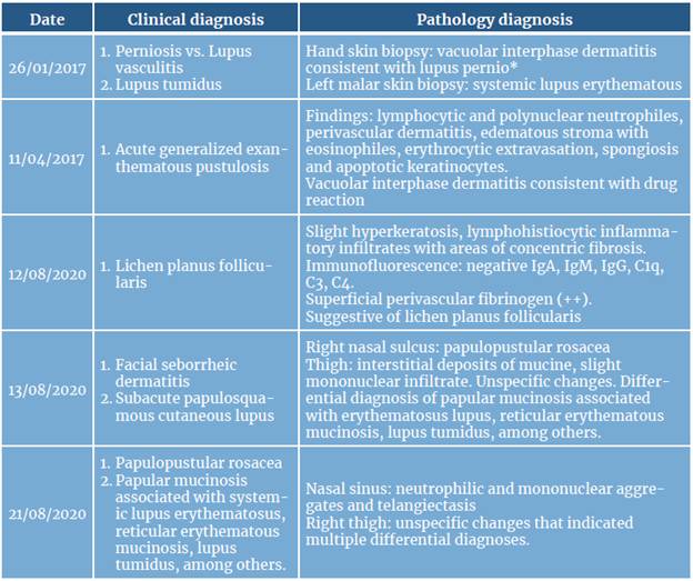
Source: Own elaboration.
* Taken directly from the biopsy report. This diagnosis, in our opinion, was the result of a terminology error, as chilblains lupus or lupus perniosis would have been more appropriate.
DISCUSSION
In medicine, the variety of terms, clinical conditions and names for diseases can lead to confusion and incorrect patient classification. An example of this situation is systemic lupus erythematosus, a disease that is associated with several symptoms that affect different organs and systems and with several manifestations such as skin disorders (malar or discoid rash, photosensitivity, alopecia, Raynaud's phenomenon, etc.) and vascular conditions (Raynaud's phenomenon, ischemic digits, digital ulcers, vasculitis, etc.) 2.
Due to the wide array of clinical manifestations, treating SLE may be challenging, especially because there are no guidelines that address lifestyle, quality of life, among other aspects 1 that are relevant for the management of complex cases, such as the one reported here.
Complex cases are of particular concern since errors in patient classification are possible, as was the case in this report. Even though the error did not alter the patient's prognosis, it is essential to explain and emphasize that there are at least three separate diseases whose names can lead to confusion, namely, perniosis, lupus pernio, and lupus perniosis (or chilblain lupus).
SLE has multiple subtypes, including chronic cutaneous LE, and chilblain lupus is, in turn, a rare form of cutaneous lupus erythematosus. The latter has different clinical and histopathological features, some of which are listed in Table 3 10.
Table 3 Subtypes of cutaneous chronic lupus erythematosus.
| Subtype | Clinical findings | Histopathological and serological findings |
|---|---|---|
| Discoid lupus erythematosus | Discoid lesions can occasionally occur on mucosal surfaces, including lips, and oral, nasal, and genital mucosa. Lesions appear as a well-demarcated, scaly, erythematous macule or papule, which gradually develops into an indurated discoid (coin-shaped) plaque with an adherent scale that is painful to remove, and can extend to the follicle causing scarring alopecia. | Hyperkeratosis dilated compact keratin-filled follicles, vacuolar degeneration of the basal keratinocytes, and an intensely inflammatory dermal infiltrate. Serologically, DLE patients have a lower incidence of ANA, dsDNA, Sm, U1RNP, and Ro/SSA antibodies, as compared to other CLE subtypes. |
| Lupus erythematosus profundus | Also known as panniculitis, it features painful firm subcutaneous nodules with occasionally overlying DLE in areas of increased fat deposition, such as the upper arms and legs, face, and breasts. | Lobular panniculitis with a dense lymphocytic infiltrate, occasionally requires the use of cell markers and gene rearrangements. |
| Chilblain lupus (synonym of lupus perniosis) | A rare form of lupus. Painful violaceous plaques and nodules in areas exposed to the cold. Central erosions or ulcerations may occur in acral surfaces (toes, fingers, heels, nose and ears) resembling frostbite. | Epidermal atrophy, interface vacuoliza-tion, and a perivascular mononuclear infiltrate |
| Lupus erythematosus tumidus | Extreme photosensitivity and benign course. Generally, in males. Lesions are erythematous, edematous, urticaria-like polycyclic plaques with elevated borders. Follicular plugging does not occur. | Dense perivascular and periadnexal infiltrates without involvement of the interface. DIF is typically negative, and ANAs are present in 10% of the patients. |
Source: Own elaboration based on Okon & Werth (10)
Regarding lupus pernio, it was first described in 1889 by Ernest Besnier (therefore, it is also known as the Besnier-Tenneson syndrome) 11,12. It is defined as a non-life-threatening manifestation of sarcoidosis 13. Its characteristic clinical presentation includes violaceous lesions or shiny nodules that appear predominantly on the nose, cheeks, and ears 11, although they can be seen in other parts of the body, such as fingers and toes, in a high proportion of patients 8,14. These nodules may present as isolated lesions or as an early presentation of systemic sarcoidosis.
Patients with this condition have a higher risk of developing pulmonary disease with more pulmonary lesions compared with other types of sarcoidosis 11. The etiology of lupus pernio is still poorly understood, as there is a complex interaction between genetic, immune deregulation, and environmental triggers. Histopathological findings include non-caseating epithelioid cell granulomas and a variety of Langhans giant cells 11.
Treatment includes topical or intralesional corticosteroids, triamcinolone, tacrolimus, or pimecrolimus. Systemic glucocorticoids can be used on skin lesions. Hydroxychloroquine, methotrexate, and minocycline are used when there is extensive skin involvement 11. Adalimumab and infliximab are indicated when lupus pernio is resistant to other types of treatments. Differential diagnosis includes fungal infections, lupus vulgaris, berylliosis, lymphoma cutis, lupus erythematosus and tuberculoid leprosy 11.
On the other hand, perniosis, known as well as idiopathic perniosis or chilblains is a rare inflammatory disease that affects acral skin and results from the idiopathic response to cold or humid conditions. Its course is acute and idiopathic, although it can also be chronic 15,16. It was first described by Hutchinson in 1888 and is characterized by cutaneous lesions located on fingers, toes, nose, ears, elbows, heels and knees, induced or aggravated by exposure to cold 15,16. Diagnosis is clinical, and biopsy is not recommended due to its unspecific histopathological changes (dermal infiltrate, edema, spongiosis, deep perieccrine inflammation, basal layer vacuolation, and necrotic keratinocytes within the epidermis). A concerning aspect of this disease is the possible development of secondary infection. Treatment includes corticosteroids and nifedipine 15,16.
Finally, the last condition is secondary perniosis, which is associated with other diseases, such as systemic lupus erythematosus 17. This condition, also known as lupus perniosis (which is a synonym of chilblain lupus) is characterized by the presence of papules or violaceous erythematous plaques than can be pruritic and painful, with a symmetric distribution on ears, fingers and toes. Histopathological findings are similar to those found in lupus or can have the characteristics stated above 7. The most frequent morphological changes, although there are no pathognomonic ones, are vacuolar degeneration of the basal layer, papillary edema, angiocentric lymphohistiocytic infiltrate, dermo-epidermal degeneration, IgM, IgA and C3 deposits, C3 and fibrinogen perivascular deposits, and mononuclear cell infiltrates 6,18. Its incidence is between 3 and 20% in patients with SLE of any age, especially women, and is commonly seen in cold climates 19.
Diagnosis is made using the Mayo Clinic diagnostic criteria, suggested by Su et al.6,18, which comprise two major criteria as follows: skin lesions in acral locations induced by exposure to cold or a drop in temperature and evidence of lupus erythematosus in the skin lesions, as determined by histopathologic examination or indirect immunofluorescence study. It also involves four minor criteria: coexistence of systemic lupus erythematosus or other skin lesion of discoid lupus erythematosus, response to anti-lupus therapy, and negative results of cryoglob-ulin and cold agglutinin studies 6,19.
As a result, the term lupus pernio is frequently confused with chilblain lupus (lupic perniosis) and perniosis, even though lupus pernio is a cutaneous manifestation of sarcoidosis that has nothing to do with the other two 6,19.
Given the differences of these diseases, the epidemiological association with cold, and the presence of systemic lupus erythematosus, the diagnosis of the reported patient is lupus perniosis (chilblain lupus) and not lupus pernio, as stated in the biopsy report and registered in her medical records, also taking into account the clinical history and characteristics of the patient, as well as the histopathologic findings incompatible with sarcoidosis.
This case has many limitations, including the relative lack of histopathologic photographs and pictures that describe the clinical progress of the patient. Likewise, biopsy descriptions and reports were lacking, and the few registered descriptions could have been better. The strengths of this report are the analysis of the case timeline, the assessment and follow-up of the patient, as well as the support given to her throughout the process.
CONCLUSION
Based on the epidemiology, clinical history and histopathologic findings, the diagnosis of the patient presented here is lupus perniosis and not lupus pernio. This case focuses on the relevance of a comprehensive approach to the patient and also highlights the importance of performing a thorough clinical assessment and the accurate use of diagnostic terms. Even though they have similar names, perniosis, lupus pernio and chilblain lupus (lupus perniosis) are different, and despite the fact that treatment can be the same for these conditions, it is crucial to prevent confusion and possible delays in diagnosis.
ETHICS STATEMENT
Written informed consent was obtained from the patient for the publication of this case and the photographs obtained during the research.
PATIENT'S PERSPECTIVE
"Since I started feeling symptoms of lupus, the fatigue, hand pain, itching and redness, as well as hair loss, have been constant. Although my diagnosis was relatively quick due to the signs on my skin, it took a year and several appointments to reach a diagnosis. I felt lost, and the answers that I received were useless. After receiving an accurate diagnosis, my symptoms have improved, especially my articular symptoms. With rituximab, the pain has somewhat subsided, and my energy levels have increased. Now, no medical treatment has taken away all the symptoms, but rituximab clearly makes me feel better.
However, my main issue is the health system, because the health promoting company (EPS, in Colombia) makes it difficult to be meticulous with the treatment, which could be better. I still have hair loss, some days my fatigue is intense but others I have more energy. The pain in my fingers that appears when I touch things is now more bearable, the face flushing is not as extreme as it was before. I am not as tired as I was before the treatment, but these symptoms reappear every 4-5 months after rituximab administration".

