


 Inglês (pdf)
 Artigo em XML
 Referências do artigo
 Enviar este artigo por email
 Citado por SciELO
  Citado por Google
  Similares em
SciELO
Similares em
SciELO  Similares em Google
 Permalink
INTRODUCTION
Bell's palsy is a facial neuropathy that has a sudden onset with loss or decrease of motor and sensory function of the facial nerve. This disorder may affect partially or completely, and unilaterally or bilaterally, facial mimicry 1,2.
Full recovery has been observed in 70% of cases, while 16% of them show moderate to severe sequelae. Convalescence time varies from 15 days to 2 months, and, in more severe cases, it can last up to 4 years 3. Patients who have experienced an episode of Bell's palsy have an 8% risk of recurrence 3,4. Different incidence rates have been reported depending on the geographic location. In most published series, incidence rates range from 11 to 40 cases per 100 000 inhabitants every year, as reported by epidemiological studies in the United States, the United Kingdom, and Mexico 3-7. The disease has a peak incidence between 15 and 45 years of age, with no sex distinction, and the following are described as risk factors: diabetes, obesity, high blood pressure, upper respiratory tract infections, immunosuppression, and pregnancy 1,5.
In 75% of the cases, the cause of this paralysis is unknown 8, but two theories have been suggested to explain its possible etiology. On the one hand, the vascular theory describes an imbalance in the extrinsic and intrinsic vascular system of the intrapetrosal facial nerve. On the other hand, the viral theory suggests that it is the consequence of a reactivation of the herpes simplex virus type 1 (HSV-1) 1,2,5. However, one of the possible hypotheses that could support the etiology of Bell's palsy is based on the neuroanatomical connection between the V-VII cranial nerves 9-11, since the maxillary branch has approximately 95% of the communication with the facial nerve, while the mandibular branch has 75%, followed by the ophthalmic branch with 34% 12,13.
Neural Therapy has its roots in the physiological current of Nervism, which emerged in the mid-nineteenth century 14. Nervism proposed that the nervous system behaved as a functional and integrative unit, playing a leading role in all the processes of the organism; this approach allowed for a radical change in the concept of the pathological origin of diseases 15,16. In this approach, disease started to be defined as a dystrophy that begins with an irritation in the nervous system that could be cumulative, reflexive, non-linear and irreversible, altering the final trophism of the tissues 15-18.
From the perspective of Nervism, it could be argued that both nervous and embryological connections are responsible for the spread of the pathologic process of the nervous system from a focus lesion point to its segmental connections 15. Thus, therapeutic and Nervism approaches are directed towards the modulation of nervous tone and irritations of the nervous system.
The present case report aims to propose that idiopathic facial paralysis or Bell's palsy is caused by a cumulative and irritative involvement of the trigeminal nerve, a hypothesis that is based on a neural therapy approach and the physiological current of Nervism.
CASE REPORT
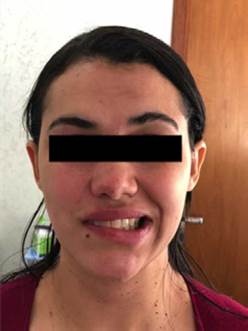
A 32-year-old woman, an early childhood educator, housewife, from a middle-class household residing in Mato Grosso do Sul, Brazil, presented in 2018, without an apparent cause, with an episode of right facial paralysis due to a decrease in frontalis muscle strength. Her symptoms at the time of consultation included incomplete closure of the right eye, decreased strength of the right eyelid, face asymmetry, and sensory alterations of the middle and lower segment of the right side of the face. She did not report pain, alteration in taste, or changes in salivary and lacrimal gland secretions (Figure 1).
At first, the patient opted for a private consultation to the neurology service at a secondary care institution in the city of Campo Grande, Mato Grosso do Sul, Brazil, where, due to her symptoms and on the basis of the physical examination, she was diagnosed with grade III right peripheral incomplete facial palsy, severity established according to the House-Brackmann score. As a result of her neurology appointment, she was prescribed treatment with oral prednisone 20 mg/day, which was administered for 5 days. During the same appointment, a brain MRI was performed in order to rule out any neurological involvement, obtaining a normal result. Given the persistence of the symptoms, the patient decided to consult the private Neural Therapy service after 10 days.
During the consultation with the Neural Therapy service, upon reviewing the patient's medical history, a number of personal medical records were found that showed previous irritations in the trigeminal nerve area that began at the age of 12 (Table 1).
Table 1 Patient's medical history in chronological order, specifying laterality, innervation, and facial nerve branches with anastomoses.
| Condition | Age | Laterality | Innervation | Anastomosis |
|---|---|---|---|---|
| Otitis | 12 years | Right | V3, X, VII | Facial nerve trunk, sensory branch of the facial nerve |
| Parotiditis | 15 years | Bilateral | Facial nerve trunk through the chorda tympani. | |
| Tonsillitis | 23 and 31 years | Bilateral | IX, X, V2 | Zygomatic branch of the facial nerve |
| Stye | 24 years | Right | V1, V2 | Temporal and zygomatic branches of the facial nerve |
Source: Own elaboration.
At the same consultation, a panoramic X-ray of the mouth was requested because of her dental history, in which an irritative focus was identified in tooth 16. With this result, the patient was referred to the Dentistry service for treatment 21 days after the onset of symptoms. At that visit, the dentist determined that there was a chronic periapical lesion in tooth 16 (Table 2).
Table 2 Dental history in chronological order specifying the intervention performed, dental piece, innervation, and facial nerve branch with anastomosis.
| Procedure | Age | Tooth | Innervation | Anastomosis |
|---|---|---|---|---|
| Restoration | 21-22 years | 16 | V2R* | Zygomatic branch of the facial nerve |
| Dental extraction | 23 years | 28, 38, 48 | V2R&L*, V3R* | Zygomatic branch of the facial nerve, facial trunk through the chorda tympani |
| Damage of restoration in tooth 16 | 30 years | 16 | V2R* | Zygomatic branch of the facial nerve |
| Restoration | 30-31 years | 16 | V2R* | Zygomatic branch of the facial nerve |
*R: right.
Source: Own elaboration.
At the Neural Therapy consultation, it was decided to start treatment from the first day of care with 1% procaine infiltrations, subject to prior informed consent. In this case, a better outcome was obtained after neural therapy stimulation with submucosal infiltration of 1% procaine in the alveolar branches of tooth 16, which was performed 20 days after the onset of symptoms. The following day, at the dental appointment, endodontics was performed on tooth 16 due to the lesion found. Table 3 shows the dental and neural therapy interventions performed in chronological order.
Table 3 Neural therapy and dental interventions correlated with patient clinical response.
| Course (days from symptom onset) | Intervention (neural therapy stimulation with 1% procaine injection) | Response |
|---|---|---|
| 10 days | Neural therapy stimulation with 1 cc of procaine in the right supraorbital and infraorbital nerve and tooth 18. | No clinical changes. |
| 13 days | Neural therapy stimulation near the right stellate ganglion with 3 cc of procaine. | No clinical changes. |
| 20 days | Neural therapy stimulation with 1 cc of procaine in tooth 16. | Irritation of the right eye conjunctiva 8 hours later. |
| 21 days | Dental care. Endodontic treatment tooth 16 | Progressive improvement of facial mimicry in 48 hours. Evolution towards grade I on the House-Brackmann score. |
| 24 days | Dental care. Extraction of tooth 18. | The improvement of facial mimicry is maintained. |
| 25 - 30 days | Control without neural therapy stimulus. | Complete recovery of facial mimicry. |
| 2 years later | Control. | Normal facial mimicry. |
Source: Own elaboration.
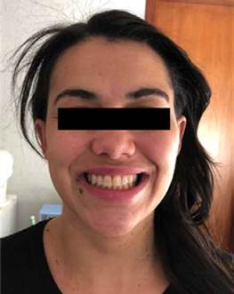
Within 48 hours of performing endodontic treatment on tooth 16, Bell's palsy improved from grade III to grade I on the House-Brackmann score, and facial mimicry symmetry was recovered. During the treatment, no concurrent interventions were performed other than those practiced by the Neural Therapy and Dentistry services (Figure 2).
Two years after undergoing the neural therapy and dental intervention, the patient returned for a follow-up, in which no adverse reactions to the treatment were reported, and no neurological sequelae were evidenced in the trigeminal and facial nerve area. During follow-up, the patient reported feeling satisfied with the treatment and authorized the publication of the case report.
DISCUSSION
The theory of Nervism defines what happened with the patient as a primary dystrophy of the trigeminal nerve, which correlates with her dental history of chronic periapical lesion in tooth 16 that caused a secondary dystrophy in the area of the facial nerve by reflex nerve mechanisms. Such dystrophy finally appeared in the form of paralysis due to the principle of Nervism as stated in Speransky's theory of the second stroke or sum of irritations 15,16.
The neuroanatomical connection between the V-VII cranial nerves enables the association of new theories with Bell's palsy:
Bell's palsy cases have been reported following dental procedures, trigeminal nerve injuries, and dental and bone infections 19-22.
Another theory refers to the vascular relationship between the middle meningeal artery that irrigates both nerves and the ischemic sympathetic reflex of the stylomastoid artery with the motor branch of the facial nerve 23,24.
According to Friedman, the proprioceptive fibers of the facial nerve are received by the trigeminal nerve at its mesencephalic nucleus 9,25.
Finally, Bell's palsy may be related to the cross-connection between the afferent nerve fibers of the intermediate nerve with the V2 fibers (maxillary nerve or pterygopalatine ganglion) 26.
From an embryological point of view, the trigeminal and facial nerves have an anastomotic association. In the fourth week of pregnancy, the first branch of the facial nerve appears and, at the end of the seventh week, the trunk of the facial nerve bifurcates into a temporal branch and a cervicofacial branch, creating an anastomosis with the V2 and V3 branches of the trigeminal nerve 27-29.
In the specific field of Neural Therapy, only cases of clinical improvement of facial paralysis have been reported, but without mentioning the pathogenic involvement of trigeminal irritation or dystrophy, nor the synergy with the Dentistry service 30-32. So far, clinical accounts have been limited to demonstrating that Neural Therapy can be a viable treatment option after conventional medical treatment has failed. However, these case reports did not take into consideration the evaluation of trigeminal nerve irritations as a possible etiologic factor. In fact, only 2 of the 7 published case reports cite the presence of dystrophic irritations in the area of the trigeminal nerve, such as dental treatments, temporomandibular joint dysfunction, tonsillitis, and chronic sinusitis. In these cases, reported in Turkey, mainly segmental Neural Therapy was performed in the head and neck area obtaining favorable outcomes 30-32.
The aim of this case report was to propose that Bell's palsy is caused by a cumulative involvement of the trigeminal nerve, a hypothesis based on the Neural Therapy approach and the physiological current of Nervism. In this case, the patient who consulted the Neural Therapy service presented with a clear history of trigeminal field irritations during her lifetime, with a predominance in the V2 branch, which were related to the interventions and dental foci described in Tables 1 and 2. Thus, a new etiologic hypothesis on the pathophysiologic relationship between the trigeminal and facial nerves is hereby proposed, which should be analyzed in further studies given the limitations of the present study as it is a case report.
Finally, this case exemplifies a situation in which a previous dystrophy or anterior alteration of the trigeminal nerve through its different branches, cumulative over time, ends up affecting via anastomosis the nervous tone or trophism of the facial nerve, which has a clear morphological and physiological support under the premise of the unity of the nervous system.
CONCLUSIONS
This case report not only highlights the role of Neural Therapy in the therapeutic support of Bell's palsy, but also represents a contribution to medical knowledge from a different physiological conceptual framework such as Nervism. It is suggested that obtaining more information on the patient's clinical history is important to determine the relationship between the facial nerve and the trigeminal nerve (Table 2), as it is a useful tool to demonstrate these findings. Furthermore, this article is the first case report showing the synergy between the Neural Therapy and Dentistry services for successful therapeutic support in Bell's Palsy.

