SciELO - Scientific Electronic Library Online

 
vol.4 número1Manifestaciones gastrointestinales de la enfermedad renal crónicaImportancia de la hiperfosfatemia en la enfermedad renal crónica, cómo evitarla y tratarla por medidas nutricionales índice de autoresíndice de materiabúsqueda de artículos
Home Pagelista alfabética de revistas  

Servicios Personalizados

Revista

Articulo

Indicadores

Links relacionados

  • En proceso de indezaciónCitado por Google
  • No hay articulos similaresSimilares en SciELO
  • En proceso de indezaciónSimilares en Google

Compartir


Revista Colombiana de Nefrología

versión On-line ISSN 2500-5006

Rev. colom. nefrol. vol.4 no.1 Bogotá ene./jun. 2017

https://doi.org/10.22265/acnef.4.1.263 

Review article

Cyclooxygenase biology in renal function - literature review

Marcella Goetz Moro1 

Paula Katherine Vargas Sánchez1 

Ana Caroline Lupepsa2 

Emeline Maria Baller2 

Gilson César Nobre Franco3  * 

1Department of Dentistry, Ponta Grossa State University, Brasil

2Department of Pharmaceutical Sciences, Ponta Grossa State University, Brasil

3Department of General Biology, Ponta Grossa State University, Brasil


Abstract

The cyclooxygenase (COX) exists in two main isoforms, COX-1 and COX-2, which are present in the renal system to ensure its homeostasis. However, in different clinical situations, these enzymes can play a physiologic role in maintaining the integrity of this organ, and also be associated with the worsening of tissue injuries/damage. In this sense, an explanation of the true biological function of the isoforms of COX enables a better understanding of the physiology and pathology of the kidney, as well as a better understanding of the consequences of its inhibition by the use of drugs. This review aimed to study the biological role of the COX enzyme in the renal system in different clinical situations.

Key words: Kidney; cyclooxygenase 1; cyclooxygenase 2; pathology; anti-Inflammatory agents; biology

Resumen

La ciclooxigenasa existe en dos isoformas principales: COX-1 y COX-2, estas se encuentran presentes en el sistema renal como parte de su homeostasis. Sin embargo, en algunas situaciones clínicas, las dos enzimas pueden desempeñar un papel en el mantenimiento de la integridad de este órgano, y en otras pueden estar asociadas a la evolución de daños y lesiones en los tejidos. En este sentido, el conocimiento de la verdadera función biológica de las isoformas de la COX permite una mejor comprensión de la fisiología y patología del riñón, así como una mejor comprensión de las consecuencias de su inhibición por el uso de medicamentos. El objetivo de esta revisión es estudiar la función biológica de la enzima COX en el sistema renal en diferentes situaciones clínicas.

Palabras clave: riñón; ciclooxigenasa 1; ciclooxigenasa 2; patología; antiinflamatorios; biología

Introduction

Cyclooxygenase (COX), the main catalyst enzyme in the prostanoid synthesis, has two main known isoforms, COX-1 and COX-2[1,2]. COX-1, which is responsible for physiological functions, is expressed mainly as a constitutive isoform[3] and, in normal conditions and in distinct regions of the kidney, generates prostaglandins that are responsible for vasodilation and diminished vascular resistance, ensuring adequate blood flow[4]. On the other hand, COX-2, initially considered pathological, is highly expressed in the presence of damaging stimuli[1]. However, this isoform is now also known to appear constitutively in some tissues, like in the kidney, where it is responsible for ensuring the tubuloglomerular feedback, contributing to the establish homeostasis[5].

In this sense, although the COX functions have been well-studied in normal situations, studies have demonstrated that, facing different clinical situations, its isoforms may assume different roles. In cases of hypovolemia and hypertension, this enzyme can develop the physiological role of maintaining the integrity of the kidney, as well as becoming associated with the aggravation of tissue lesion/damage, as observed in the polycystic kidney disease[4,6,10].

Thus, clarifying the true biological functioning of the COX isoforms enables a better comprehension of the kidney physiology and pathology, as well as a better understanding of the consequences of the isoforms' inhibition by medication. This review intended to study the biological role of the COX enzyme in the renal system in different clinical situations.

Cyclooxygenase

The cyclooxygenase (COX) is the main enzyme that catalyses the prostaglandin synthesis from arachidonic acid. This way, it originates different prostanoids with different functions in the organism[1].

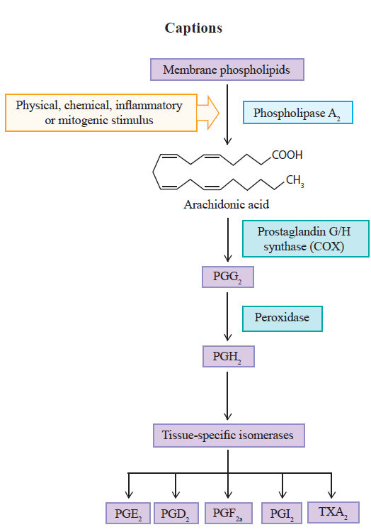
This process is initiated with the archidonic acid, present in the lipid bilayer of the cell membrane, triggering the prostanoid biosynthesis pathway due to the presence of different stimuli[11,16]. The pathway involves a sequence of three main steps. Initially, the arachidonic acid is liberated, by hydrolysis, through the action of the phospholipase A2 enzyme (PLA2). Once liberated, the second step begins, with oxygenation by the prostaglandin G/H synthase, or cyclooxygenase, resulting in the prostaglandin endoperoxide G2 (PGG2). In sequence, the same COX enzymes reduce the PGG2 into prostaglandin H2 (PGH2)[15,17]. The prostaglandin G/H synthase has the cyclooxygenase and peroxidase catalytic sites. The former converts arachidonic acid into PGG2, which is acted upon by the peroxidase enzyme and reduced to the unstable intermediate, PGH2, responsible for the formation of different kinds of prostanoids such as prostacyclin (PGI2), prostaglandin D2 (PGD2), prostaglandin E2 (PGE2), prostaglandin F2a (PGF2a) and thromboxane A2 (TXA2), through its conversion by the tissue-specific isomerases[13,18,20]. These prostanoids provoke different actions in the organism through specific receptors, which depend on the type of cell and tissue involved in the process (Figure 1) [20,22].

Figure 1 Prostanoid biosynthesis pathway. Source: author 

There are different isoforms of COX, but the most studied are COX-1 and COX-2[1]. COX-1 was initially named physiological or constitutive for being present in the majority of tissues and being expressed in an almost steadily fashion[13,19,23]. Its main role consists of maintaining a basal rate of the biosynthesis of prostanoids in the body, in order to promote their rapid, short-duration increase. On the other hand, COX-2 is hardly found in most tissues, but its production increases by approximately 20 times in the presence of inflammatory stimuli. For this reason, it has been called the pathological or induced enzyme[13,19,23,24]. However, recent studies have demonstrated that it is also found constitutively in some tissues, being responsible for the formation of prostanoids that maintain homeostasis in different organs and systems of the body[1,24,26].

Cyclooxygenase in renal function Normal Function

COX-1 is found in the glomeruli, in the medullary and cortical region of the collecting ducts and in the afferent and efferent arterioles[27], ensuring the maintenance of the kidney's physiological functions, such as hemodynamic regulation and glomerular filtration rate (GFR)[13,28,30]. This isoform produces PGE2 and PGD2, which antagonize the vasoconstrictive action of angiotensin II (Ang II), and inhibit the release of norepinephrine, respectively[31].

In this way, these prostanoids promote vasodilation, increasing the perfusion of the organ and causing redistribution in the blood flow from the renal cortex to the nephrons in the intramedullary region[31]. Additionally, the PGE2, alongside the PGF2a, possess the diuretic and natriuretic effects and the PGE2, like the PGI2, antagonize the action of vasopressin[31].

Although COX-2 has been initially considered pathological, is also found constitutively in the kidneys[29,32]. Studies in COX-2 knockout rats demonstrate a grave defect in the renal formation, suggesting this isoform performs an important task in the development of the kidneys[33].

COX-2 is situated in particularly important regions for the renal function, concentrating mainly in the medullary region, and in the cortical region to a lesser extent[32]. Specifically, it can be found in smooth muscle cells of the afferent and efferent arterioles, in the endothelium (straight venules and vasa recta), renal artery, interstitial fibroblasts, and in podocytes in healthy kidneys[34]. It is also expressed in the thick ascending limb of the Henle loop, in addition to being found in the macula densa, which is responsible for mediating the interaction between glomerular filtration and proximal reabsorption, and for controlling the levels of sodium and potassium ions in the lumen of the distal tubule through the renin-angiotensin-aldosterone system[7,8,27,28,35].

Like in COX-1, prostaglandins (PGs) also derive from COX-2, being important physiological modulators of vascular tone and hydric balance in the kidneys[13,35]. The prominent PGs in renal tissue are PGE2 and PGI2, where PGE2 is synthesized by the tubular epithelium and interstitial cells, expressed in the renal tubules regulating the transport of chloride and sodium in the Henle loop, besides auxiliating water transport and blood flow. PGI2 is located in the renal cortex, controlling the glomerular filtration rate and renin secretion[7,29].

Hypovolemia

Hypovolemia is characterized by a decrease in blood volume, being mainly the result of excessive loss of water in the kidney or hemorrhage. As a consequence, arterial pressure drops, with the possibility of hypovolemic shock and death[13,31].

In a state of hypovolemia with consequent depletion of sodium levels, there is an increase of COX-2 in the cortical region, and a decrease of its expression in the medullary region, causing the renin-angiotensin-aldosterone system to be activated. The juxtaglomerular cells secrete the hormone renin, which contributes to the formation of Ang II, responsible for the vasoconstriction of the efferent arteriole together with norepinephrine, guaranteeing the increase of the intraglomerular hydrostatic pressure. The hormone also stimulates the secretion of aldosterone, responsible for promoting the reabsorption of sodium and, ultimately, inducing the liberation of antidiuretic hormone (ADH), which increases water and urea reabsorption. At the same time, angiotensin stimulates the synthesis of renal vasodilator prostaglandins (afferent arteriole), which guarantees the tubuloglomerular feedback, reestablishing the glomerular filtration rate, and ensuring normal blood flow to protect the kidney from acute functional deterioration[5,13,31,35,37].

The administration of selective or non-selective COX-2 non-steroidal anti-inflammatory drugs (NSAIDs) prevents the release of renin and, consequently, the restoration of renal function[38].

Hypotension

Hypotension leads to low effective filtration pressure in the glomeruli, which can become a state of chronic kidney disease. In these situations, the function of compensatory vasoconstrictor and vasodilator prostaglandins (PGE2 e PGI2), provenient from COX-2, is amplified in an attempt to maintain adequate organ perfusion[39,40].

This action favors the resorption of sodium/ water, which maintains a renal homeostasis by the GFR. In agreement, the thromboxane (TX), produced by COX-1, provokes vasoconstriction. Thus, NSAIDs may be use carefully because they can act blocking the COX-1 and COX-2 enzymes and, consequently this mechanism[41]. In the study of Calistro Neto and group (2015), animals that use Parecoxib, a selective inhibitor of COX-2, had renal detrimental effect by the non-activation of renin-angioten-sin-aldosterone system[41].

In disagreement with the results, Tunctan and group (2013) declare that COX-2 induces hypotension, because their vasodilatory capacity. This study concludes that the use of a COX-2 inhibitor is able to restore blood pressure[42].

Renal ischemia/reperfusion injury

The pathology of this injury is marked by the presence of an inflammatory response and oxidative stress[43]. The COX-2 is highly expressed and is able to aggravate the inflammation. So, the use of a NSAID that selectively inhibits COX-2, also called coxib, can be benefic to the renal function by prevention of oxidative injury[27,43,44].

More specifically, the use of parecoxib, a coxib, resulted in renal protection[41]. A similar study that uses indomethacin, a non-selective NSAID, and rofecoxib, a coxib, showed that both drugs were associated with improved renal function and a reduction of proinflammatory cytokine levels[45].

To finish, the use of nimesulida, a COX-2 selective inhibitor drug, also prevented the renal damage[46].

Hypertension

In contrast, in a state of hypertension, COX-2 expression will also be increased. However, unlike in hypotension, this increase will occur in the medullary tissues in an attempt to ensure that the vasoconstrictive and hypertensinogenic effects of Ang II be softened, aiming then to reestablish sodium and water regulation, as well as the maintenance of medullary blood flow[47]. In this sense, the use of any NSAIDs aggravates the hypertension and it must be avoided. The block of PGE2 by drugs lead to sodium retention which exacerbate the deleterious process[38].

Fibrosis

Another alteration of the renal system is fibrosis. It is characterized by the development of glomerular sclerosis and interstitial fibrosis, being one of the consequences of the final stages of several chronic kidney diseases[48]. The COX-2 isoform guarantees protection against fibroses and, by virtue of that, the high quantity of PGE2 generated can hinder the development of the condition[48].

Polycystic kidney disease

As well as renal ischemia/reperfusion injury, the COX enzyme can also present deleterious effects in other situations in the renal system. In the presence of polycystic kidney disease, a autosomal dominant renal disorder frequently hereditary in nature characterized by the continuous growth of cysts, there is an increase in activity of PGE2 and PGI2 (derived from both isoforms of COX), responsible for stimulating cellular proliferation, secretion of fluid and cyst formation[49].

In a humam study, by the blood collection, it was found that patients with autosomal polycystic kidney disease (APKD) have high levels of COX compared to the health group[9]. Agreeing with these authors, by culture of epithelial cells from cysts of patients with APKD, Xu et al. (2012)[50] concluded that the use of a COX-2 inhibitor is capable to reducing the epithelial proliferation, promoting apoptosis of these, and decrease the PGE2 synthesis[50].

Thus, the inhibition of COX-1 or COX-2 by NSAIDs may reduce the progression of the disease[10].

Diabetes mellitus

Diabetes mellitus is a chronic disease that leads to kidney damage. In this context, it has been found that inducing COX-2 in the proximal tubules may lead to an increase in PGE2 production and in protein excretion in diabetic mice[51]. Even so, studies are showing that COX-2 and PGE2 production were increased in patients with diabetes mellitus, leading to the use of a COX-2 inhibition which can attenuate different renal diseases[52].

In agreement with these findings, a study conducted in animal models has determined that, when administering rofecoxib, a selective COX-2 inhibitor, protenuria levels in Zucker rats normalized[53]. In contrast, there are reports in the literature that in Zucker rats (obese) and db/db mice (insulin resistant), renal COX-2 has been positively regulated[54,55].

Diabetic patients have an excessive polyuria and urinary concentration of PGE2. So, the use of a NSAID must be used to control the levels of PGE2 and adequate the urine concentration[27].

In the study of Liu and group (2016), Ibuprofen was administered in rats with renal impairment and type 1 diabetes. The use of ibuprofen was able to decrease urinary protein excretion, blood urea nitrogen, glomerular basement membrane thickening, renal fibrosis, COX-2, and interleukin 1(3 (IL-1P) level, suggesting that this drug can prevent the development of renal damage in the diabetic group[56].

Kidney inflammation

The expression of COX-2 enzyme is considerably increased in response to renal tissue inflammation[57]. When activated in pathological processes, there is the formation of proinflammatory cytokines (interleukin-1 and TNF-a) and reactive oxygen species (ROS), which play a critical role in the renal physiopathology[40,58]. There is also a positive feedback through the signaling pathways that promote the development and progression of the disease[40,58,59] .

Authors state that inflammation is the causal factor that collaborates with renal lesion, especially with PGE2 which, although in physiological conditions contributes to the maintenance of renal homeostasis[40,60,61], in pathological situations might be responsible for unleashing a deleterious process[40].

This difference in the PGE2 action in various clinical situations is due to its attachment to four receptors, EP1, EP2, EP3 and EP4[40,62]. EP1 is responsible for natriuresis and diuresis and regulation of blood pressure. Receptors EP2 and EP4 have been reported to activate production of adenylate cyclase and increase (cAMP) and to increase water reabsorption, on the other hand, EP3 have the opposite effects[27,61,63,64].

A study conducted in rats subjected to nephrectomy has found that, after 4 weeks, the animals presented increased levels of urea and creatinine, reduction of creatinine clearance, and disorders in the metabolism of calcium and phosphorus, highlighting significant kidney failure[40]. However, in rats where PGE2 production was suppressed, there was a notable and stable improvement in all those alterations, suggesting that the PGE2-induced inflammation may be involved in the progression of renal lesion[40].

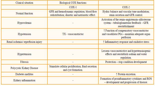
The table 1 presents a summary of the COX (COX-1 and COX-2) system roles in the different clinical situations mentioned.

Table: 1 Biological functions of the COX enzyme in different clinical situations. 

Conclusion

Both COX isoforms play important roles in the regulation of renal homeostasis. However, in different clinical situations, their functions might be altered. Thus, the exactly role of COX-1 and COX-2 and their inhibition by the use of NSAIDs still remains controversial. So, it is important that the health care professional be aware of these possible alterations in order to provide the best clinical course of action.

References

1. Akyazi I, Eraslan E, Gulcubuk A, et al. Long-term aspirin pretreatment in the prevention of cerulein-induced acute pancreatitis in rats. World journal of gastroenterology : WJG 2013;19:2894-2903. [ Links ]

2. Chikazu D, Tomizuka K, Ogasawara T, et al. Cyclooxygenase-2 activity is essential for the osseointegration of dental implants. International journal of oral and maxillofacial surgery 2007; 36:441-446. [ Links ]

3. Troxler M, Dickinson K, Homer-Vanniasinkam S. Platelet function and antiplatelet therapy. The British journal of surgery 2007; 94:674-682. [ Links ]

4. Sanchez PL, Salgado LM, Ferreri NR, Escalante B. Effect of cyclooxygenase-2 inhibition on renal function after renal ablation. Hypertension 1999; 34:848-853. [ Links ]

5. Lomas AL, Grauer GF. The renal effects of NSAIDs in dogs. Journal of the American Animal Hospital Association 2015; 51:197-203. [ Links ]

6. Rouzer CA, Marnett LJ. Cyclooxygenases: structural and functional insights. Journal of lipid research 2009;50 Suppl:S29-34. [ Links ]

7. Hao S, Hernandez A, Quiroz-Munoz M, Cespedes C, Vio CP, Ferreri NR. PGE(2) EP(3) receptor downregulates COX-2 expression in the medullary thick ascending limb induced by hypertonic NaCl. American journal of physiology Renal physiology 2014; 307:F736-746. [ Links ]

8. Kaminska K, Szczylik C, Lian F, Czarnecka AM. The role of prostaglandin E2 in renal cell cancer development: future implications for prognosis and therapy. Future oncology (London, England) 2014; 10:2177-2187. [ Links ]

9. Klawitter J, Klawitter J, McFann K, et al. Bioactive lipid mediators in polycystic kidney disease. Journal of lipid research 2014; 55:1139-1149. [ Links ]

10. Liu Y, Rajagopal M, Lee K, et al. Prostaglandin E(2) mediates proliferation and chloride secretion in ADPKD cystic renal epithelia. American journal of physiology Renal physiology 2012; 303:F1425-1434. [ Links ]

11. Vane JR. Inhibition of prostaglandin synthesis as a mechanism of action for aspirin-like drugs. Nature: New biology 1971; 231:232-235. [ Links ]

12. Marnett LJ, Rowlinson SW, Goodwin DC, Kalgutkar AS, Lanzo CA. Arachidonic acid oxygenation by COX-1 and COX-2. Mechanisms of catalysis and inhibition. The Journal of biological chemistry 1999; 274:22903-22906. [ Links ]

13. Kummer CL, Coelho TC. [Cycloxygenase-2 inhibitors nonsteroid anti-inflammatory drugs: current issues.]. Revista brasileira de anestesiologia 2002;52:498-512. [ Links ]

14. Miller SB. Prostaglandins in health and disease: an overview. Seminars in arthritis and rheumatism 2006; 36:37-49. [ Links ]

15. Patrono C, Rocca B. Nonsteroidal antiinflammatory drugs: past, present and future. Pharmacological research : the official journal of the Italian Pharmacological Society 2009;59:285-289. [ Links ]

16. Smith JW, Al-Khamees O, Costall B, Naylor RJ, Smythe JW. Chronic aspirin ingestion improves spatial learning in adult and aged rats. Pharmacology, biochemistry, and behavior 2002; 71:233-238. [ Links ]

17. Moncada S, Ferreira SH, Vane JR. Bioassay of prostaglandins and biologically active substances derived from arachidonic acid. Advances in prostaglandin and thromboxane research 1978; 5:211-236. [ Links ]

18. Rimon G, Sidhu RS, Lauver DA, et al. Coxibs interfere with the action of aspirin by binding tightly to one monomer of cyclooxygenase-1. Proceedings of the National Academy of Sciences of the United States of America 2010; 107:28-33. [ Links ]

19. Kirkby NS, Chan MV, Lundberg MH, et al. Aspirin-triggered 15-epilipoxin A4 predicts cyclooxygenase-2 in the lungs of LPS-treated mice but not in the circulation: implications for a clinical test. FA-SEB journal : official publication of the Federation of American Societies for Experimental Biology 2013;27:3938-3946. [ Links ]

20. Patrignani P, Patrono C. Cyclooxygenase inhibitors: From pharmacology to clinical read-outs. Biochimica et biophysica acta 2015; 1851:422-432. [ Links ]

21. Aldrovandi M, Hammond VJ, Podmore H, et al. Human platelets generate phospholipid-esterified prostaglandins via cyclooxygenase-1 that are inhibited by low dose aspirin supplementation. Journal of lipid research 2013; 54:3085-3097. [ Links ]

22. Kawahara K, Hohjoh H, Inazumi T, Tsuchiya S, Sugimoto Y. Prostaglandin E-induced inflammation: Relevance of prostaglandin E receptors. Biochimica et biophysica acta 2015; 1851:414-421. [ Links ]

23. Hilario MO, Terreri MT, Len CA. Nonsteroidal anti-inflammatory drugs: cyclooxygenase 2 inhibitors. Jornal de pediatria 2006; 82:S206-212. [ Links ]

24. Barudzic N, Turjacanin-Pantelic D, Zivkovic V, et al. The effects of cyclooxygenase and nitric oxide synthase inhibition on oxidative stress in isolated rat heart. Molecular and cellular biochemistry 2013; 381:301-311. [ Links ]

25. Cairns JA. The coxibs and traditional nonsteroidal anti-inflammatory drugs: a current perspective on cardiovascular risks. The Canadian journal of cardiology 2007; 23:125-131. [ Links ]

26. Murtaza G, Karim S, Najamul-Haq M, et al. Interaction analysis of aspirin with selective amino acids. Acta poloniae pharmaceutica 2014; 71:139-143. [ Links ]

27. Norregaard R, Kwon TH, Frokiaer J. Physiology and pathophysiology of cyclooxygenase-2 and prostaglandin E2 in the kidney. Kidney research and clinical practice 2015; 34:194-200. [ Links ]

28. Nantel F, Meadows E, Denis D, Connolly B, Metters KM, Giaid A. Immunolocalization of cyclooxygenase-2 in the macula densa of human elderly. FEBS letters 1999; 457:475-477. [ Links ]

29. DeMaria AN, Weir MR. Coxibs--beyond the GI tract: renal and cardiovascular issues. Journal of pain and symptom management 2003; 25:S41-49. [ Links ]

30. Moore N, Pollack C, Butkerait P. Adverse drug reactions and drug-drug interactions with over-the-counter NSAIDs. Therapeutics and clinical risk management 2015; 11:1061-1075. [ Links ]

31. Batlouni M. [Nonsteroidal anti-inflammatory drugs: cardiovascular, cerebrovascular and renal effects]. Arquivos brasileiros de cardiologia 2010; 94:556-563. [ Links ]

32. Ahmetaj-Shala B, Kirkby NS, Knowles R, et al. Evidence that links loss of cyclooxygenase-2 with increased asymmetric dimethylarginine: novel explanation of cardiovascular side effects associated with anti-inflammatory drugs. Circulation 2015; 131:633-642. [ Links ]

33. Zhang MZ, Wang JL, Cheng HF, Harris RC, McKanna JA. Cyclooxygenase-2 in rat nephron development. The American journal of physiology 1997; 273:F994-1002. [ Links ]

34. Khan KN, Venturini CM, Bunch RT, et al. Interspecies differences in renal localization of cyclooxygenase isoforms: implications in nonsteroidal antiinflammatory drug-related nephrotoxicity. Toxicologic pathology 1998; 26:612-620. [ Links ]

35. Dubois RN, Abramson SB, Crofford L, et al. Cyclooxygenase in biology and disease. FASEB journal : official publication of the Federation of American Societies for Experimental Biology 1998;12:1063-1073. [ Links ]

36. Lipsky PE. Defining COX-2 inhibitors. The Journal of rheumatology Supplement 2000; 60:13-16. [ Links ]

37. Curiel RV, Katz JD. Mitigating the cardiovascular and renal effects of NSAIDs. Pain medicine (Malden, Mass) 2013; 14 Suppl 1:S23-28. [ Links ]

38. Harris RC. Physiologic and pathophysiologic roles of cyclooxygenase-2 in the kidney. Transactions of the American Clinical and Climatological Association 2013; 124:139-151. [ Links ]

39. Wang C, Luo Z, Kohan D, et al. Thromboxane prostanoid receptors enhance contractions, endothelin-1, and oxidative stress in microvessels from mice with chronic kidney disease. Hypertension 2015; 65:1055-1063. [ Links ]

40. Jia Z, Zhang Y, Ding G, Heiney KM, Huang S, Zhang A. Role of COX-2/mPGES-1/prostaglandin E2 cascade in kidney injury. Mediators of inflammation 2015; 2015:147894. [ Links ]

41. Calistro Neto JP, Torres Rda C, Goncalves GM, et al. Parecoxib reduces renal injury in an ischemia/ reperfusion model in rats. Acta cirurgica brasileira / Sociedade Brasileira para Desenvolvimento Pesquisa em Cirurgia 2015; 30:270-276. [ Links ]

42. Tunctan B, Korkmaz B, Sari AN, et al. Contribution of iNOS/sGC/PKG pathway, COX-2, CYP4A1, and gp91(phox) to the protective effect of 5,14-HEDGE, a 20-HETE mimetic, against vasodilation, hypotension, tachycardia, and inflammation in a rat model of septic shock. Nitric oxide : biology and chemistry / official journal of the Nitric Oxide Society 2013;33:18-41. [ Links ]

43. Liu HB, Meng QH, Huang C, Wang JB, Liu XW. Nephroprotective Effects of Polydatin against Is-chemia/Reperfusion Injury: A Role for the PI3K/Akt Signal Pathway. Oxidative medicine and cellular longevity 2015; 2015:362158. [ Links ]

44. Wang ZS, Liu XH, Wang M, et al. Metformin attenuated the inflammation after renal ischemia/reperfusion and suppressed apoptosis of renal tubular epithelial cell in rats. Acta cirurgica brasileira / Sociedade Brasileira para Desenvolvimento Pesquisa em Cirurgia 2015; 30:617-623. [ Links ]

45. Feitoza CQ, Goncalves GM, Semedo P, et al. Inhibition of COX 1 and 2 prior to renal ischemia/ reperfusion injury decreases the development of fibrosis. Molecular medicine (Cambridge, Mass) 2008; 14:724-730. [ Links ]

46. Suleyman Z, Sener E, Kurt N, Comez M, Yapanoglu T. The effect of nimesulide on oxidative damage inflicted by ischemia-reperfusion on the rat renal tissue. Renal failure 2015; 37:323-331. [ Links ]

47. Gonzalez AA, Green T, Luffman C, Bourgeois CR, Gabriel Navar L, Prieto MC. Renal medullary cyclooxygenase-2 and (pro)renin receptor expression during angiotensin II-dependent hypertension. American journal of physiology Renal physiology 2014; 307:F962-970. [ Links ]

48. Kamata M, Hosono K, Fujita T, Kamata K, Majima M. Role of cyclooxygenase-2 in the development of interstitial fibrosis in kidneys following unilateral ureteral obstruction in mice. Biomedicine & pharmacotherapy = Biomedecine & pharmacotherapie 2015; 70:174-180. [ Links ]

49. Ibrahim NH, Gregoire M, Devassy JG, et al. Cyclooxygenase product inhibition with acetylsalicylic acid slows disease progression in the Han:SPRD-Cy rat model of polycystic kidney disease. Prostaglandins & other lipid mediators 2015; 116-117:19-25. [ Links ]

50. Xu T, Wang NS, Fu LL, Ye CY, Yu SQ, Mei CL. Celecoxib inhibits growth of human autosomal dominant polycystic kidney cystlining epithelial cells through the VEGF/Raf/MAPK/ERK signaling pathway. Molecular biology reports 2012; 39:7743-7753. [ Links ]

51. Mohamed R, Jayakumar C, Ranganathan PV, Ganapathy V, Ramesh G. Kidney proximal tubular epithelial-specific overexpression of netrin-1 suppresses inflammation and albuminuria through suppression of COX-2-mediated PGE2 production in streptozotocin-induced diabetic mice. The American journal of pathology 2012; 181:1991-2002. [ Links ]

52. Jia Z, Sun Y, Liu S, Liu Y, Yang T. COX-2 but not mPGES-1 contributes to renal PGE2 induction and diabetic proteinuria in mice with type-1 diabetes. PloS one 2014; 9:e93182. [ Links ]

53. Dey A, Williams RS, Pollock DM, et al. Altered kidney CYP2C and cyclooxygenase-2 levels are associated with obesity-related albuminuria. Obesity research 2004; 12:1278-1289. [ Links ]

54. Sun Y, Jia Z, Liu G, et al. PPARgamma Agonist Rosiglitazone Suppresses Renal mPGES-1/PGE2 Pathway in db/db Mice. PPAR research 2013; 2013:612971. [ Links ]

55. Komers R, Zdychova J, Cahova M, Kazdova L, Lindsley JN, Anderson S. Renal cyclooxygenase-2 in obese Zucker (fatty) rats. Kidney international 2005; 67:2151-2158. [ Links ]

56. Liu YW, Zhu X, Cheng YQ, et al. Ibuprofen attenuates nephropathy in streptozotocininduced diabetic rats. Molecular medicine reports 2016; 13:5326-5334. [ Links ]

57. Song KI, Park JY, Lee S, et al. Protective effect of tetrahydrocurcumin against cisplatin-induced renal damage: in vitro and in vivo studies. Planta medica 2015; 81:286-291. [ Links ]

58. Feng L, Xia Y, Garcia GE, Hwang D, Wilson CB. Involvement of reactive oxygen intermediates in cyclooxygenase-2 expression induced by interleukin-1, tumor necrosis factor-alpha, and lipopolysaccharide. The Journal of clinical investigation 1995; 95:1669-1675. [ Links ]

59. Fujihara CK, Antunes GR, Mattar AL, et al. Cyclooxygenase-2 (COX-2) inhibition limits abnormal COX-2 expression and progressive injury in the remnant kidney. Kidney international 2003; 64:2172-2181. [ Links ]

60. Kinsey GR, Sharma R, Okusa MD. Regulatory T cells in AKI. Journal of the American Society of Nephrology : JASN 2013;24:1720-1726. [ Links ]

61. Hao CM, Breyer MD. Physiologic and pathophysiologic roles of lipid mediators in the kidney. Kidney international 2007; 71:1105-1115. [ Links ]

62. Nasrallah R, Hassouneh R, Hebert RL. Chronic kidney disease: targeting prostaglandin E2 receptors. American journal of physiology Renal physiology 2014; 307:F243-250. [ Links ]

63. Zhang Y, Guan Y, Schneider A, Brandon S, Breyer RM, Breyer MD. Characterization of murine vasopressor and vasodepressor prostaglandin E(2) receptors. Hypertension 2000; 35:1129-1134. [ Links ]

64. Breyer MD, Breyer RM. Prostaglandin E receptors and the kidney. American journal of physiology Renal physiology 2000; 279:F12-23. [ Links ]

Referenciar este artículo: Goetz Moro M, Vargas Sánchez PK, Lupepsa AC, Baller EM, Nobre Franco GC. Cyclooxygenase biology in renal function - literature review. Rev. Colomb. Nefrol. 2017;4(1): 27 - 37

Ethical disclosures Protection of human and animal subjects The authors declare that no experiments were performed on humans or animals for this study

Confidentiality of data The authors declare that no patient data appear in this article

Right to privacy and informed consent The authors declare that no patient data appear in this article

Funding The authors expressly state that there is no conflict of interest

Received: May 12, 2016; Accepted: February 12, 2016; other: February 07, 2017

* Corresponding author: Gilson Cesar Nobre Franco. Universidade Estadual de Ponta Grossa - Departamento de Odontologia - Pós Graduação Stricto Sensu em Odontologia. Av. General Carlos Cavalcanti, n° 4748, Campus de Uvaranas, Ponta Grossa, PR, Brazil. ZIP code: 84030-900, e-mail: gilsoncnf@gmail.com.

Creative Commons License This is an open-access article distributed under the terms of the Creative Commons Attribution License