<?xml version="1.0" encoding="ISO-8859-1"?><article xmlns:mml="http://www.w3.org/1998/Math/MathML" xmlns:xlink="http://www.w3.org/1999/xlink" xmlns:xsi="http://www.w3.org/2001/XMLSchema-instance">
<front>
<journal-meta>
<journal-id>0012-7353</journal-id>
<journal-title><![CDATA[DYNA]]></journal-title>
<abbrev-journal-title><![CDATA[Dyna rev.fac.nac.minas]]></abbrev-journal-title>
<issn>0012-7353</issn>
<publisher>
<publisher-name><![CDATA[Universidad Nacional de Colombia]]></publisher-name>
</publisher>
</journal-meta>
<article-meta>
<article-id>S0012-73532015000100015</article-id>
<article-id pub-id-type="doi">10.15446/dyna.v82n189.42364</article-id>
<title-group>
<article-title xml:lang="en"><![CDATA[A viable wireless PC assisted alternative to studies of vectorcardiography]]></article-title>
<article-title xml:lang="es"><![CDATA[Una viable alternativa para estudios de vectorcardiografía de manera inalámbrica asistida por computadora]]></article-title>
</title-group>
<contrib-group>
<contrib contrib-type="author">
<name>
<surname><![CDATA[Estrada-Gutiérrez]]></surname>
<given-names><![CDATA[Juan Carlos]]></given-names>
</name>
<xref ref-type="aff" rid="A01"/>
</contrib>
<contrib contrib-type="author">
<name>
<surname><![CDATA[Hernández-Sámano]]></surname>
<given-names><![CDATA[Alfonso]]></given-names>
</name>
<xref ref-type="aff" rid="A01"/>
</contrib>
<contrib contrib-type="author">
<name>
<surname><![CDATA[Mazón-Valadez]]></surname>
<given-names><![CDATA[Ernesto Edgar]]></given-names>
</name>
<xref ref-type="aff" rid="A01"/>
</contrib>
<contrib contrib-type="author">
<name>
<surname><![CDATA[Ávila-Paz]]></surname>
<given-names><![CDATA[José]]></given-names>
</name>
<xref ref-type="aff" rid="A01"/>
</contrib>
<contrib contrib-type="author">
<name>
<surname><![CDATA[González-Vega]]></surname>
<given-names><![CDATA[Arturo]]></given-names>
</name>
<xref ref-type="aff" rid="A02"/>
</contrib>
<contrib contrib-type="author">
<name>
<surname><![CDATA[Leyva-Cruz]]></surname>
<given-names><![CDATA[Juan Alberto]]></given-names>
</name>
<xref ref-type="aff" rid="A03"/>
</contrib>
<contrib contrib-type="author">
<name>
<surname><![CDATA[Cano-González]]></surname>
<given-names><![CDATA[Mario Eduardo]]></given-names>
</name>
<xref ref-type="aff" rid="A01"/>
</contrib>
</contrib-group>
<aff id="A01">
<institution><![CDATA[,Universidad de Guadalajara Centro Universitario de la Ciénega ]]></institution>
<addr-line><![CDATA[Guadalajara ]]></addr-line>
<country>México</country>
</aff>
<aff id="A02">
<institution><![CDATA[,Universidad de Guanajuato División de Ciencias e Ingenierías ]]></institution>
<addr-line><![CDATA[León ]]></addr-line>
<country>México</country>
</aff>
<aff id="A03">
<institution><![CDATA[,Universidade de Estadual de Feira de Santana Dpto. de Física ]]></institution>
<addr-line><![CDATA[Bahía ]]></addr-line>
<country>Brasil</country>
</aff>
<pub-date pub-type="pub">
<day>00</day>
<month>02</month>
<year>2015</year>
</pub-date>
<pub-date pub-type="epub">
<day>00</day>
<month>02</month>
<year>2015</year>
</pub-date>
<volume>82</volume>
<numero>189</numero>
<fpage>118</fpage>
<lpage>126</lpage>
<copyright-statement/>
<copyright-year/>
<self-uri xlink:href="http://www.scielo.org.co/scielo.php?script=sci_arttext&amp;pid=S0012-73532015000100015&amp;lng=en&amp;nrm=iso"></self-uri><self-uri xlink:href="http://www.scielo.org.co/scielo.php?script=sci_abstract&amp;pid=S0012-73532015000100015&amp;lng=en&amp;nrm=iso"></self-uri><self-uri xlink:href="http://www.scielo.org.co/scielo.php?script=sci_pdf&amp;pid=S0012-73532015000100015&amp;lng=en&amp;nrm=iso"></self-uri><abstract abstract-type="short" xml:lang="en"><p><![CDATA[The development of an analog device to wirelessly modulate and record twelve cardiac leads, PC assisted is reported. The system, which has been developed using low cost components, contains only one signal conditioning stage and one stage for amplitude modulation with FM transmission/reception. Also it contains an interface to select the cardiac leads remotely, which is based in the transmission/decoding of audible dual frequency tones. The device has been tested on a group of volunteers simultaneously using a registered trademark electrocardiograph, to assess the performance of the new device. Furthermore, the experimental results indicate that the device is a tool suitable to conduct studies of vector-cardiography with maximum uncertainties of 20 % in the cardiac axis orientation measurement.]]></p></abstract>
<abstract abstract-type="short" xml:lang="es"><p><![CDATA[Se presenta el desarrollo de un dispositivo analógico, para modular y registrar de manera inalámbrica las doce derivaciones cardiacas con una PC. El sistema que ha sido desarrollado con componentes de bajo costo, contiene una sola etapa de acondicionamiento de señales, una etapa de modulación de las señales en AM con transmisión/recepción en FM, más un medio de selección de las derivaciones de manera remota, basado en la transmisión/decodificación de tonos audibles de doble frecuencia. El dispositivo ha sido probado en un grupo de voluntarios de manera simultánea empleando un electrocardiógrafo de marca con el fin de evaluar su funcionamiento. Los resultados experimentales indican que el sistema es una herramienta capaz de conducir estudios de vector-cardiografía con una incertidumbre máxima del 20 % en la medición de la orientación del eje cardiaco]]></p></abstract>
<kwd-group>
<kwd lng="en"><![CDATA[wireless]]></kwd>
<kwd lng="en"><![CDATA[cardiac]]></kwd>
<kwd lng="en"><![CDATA[audio]]></kwd>
<kwd lng="en"><![CDATA[modulation]]></kwd>
<kwd lng="es"><![CDATA[inalámbrico]]></kwd>
<kwd lng="es"><![CDATA[cardiaco]]></kwd>
<kwd lng="es"><![CDATA[audio]]></kwd>
<kwd lng="es"><![CDATA[modulación]]></kwd>
</kwd-group>
</article-meta>
</front><body><![CDATA[ <p><font size="2" face="Verdana, Arial, Helvetica, sans-serif"><a href="http://dx.doi.org/10.15446/dyna.v82n189.42364" target="_blank">http://dx.doi.org/10.15446/dyna.v82n189.42364</a></font></p>     <p align="center"><font size="4" face="Verdana, Arial, Helvetica, sans-serif"><b>A viable wireless PC assisted alternative to  studies of vectorcardiography</b></font></p>     <p align="center"><i><b><font size="3" face="Verdana, Arial, Helvetica, sans-serif">Una  viable alternativa para estudios de vectorcardiograf&iacute;a de manera inal&aacute;mbrica  asistida por computadora</font></b><font size="3" face="Verdana, Arial, Helvetica, sans-serif"></font></i></p>     <p align="center">&nbsp;</p>     <p align="center"><font size="2" face="Verdana, Arial, Helvetica, sans-serif"><b>Juan Carlos Estrada-Guti&eacute;rrez <i><sup>a</sup></i>, Alfonso Hern&aacute;ndez-S&aacute;mano <i><sup>a</sup></i>, Ernesto Edgar   Maz&oacute;n-Valadez <i><sup>a</sup></i>, Jos&eacute; &Aacute;vila-Paz <i><sup>a</sup></i>, Arturo   Gonz&aacute;lez-Vega <i><sup>b</sup></i>, Juan   Alberto Leyva-Cruz <i><sup>c</sup></i> &amp;   Mario Eduardo Cano-Gonz&aacute;lez <i>*<sup>a</sup></i></b></font></p>     <p align="center">&nbsp;</p>     <p align="center"><font size="2" face="Verdana, Arial, Helvetica, sans-serif"><sup><i>a </i></sup><i>Centro Universitario de la Ci&eacute;nega, Universidad de Guadalajara,   Guadalajara, M&eacute;xico, <a href="mailto:jcarlosredes@gmail.com">jcarlosredes@gmail.com</a>, <a href="mailto:h.s.alfonso@gmail.com">h.s.alfonso@gmail.com</a>, <a href="mailto:mazon_valadez@hotmail.com">mazon_valadez@hotmail.com</a>, <a href="mailto:jocmos@hotmail.com">jocmos@hotmail.com</a>, <a href="mailto:meduardo2001@hotmail.com">meduardo2001@hotmail.com</a>*.    <br>   <sup>b </sup>Divisi&oacute;n de Ciencias e Ingenier&iacute;as, Universidad de Guanajuato,  Le&oacute;n, M&eacute;xico, <a href="mailto:ar.glz.ve@gmail.com">ar.glz.ve@gmail.com</a>.    <br>  <sup>c</sup> Dpto. de F&iacute;sica, Universidade de Estadual de Feira de Santana,  Bah&iacute;a, Brasil, <a href="mailto:jalbertoleyva@yahoo.com.br">jalbertoleyva@yahoo.com.br</a>. </i></font></p>     <p align="center">&nbsp;</p>     ]]></body>
<body><![CDATA[<p align="center"><font size="2" face="Verdana, Arial, Helvetica, sans-serif"><b>Received: February 28<sup>th</sup>, 2014. Received in revised form:   October 28<sup>th</sup>, 2014. Accepted: November 11<sup>th</sup>, 2014.</b></font></p>     <p align="center">&nbsp;</p> <hr>     <p><font size="2" face="Verdana, Arial, Helvetica, sans-serif"><b>Abstract    <br> </b></font><font size="2" face="Verdana, Arial, Helvetica, sans-serif">The development of an analog device to wirelessly modulate and record  twelve cardiac leads, PC assisted is reported. The system, which has been  developed using low cost components, contains only one signal conditioning  stage and one stage for amplitude modulation with FM transmission/reception.  Also it contains an interface to select the cardiac leads remotely, which is  based in the transmission/decoding of audible dual frequency tones. The device  has been tested on a group of volunteers simultaneously using a registered  trademark electrocardiograph, to assess the performance of the new device.  Furthermore, the experimental results indicate that the device is a tool  suitable to conduct studies of vector-cardiography with maximum uncertainties of 20 % in the cardiac axis orientation measurement.</font></p>     <p><font size="2" face="Verdana, Arial, Helvetica, sans-serif"><i>Keywords</i>: wireless; cardiac;  audio; modulation.</font></p>     <p><font size="2" face="Verdana, Arial, Helvetica, sans-serif"><b>Resumen    <br> </b></font><font size="2" face="Verdana, Arial, Helvetica, sans-serif">Se presenta el  desarrollo de un dispositivo anal&oacute;gico, para modular y registrar de manera  inal&aacute;mbrica las doce derivaciones cardiacas con una PC. El sistema que ha sido  desarrollado con componentes de bajo costo, contiene una sola etapa de  acondicionamiento de se&ntilde;ales, una etapa de modulaci&oacute;n de las se&ntilde;ales en AM con  transmisi&oacute;n/recepci&oacute;n en FM, m&aacute;s un medio de selecci&oacute;n de las derivaciones de  manera remota, basado en la transmisi&oacute;n/decodificaci&oacute;n de tonos audibles de  doble frecuencia. El dispositivo ha sido probado en un grupo de voluntarios de  manera simult&aacute;nea empleando un electrocardi&oacute;grafo de marca con el fin de  evaluar su funcionamiento. Los resultados experimentales indican que el sistema  es una herramienta capaz de conducir estudios de vector-cardiograf&iacute;a con una incertidumbre m&aacute;xima del 20 % en la medici&oacute;n de la orientaci&oacute;n del eje cardiaco.</font></p>     <p><font size="2" face="Verdana, Arial, Helvetica, sans-serif"><i>Palabras clave</i>: inal&aacute;mbrico; cardiaco; audio; modulaci&oacute;n.</font></p> <hr>     <p>&nbsp;</p>     <p><font size="3" face="Verdana, Arial, Helvetica, sans-serif"><b>1. Introduction</b></font></p>     ]]></body>
<body><![CDATA[<p><font size="2" face="Verdana, Arial, Helvetica, sans-serif">The  electrocardiographic devices designed to perform diagnostics use 6 electrodes  connected on the chest, 3 electrodes on the Einthoven triangle and another one  as neutral reference. These electrodes are connected to a stage of conditioning  and amplification which allow the detection of signals in the bandwidth from  0.05 to 150 Hz. This is in agreement with the AHA (American Heart Association)  to diagnose adolescents and adults &#91;1,2&#93;, and with the frequency spectrum of  the ECG &#91;3&#93;. Other researchers suggest a bandwidth 0.01 to 100 Hz &#91;4&#93;. These  devices are also composed of a cardiac leads selector and a mean for display  the electrocardiograms.</font></p>     <p><font size="2" face="Verdana, Arial, Helvetica, sans-serif">Currently are observed several researches about new  portables devices to the monitoring of the cardiac beats using smartphones or  PC assisted with one or more means of data transmission &#91;5-7&#93;. These kind of  interfaces are USB &#91;8&#93;, Blue Tooth modules &#91;9&#93;, Wi-fi &#91;10&#93; or Zigbee &#91;11&#93;.  Other devices possesses a small size and low power consumption &#91;12,13&#93;.  Nevertheless, some of these alternatives which work <i>in situ</i> or remotely to obtain the twelve cardiac leads use more  than one stage of amplification. This fact increases the cost and volume of the  devices. Furthermore, in the last works is not sufficiently explained the stage  of selection for the cardiac leads, neither the electrocardiograms presented  are compared with others measured simultaneously using a trademark device, in  order to qualitatively contrast their working.</font></p>     <p><font size="2" face="Verdana, Arial, Helvetica, sans-serif">In agreement with the  AHA, in order to make a more accurate diagnosis of heart function is necessary  to measure their activity in a horizontal and vertical plane. Regarding the  dipolar-electric approximation to model the heart &#91;14&#93;, the electrodes are  distributed following different equipotential lines and only 10 electrodes are  needed to obtain 12 cardiac leads. Therefore, by analyzing the sense (up or  down) of the QRS complex in the derivations D1 and AVF is obtained the quadrant  of the axial orientation <i><font face="Symbol">q</font></i> of  the cardiac axis. Similarly, analyzing the sense in the precordial V2 we can  obtain the sign of the azimuthal orientation <i><font face="Symbol">j</font></i>. The cardiac axis is very important because their variations  are related with many cardiac diseases &#91;15&#93;. By analyzing the rate <i>r</i> between the amplitudes R and S from  the base line (iso-electric line), is possible to approximate the calculus of <i><font face="Symbol">q</font></i> and <i><font face="Symbol">j</font></i>. For example: if in D1 is observed <i>r = 1,</i> the corresponding angle is <i><font face="Symbol">q</font></i> = <i>90°</i> with the X  axis, therefore the values <i>r = 1.5</i> and <i>0.5</i> corresponds with <i><font face="Symbol">q</font></i> = <i>45°</i> and <i>135°</i>. The sign of <i><font face="Symbol">q</font></i> depends of the sense of R in  AVF &#91;15&#93;. </font></p>     <p><font size="2" face="Verdana, Arial, Helvetica, sans-serif">Recently our research group has performed a low cost  device to convert a bipolar cardiac lead into an audible signal (AM modulated),  this signal is transmitted in FM by a transmitter/receiver pair, to be stored  trough the audio input of a PC &#91;16&#93;. Although analog transmission could be  thought as outdated and has to bear with transmission impairments (Attenuation,  Distortion and Noise) as well as the digital transmission does, it flaunts  quite a useful characteristic unlike its most modern digital counterpart, the  analog transmission inherently bypasses the critical issue of data packet  frames losses due to conversion-transmission delays and bottlenecks  in the communication protocols which is of utmost importance for the  trusty representation of the received signal &#91;17,18&#93;. For this reason the aim  of this work is to extend the working of this device by performing the  instrumentation to wirelessly obtain the 12 cardiac leads for diagnostic or  telemedicine purposes. Moreover, all the measurements are obtained  simultaneously with our prototype and a trademark device, which satisfies the  standard of the Association for the Advancement of Medical Instrumentation  (AAMI). In order to evaluate the working of our new system and their ability to  be used in vectorcardiography. Furthermore the device shall require a low cost  for their construction using only one stage of conditioning and amplification.</font></p>     <p><font size="2" face="Verdana, Arial, Helvetica, sans-serif">As the measurement of the cardiac leads is carried out  differentially, there are two voltage poles V+ and V-, where the first one is  more positive with respect to a same neutral reference (connected at the right  leg of the patient). Both poles feed an instrumentation operational amplifier.
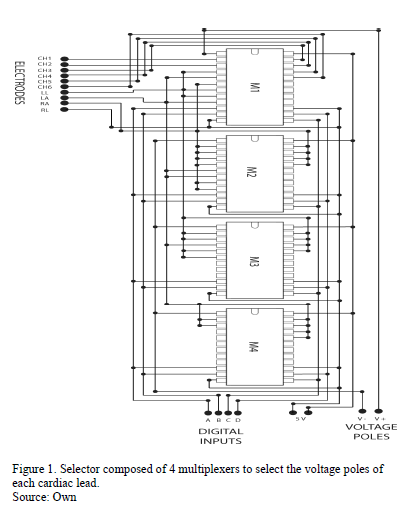
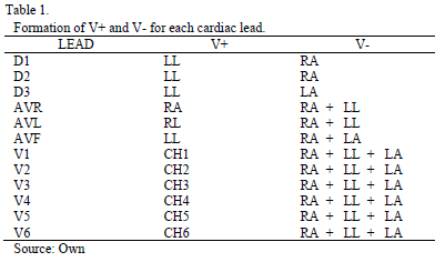
</font></p>     <p><font size="2" face="Verdana, Arial, Helvetica, sans-serif">The     <a href="#tab01">Table 1</a> &#91;15,16,19,20&#93; summarizes the procedure to interconnect the electrodes  in order to obtain poles of the 12 cardiac leads using an electronic selector.  The notation is: left arm (LA); right arm (RA); left leg (LL); right leg (RL); fourth intercostal space, just to  the right of the sternum (CH1); fourth intercostal space, just to the left of  the sternum (CH2); between CH2 and CH4 (CH3); fifth intercostal space in the  left mid-clavicular line (CH4); horizontally even with CH4 in the left anterior  axilary line (CH5); horizontally even with CH4 and CH5 in the mid-axilary line  (CH6); the &quot;+&quot; sign mean electrodes connected.</font></p>     <p align="center"><font size="2" face="Verdana, Arial, Helvetica, sans-serif"><a name="tab01"></a></font><img src="/img/revistas/dyna/v82n189/v82n189a15tab01.gif"></p>     <p>&nbsp;</p>     <p><font size="3" face="Verdana, Arial, Helvetica, sans-serif"><b>2. Materials and  methods</b></font></p>     <p><b><font size="2" face="Verdana, Arial, Helvetica, sans-serif">2.1. Remote  selector of the 12 cardiac leads
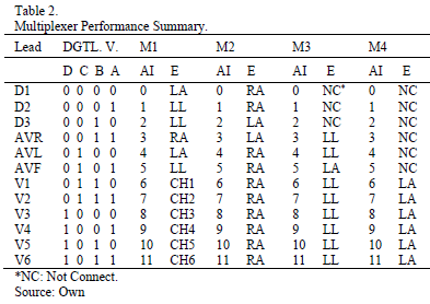
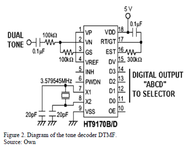
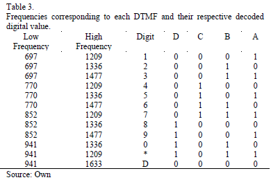
</font></b></p>     ]]></body>
<body><![CDATA[<p><font size="2" face="Verdana, Arial, Helvetica, sans-serif">The cardiac leads selector is performed using an array of 4 analogs  multiplexers (HEF4067B) and it is detailed in the <a href="#fig01">Fig. 1</a>. In the bottom the  electrodes are connected to the multiplexers (following the notation of <a href="#fig01">Table  1</a>) whereas on the right side are observed the digital inputs A, B, C, D; also,  the output voltage V+ y V- for each cardiac derivation. This kind of  multiplexers are characterized for the selection of the 16 possible analog  inputs (AI), using a combination of values ABCD. They have a very low total  harmonic distortion (THD) and the capability of switching electric current in  the range of <font face="Symbol">m</font>A at high speed. Our selector just needs 4 digital inputs in  total, which feed at the same time all the multiplexers in order to obtain the  pair of voltage poles of any chosen cardiac lead. The <a href="#tab02">Table 2</a> shows the  relationship between the cardiac leads and the digital values ABCD for each  multiplexer, also the connections of the electrodes with their analog inputs.  This selector is able of generate all the combinations detailed in Table 1.</font></p>     <p align="center"><font size="2" face="Verdana, Arial, Helvetica, sans-serif"><a name="fig01"></a></font><img src="/img/revistas/dyna/v82n189/v82n189a15fig01.gif"></p>     <p align="center"><font size="2" face="Verdana, Arial, Helvetica, sans-serif"><a name="tab02"></a></font><img src="/img/revistas/dyna/v82n189/v82n189a15tab02.gif"></p>     <p><font size="2" face="Verdana, Arial, Helvetica, sans-serif">On the other hand, the values ABCD are obtained using a  dual tone decoder, which is designed to decode audible tones of dual frequency  (DTMF) HT9170B/D. When this chip receives a dual tone it allows four digital  outputs, which could be connected directly to the selector. In the <a href="#fig02">Fig. 2</a> is  shown the diagram of the DTMF, and the <a href="#tab03">Table 3</a> show the frequencies which  compound a tone and their corresponding digital outputs, for each digit  employed.</font></p>     <p align="center"><font size="2" face="Verdana, Arial, Helvetica, sans-serif"><a name="fig02"></a></font><img src="/img/revistas/dyna/v82n189/v82n189a15fig02.gif"></p>     <p align="center"><font size="2" face="Verdana, Arial, Helvetica, sans-serif"><a name="tab03"></a></font><img src="/img/revistas/dyna/v82n189/v82n189a15tab03.gif"></p>     <p><font size="2" face="Verdana, Arial, Helvetica, sans-serif">The DTMF of <a href="#tab03">Table 3</a>,  are superimposed sine waves and correspond to the sound heard when the digit of  some telephonic devices is pressed. For instance: when a sinusoidal pulse  composed of 697 Hz and 1209 Hz signal is received (corresponding to the digit 1),  their correct decoding allow us the 4 digital values 0001,  which, in accord with <a href="#tab02">Table 2</a> correspond to the D2 lead. In our design, the  tones corresponding to each digit are generated from a computer via the audio  output, with maximum amplitude of 300 mV. This switching interface can operate  in full wired way, nevertheless it is very easy to adapt to the digital outputs  of a microcontroller to other applications. However, since the goal is to work  wirelessly, the tones are transmitted using an FM audio-transmitter (ideal for  microphone). To demodulate the FM signal, the tone must be amplified up to a  value close to 1 V, therefore the FM receiver is connected to a non-inverting  amplification stage using an operational amplifier with gain <i>G = 11, in order to feed the DTMF and avoid decoding errors. </i></font></p>     <p><font size="2" face="Verdana, Arial, Helvetica, sans-serif">In summary, the procedure for remotely selection of a cardiac lead is the following: at first,  from the PC audio output, the dual tone are generated and transmitted as FM,  the receiver output is amplified and introduced to the DTMF which generate the  four digital signals to feed the lead selector, where the two poles that feed  the amplifier are obtained, the output is then modulated in amplitude and  transmitted by a second FM transmitter, to then be acquired through the audio  card and digitally demodulated on a PC.</font></p>     <p><font size="2" face="Verdana, Arial, Helvetica, sans-serif"><b><i>2.2 Heart signal amplifier and conditioner</i></b></font></p>     <p><font size="2" face="Verdana, Arial, Helvetica, sans-serif">A schematic diagram of the full system is shown in the     <a href="#fig03">Fig. 3(A)</a>, where it can see the electrodes on the patient and connected to the  selector, which feeds the instrumentation amplifier, connected to the AM  modulator and then to the (FM2) transmitter. Also the DTMF connected to the  multiplexer and to the receiver of the (FM1) transmitter are displayed. In the  same way, it can be observed the audio card output of a laptop connected to the  FM1 to transmit the tone, and the FM2 receiver connected to the audio card  input, where the AM signals are demodulated. While the <a href="#fig03">Fig. 3(B)</a> shows the  corresponding block diagram. To avoid potential interference, care should be  taken that the FM1 modulation frequency is not an integer multiple of FM2.</font></p>     ]]></body>
<body><![CDATA[<p align="center"><font size="2" face="Verdana, Arial, Helvetica, sans-serif"><a name="fig03"></a></font><img src="/img/revistas/dyna/v82n189/v82n189a15fig03.gif"></p>     <p><font size="2" face="Verdana, Arial, Helvetica, sans-serif">
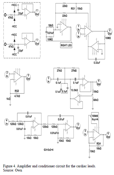
The amplification consists of six stages built with mostly  inexpensive electronic components. As shown in <a href="#fig04">Fig. 4</a>, the first stage is used  to increase the CMRR of the instrumentation amplifier and is connected to the  right leg of the patient, then is used an AD620 instrumentation amplifier with  gain given by eq. (1).</font></p>     <p><img src="/img/revistas/dyna/v82n189/v82n189a15eq01.gif"></p>     <p><font size="2" face="Verdana, Arial, Helvetica, sans-serif">which is feedback to the reference input by means of an  operational amplifier on integrating configuration, with this step, the base  line signal is stabilized, since it has a high pass filter behavior. Then there  is a non-inverting amplifier stage with gain given by eq. (2).</font></p>     <p><img src="/img/revistas/dyna/v82n189/v82n189a15eq02.gif"></p>     <p><font size="2" face="Verdana, Arial, Helvetica, sans-serif">followed by a rejection stage for the 60 Hz frequency and  an active four order butterwort type low pass filter with gain <i>G<sub>3</sub></i> = 4; and finally a last  non-inverting amplification with a gain<i> G<sub>4</sub></i> = 2, plus the option to regulating the DC level to take control of the voltage  levels that will be modulated in amplitude. Except for the feedback stage,  rejection of 60 Hz and stabilization of the baseline stage, all other steps are  explained in &#91;16&#93;. With this amplifier and selector it is achieved: A bandwidth  of 0.034 to 150 Hz with a signal/noise ratio about to 40 dB, total gain G<sub>t </sub>= 195.62, input impedance of 1 G<font face="Symbol">W</font> (according to the datasheet for  the AD620), common mode rejection CMRR close to 100 dB since all the used  amplifiers have a CMRR of about 110 dB, restoration time for base line minor  than 3 s and adjustable DC level. Prior to the differential amplification  stage, each electrode are connected to a voltage follower using an operational  amplifier for the correct coupling of the electrodes and the subsequent  circuits (See dotted line in <a href="#fig04">Fig. 4)</a>. At the output of each follower a 47  k<font face="Symbol">W</font> resistor are connected to decrease any potential leakage currents and  clipping diodes that discharge overvoltage to the power supply. The leakage  currents sensed between terminals (lead-lead) and between terminals and ground  (lead-ground) are less than 10 <font face="Symbol">m</font>A (in good agreement with &#91;20,21&#93;).  Additionally, to avoid exposing patients to possible variations of the line voltage,  the device is powered by a 12v battery connected to a regulating circuit for a  dual voltage output of ± 5 V.
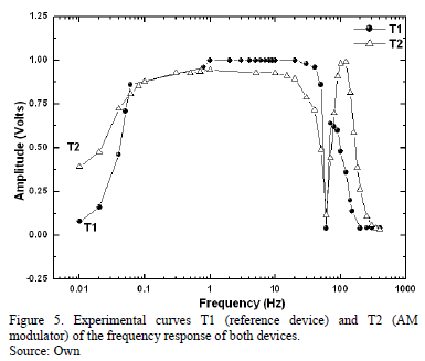
</font></p>     <p align="center"><font size="2" face="Verdana, Arial, Helvetica, sans-serif"><a name="fig04"></a></font><img src="/img/revistas/dyna/v82n189/v82n189a15fig04.gif"></p>     <p><font size="2" face="Verdana, Arial, Helvetica, sans-serif">In the graphs of <a href="#fig05">Fig. 5</a>, are displayed two curves  experimentally determined (T1 y T2). The one of a Fukuda Denshi Cardimax  FX-1201 electrocardiograph used as reference pattern (T1) and the signal  obtained from our new device (T2), respectively. In this experiment the curves  are obtained using a function generator Stanford Research mod. DS335 and an  oscilloscope Tektronix DPO7104. It is observed that the curve obtained with the  reference device T1 has a minimum cutoff frequency 0.05 Hz followed by a  rejection of the 60 Hz band with an attenuation of -30 dB and a maximum cutoff  frequency of 100 Hz. Whereas T2 has a minimum cutoff frequency of 0.034 Hz,  then a rejection to the 60 Hz frequency of -19 dB and a maximum cutoff  frequency of 150 Hz. Our observations indicate that increasing the attenuation  of the 60 Hz signal with the notch filter lead to a reduction of components of  the signals which may be important for a possible diagnostic, for instance:  between the range of 30 a 40 Hz &#91;3&#93;. It is also  noted that for T1 the attenuation is higher than in T2 for frequencies between  80 Hz and 100 Hz. Even so frequencies between 100 Hz and 110 Hz have a slightly  higher intensity than all other (around 0.8dB), due to the established gain in  the low-pass and to its transfer function &#91;22&#93;.</font></p>     <p align="center"><font size="2" face="Verdana, Arial, Helvetica, sans-serif"><a name="fig05"></a></font><img src="/img/revistas/dyna/v82n189/v82n189a15fig05.gif"></p>     <p><font size="2" face="Verdana, Arial, Helvetica, sans-serif"><b><i>2.3. Amplitude  modulator of the cardiac leads
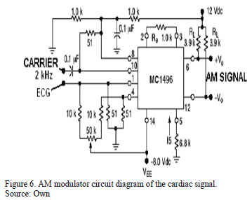
</i></b></font></p>     ]]></body>
<body><![CDATA[<p><font size="2" face="Verdana, Arial, Helvetica, sans-serif">To  the AM modulation an integrated circuit of the family MC1496BPG specially  designed this purpose is employed. This circuit provides a lower total harmonic  distortion (THD) than the modulation method based on a simple arrangement of  differential transistors as used in &#91;16&#93;, while the 2 kHz carrier signal is  generated with a double integrator arrangement based on an operational  amplifier as in &#91;16&#93;. The <a href="#fig06">Fig. 6</a> shows the electronic circuit diagram of the  modulator. Once the amplitude is modulated, the cardiac signal is converted into audible signal which are  transmitted using a commercial microphone FM transmitter-receiver system. The acquisition  of the &quot;audible cardiac signals&quot; is performed using the audio input of a PC  with a maximum sampling capacity of 192000 kS/s, 16 bits resolution, THD lower  than 0.1% and bandwidth from 4 Hz to18 kHz in the Lab-View 8.2 environment. A  similar setup is realized in &#91;23&#93;</font></p>     <p align="center"><font size="2" face="Verdana, Arial, Helvetica, sans-serif"><a name="fig06"></a></font><img src="/img/revistas/dyna/v82n189/v82n189a15fig06.gif"></p>     <p><font size="2" face="Verdana, Arial, Helvetica, sans-serif"><b><i>2.4. Demodulation  of the amplitude modulated leads</i></b></font></p>     <p><font size="2" face="Verdana, Arial, Helvetica, sans-serif">For the signals demodulation, the National Instruments  demodulation toolkit is used. As the cardiac signals of each person may vary in  amplitude, moreover the amplitudes of the precordial signals are usually  greater than the bipolar leads in the same subject, then the modulation factor  of each measure lead changes considerably which could cause changes to the  original signal, if is not properly demodulated. However, in this work a  coherent type AM digital demodulation is used, which takes the carrier signal as  an input parameter and it is independent of the modulation factor, as is  described in below.</font></p>     <p><font size="2" face="Verdana, Arial, Helvetica, sans-serif">Initially the envelope signal<i> m(t)</i> becomes the signal described in eq. (3).</font></p>     <p><img src="/img/revistas/dyna/v82n189/v82n189a15eq03.gif"></p>     <p><font size="2" face="Verdana, Arial, Helvetica, sans-serif">where <i>C</i> is a  constant that depends on the amplitude of the carrier signal. Multiplying the eq.  (3) by <i>cos(wt)</i> and using the  trigonometric identity <i>cos<sup>2</sup>(<font face="Symbol">q</font>)  = ½ + cos(2<font face="Symbol">q</font>)/2</i> is obtained the eq. (4)</font></p>     <p><img src="/img/revistas/dyna/v82n189/v82n189a15eq04.gif"></p>     <p><font size="2" face="Verdana, Arial, Helvetica, sans-serif">Subsequently, to  recover the<i> m(t)</i> signal, the <i>y(t)</i> signal  is filtered using a band-pass filter tuned to the  frequency <i>w,</i> in order to remove the  DC component and higher frequencies than the carrier signal. Finally the result  is multiplied by 2.</font></p>     <p>&nbsp;</p>     ]]></body>
<body><![CDATA[<p><font size="3" face="Verdana, Arial, Helvetica, sans-serif"><b>3. Measurements  and discussion</b></font></p>     <p><b><font size="2" face="Verdana, Arial, Helvetica, sans-serif">3.1. Measurements  of 12 leads modulated</font></b><font size="2" face="Verdana, Arial, Helvetica, sans-serif"></font></p>     <p><font size="2" face="Verdana, Arial, Helvetica, sans-serif">
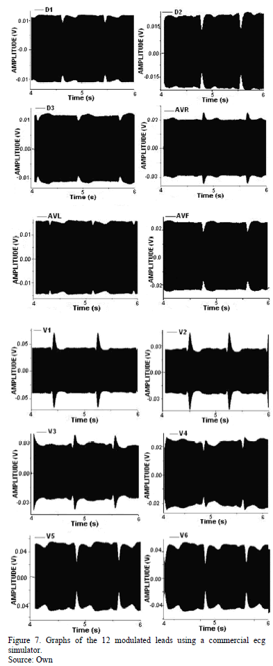
After all the design, simulation and assembly work of the  electronic device, the execution stage of experiments began, in order to test  the ability of detection and acquisition using a commercial ecg signal  simulator. In the <a href="#fig07">Fig. 7</a> is show a typical measurement of the 12-lead  amplitude-modulated. By analyzing the graphs, are evidenced higher modulation  factors in the precordial leads. In these measurements the maximum THD observed  oscillates around 4%, this is due to nonlinear behavior of the semiconductor  components of the transmission/reception of signals system. However, the  demodulated signals shows THD values lowers than 1% due to the lowpass filter  of high order implicit in the digital demodulate method.</font></p>     <p align="center"><font size="2" face="Verdana, Arial, Helvetica, sans-serif"><a name="fig07"></a></font><img src="/img/revistas/dyna/v82n189/v82n189a15fig07.gif"></p>     <p><font size="2" face="Verdana, Arial, Helvetica, sans-serif"><b><i>3.2. Measurements  with volunteers</i></b></font></p>     <p><font size="2" face="Verdana, Arial, Helvetica, sans-serif">Thereafter,  measurements of 12 cardiac leads (intervals of 3 seconds per lead) are obtained  from a group of 12 volunteers with no history of heart disease,  as follows: the electrodes are connected to our device and simultaneously  to the inputs of the electrocardiograph of reference ECG<sub>ref</sub>. The  signals of ECG<sub>ref</sub> are acquired of wired manner from a laptop over a  distance of <i>10 m</i>, using an  analog acquisition interface USB-6805 from National Instruments, with 5 kS/s of  sampling frequency and a shielded USB cable. While the same cardiac lead is  modulated in AM and transmitted by </font></p>     <p><font size="2" face="Verdana, Arial, Helvetica, sans-serif">FM2 (trademark ROMMS mod. MC-100),  to the audio input of the same computer. The data are acquired with sampling  frequency of 50 kS/s, in order to demodulate, store and graph them in real  time; on a same graphical interface developed in LabView environment. Each lead  is selected wirelessly from the same distance ( 10 m) using the FM1  transmitter (trademark steren, mod. Mic-280).
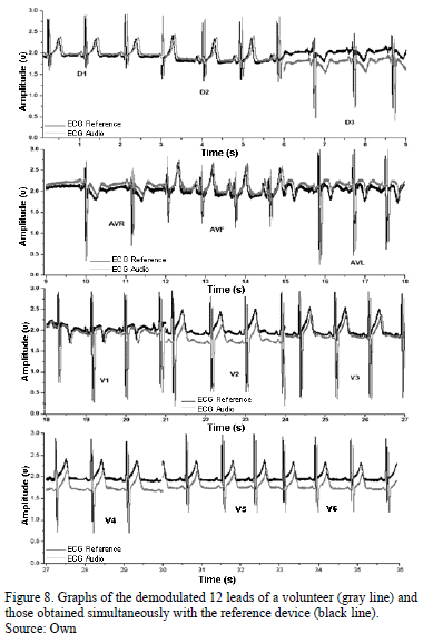
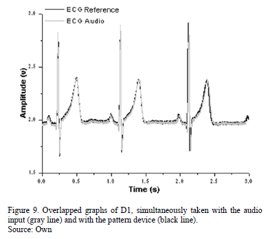
 </font></p>     <p><font size="2" face="Verdana, Arial, Helvetica, sans-serif">In the <a href="#fig08">Fig. 8</a> is shown the measurements of the 12 leads of  the same volunteer, using both devices. Plotting the measured signals by the  two methods is generally observed a shift in the amplitude and time. This  observed displacement in the amplitudes can be explained by the different DC  levels (offset) found in the signals. Furthermore the horizontal shift is due  to the period to the acquisition, digitization, demodulation and sampling.  Moreover, in <a href="#fig09">Fig. 9</a> are displayed the ECG signals of D1 acquired with both  devices, they are shown overlapped deliberately, in order to visibly compare  the measurements more clearly. As can be observed the shape of the undulations,  intervals and segments which compound the signals are maintained. This fact is  very important because the iso-electric lines and amplitudes using both devices  must be conserved, in order to carry out a correct determination of <i><font face="Symbol">q</font></i> and <i><font face="Symbol">j</font></i>. Nevertheless, a further analysis is also necessary.</font></p>     <p align="center"><font size="2" face="Verdana, Arial, Helvetica, sans-serif"><a name="fig08"></a></font><img src="/img/revistas/dyna/v82n189/v82n189a15fig08.gif"></p>     <p align="center"><font size="2" face="Verdana, Arial, Helvetica, sans-serif"><a name="fig09"></a></font><img src="/img/revistas/dyna/v82n189/v82n189a15fig09.gif"></p>     ]]></body>
<body><![CDATA[<p><font size="2" face="Verdana, Arial, Helvetica, sans-serif"><b><i>3.3. Quantitative comparison between both devices</i></b></font></p>     <p><font size="2" face="Verdana, Arial, Helvetica, sans-serif">Actually there are several investigations &#91;15,24-27&#93;,  which employee different alternatives to the comparison of two signals. They  are based in the direct measurement of the parameters of cardiology, or by  calculating those parameters using signal processing based on Wavelets. </font></p>     <p><font size="2" face="Verdana, Arial, Helvetica, sans-serif">
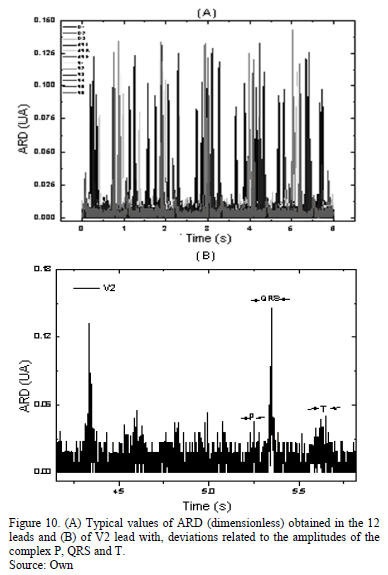
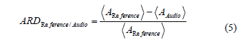
For this purpose,  initially we have done a subroutine in the Labview acquisition program to set  the base lines (offset) at the same level and subtract the time shifting  between them, in order to start at the same point. Likewise the acquired  signals through the USB-6508 card are interpolated to match the number of  samples acquired via the audio input. In this way are evaluated the absolute  relative deviations (ARD) between the two signals point to point, following the  definition of the eq. (5). In the graph of the <a href="#fig10">Fig. 10(A)</a> it can see the  typical values of ARD of the volunteer 12 leads, whereas the <a href="#fig10">Fig. 10(B)</a> shows  the typical values of ARD of the V2 lead, where are displayed the  deviations to the amplitudes of the complex P, QRS and T. After a careful  analysis of the ARD values in the leads of each volunteer, it is observed that  the largest differences occur in the peak values of the QRS (mainly those  associated with the amplitudes of the R and S), becoming more evident in leads  of higher amplitude as V2, V4 and V5 and reaching differences up to 15 %.</font></p>     <p align="center"><font size="2" face="Verdana, Arial, Helvetica, sans-serif"><a name="fig10"></a></font><img src="/img/revistas/dyna/v82n189/v82n189a15fig10.gif"></p>     <p><font size="2" face="Verdana, Arial, Helvetica, sans-serif">
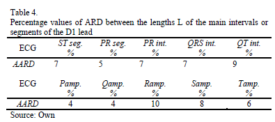
To determine the differences between the lengths <i>L</i> of the main intervals or segments of  both kind of signals, the graph of D1 was analyzed in Origin 6.0 (the leads D1  or D2 are normally used for the reference device) of each volunteers, using the  peak detection tool (peak detection). Next, the average value of the ARD is  calculated following eq. (6) and the percentage values obtained  are shown in the second row of <a href="#tab04">Table 4</a>. The same criterion is employed to  determine the AARD values of the undulations P, Q, R, S and T, in the lead D2  as is shown in the last row of Table 4.</font></p>     <p><img src="/img/revistas/dyna/v82n189/v82n189a15eq05.gif"></p>     <p><img src="/img/revistas/dyna/v82n189/v82n189a15eq06.gif"></p>     <p align="center"><font size="2" face="Verdana, Arial, Helvetica, sans-serif"><a name="tab04"></a></font><img src="/img/revistas/dyna/v82n189/v82n189a15tab04.gif"></p>     <p><font size="2" face="Verdana, Arial, Helvetica, sans-serif">These ARD and AARD values experimentally found in the  interval and segments of the signals, may be due to small variatios of the  carrier AM signal, wereas the AARD values of the amplitudes are due to the  differences in the frequency response of both devices, as shown in <a href="#fig05">Fig. 5</a>.  Indeed, analyzing the <i>AARD</i> values of <i>Ramp.</i> and <i>Samp.</i> of the <a href="#tab04">Table 4</a> and regarding that the cardiac axis is related  with the ratio of the amplitudes <i>r = R/S</i>,  the maximum expected error of cardiac axis orientation (eq. (7)) could be close  to 20 %. This finding shows the importance of comparing all the alternatives of  electrocardiographic devices, against a pattern device, for example &#91;5-13&#93;.</font></p>     <p><img src="/img/revistas/dyna/v82n189/v82n189a15eq07.gif"></p>     ]]></body>
<body><![CDATA[<p>&nbsp;</p>     <p><font size="3" face="Verdana, Arial, Helvetica, sans-serif"><b>4. Conclusions</b></font></p>     <p><font size="2" face="Verdana, Arial, Helvetica, sans-serif">In this work is presented in detail a low cost viable  alternative PC assisted to determine 12-lead electrocardiograms (one by one),  using a single stage of amplification and filtering, to conduct studies of  vector-cardiography. The leads can be selected and stored using the audio input  and output of a computer respectively; and a pair of inexpensive audio  transmitter/receiver devices. </font></p>     <p><font size="2" face="Verdana, Arial, Helvetica, sans-serif">Using an AM modulator with audible carrier, each lead  becomes an audio signal, and could be transmitted through the microphone input  of an economic mobile phone device. In the same way, using the telephone keypad  and headphone output the desired cardiac lead can be selected. Nevertheless the  module of analog transmission/reception can be easily replaced by a modern  digital transmission system, in order to obtain the twelve cardiac leads  through a peripheral port, like USB or COM.</font></p>     <p><font size="2" face="Verdana, Arial, Helvetica, sans-serif">We have developed an alternative to the selection of  cardiac leads using an arrangement of commercial analog multiplexers, which can  be controlled wired or wirelessly from the audio output of a PC, using dual  frequency tones and a tone decoder. However, the selector can be used to easily  switch between cardiac leads via digital outputs from another device such as: a  parallel port, a microcontroller, a digital signal transmitter zigbee or  manually using a 4-digit keypad. So that, many devices reported for evaluation  the vertical plane of the heart, could be extended by adapting this selector in  order to perform studies of vectorcardiography and the criterions of eq. (7) to  evaluate the viability of the devices could be used.</font></p>     <p><font size="2" face="Verdana, Arial, Helvetica, sans-serif">The system can be reproduced by an approximate cost of  US$90 and exhibits a good performance according to the trademark pattern  device, which meets the international standards for electrocardiography.  Therefore it shown to be a viable tool to measure cardiac bio-electric  potential, for telemedicine applications &#91;28&#93;, research in biomedical  engineering or in teaching activities. Due to their low cost, the device  represents a suitable tool to be used in developing countries, similar to &#91;29&#93;.</font></p>     <p>&nbsp;</p>     <p><font size="3" face="Verdana, Arial, Helvetica, sans-serif"><b>Acknowledgements</b></font></p>     <p><font size="2" face="Verdana, Arial, Helvetica, sans-serif">The authors wish to thank the facilities provided for  obtaining measurements to the staff of the regional hospital of the IMSS in  Ocotl&aacute;n Jalisco and to CONACyT by the support given to students.</font></p>     <p>&nbsp;</p>     ]]></body>
<body><![CDATA[<p><font size="3" face="Verdana, Arial, Helvetica, sans-serif"><b>References</b></font></p>     <!-- ref --><p><font size="2" face="Verdana, Arial, Helvetica, sans-serif"><b>&#91;1&#93;</b> Kligfield, P., Gettes, L.S., Bailey,  J.J., Childers, R., Deal, B.J., Hancock, E.W. et al. Recommendations for the  standardization and interpretation of the electrocardiogram: Part I: The  electrocardiogram and its technology: A scientific statement from the American  Heart Association Electrocardiography and Arrhythmias Committee, Council on  Clinical Cardiology; the American College of Cardiology Foundation; and the  Heart Rhythm Society. Endorsed by the International Society for Computerized  Electrocardiology. Heart Rhythm, 4 (3), pp. 394-412, 2007. <a href="http://dx.doi.org/10.1016/j.jacc.2007.01.024" target="_blank">http://dx.doi.org/10.1016/j.jacc.2007.01.024</a> </font>&nbsp;&nbsp;&nbsp;&nbsp;&nbsp;&nbsp;&nbsp;&nbsp;[&#160;<a href="javascript:void(0);" onclick="javascript: window.open('/scielo.php?script=sci_nlinks&ref=000094&pid=S0012-7353201500010001500001&lng=','','width=640,height=500,resizable=yes,scrollbars=1,menubar=yes,');">Links</a>&#160;]<!-- end-ref --><!-- ref --><p><font size="2" face="Verdana, Arial, Helvetica, sans-serif"><b>&#91;2&#93;</b> Pipberger, H.V. et al., Recommendations  for standardization of leads and specifications for instruments in  electrocardiography and vector cardiography. Circulation, 52, pp. 11-31, 1975.    &nbsp;&nbsp;&nbsp;&nbsp;&nbsp;&nbsp;&nbsp;&nbsp;[&#160;<a href="javascript:void(0);" onclick="javascript: window.open('/scielo.php?script=sci_nlinks&ref=000095&pid=S0012-7353201500010001500002&lng=','','width=640,height=500,resizable=yes,scrollbars=1,menubar=yes,');">Links</a>&#160;]<!-- end-ref --></font></p>     <!-- ref --><p><font size="2" face="Verdana, Arial, Helvetica, sans-serif"><b>&#91;3&#93;</b> Ferrero, C.A. and Hall, E.J., Textbook of  Medical Physiology, 11 ed., Mc Graw-Hill, Spain, 2001. pp. 103-114,    &nbsp;&nbsp;&nbsp;&nbsp;&nbsp;&nbsp;&nbsp;&nbsp;[&#160;<a href="javascript:void(0);" onclick="javascript: window.open('/scielo.php?script=sci_nlinks&ref=000097&pid=S0012-7353201500010001500003&lng=','','width=640,height=500,resizable=yes,scrollbars=1,menubar=yes,');">Links</a>&#160;]<!-- end-ref --> </font></p>     <!-- ref --><p><font size="2" face="Verdana, Arial, Helvetica, sans-serif"><b>&#91;4&#93;</b> Henneberg, K.A., Biopotentials and  electrophysiology measurement, in Aller Mauren. Measurements instrumentation  and sensors Handbook, CRC press, 2a ed., Boca Raton, Florida, 1999, pp.  2019-2037.    &nbsp;&nbsp;&nbsp;&nbsp;&nbsp;&nbsp;&nbsp;&nbsp;[&#160;<a href="javascript:void(0);" onclick="javascript: window.open('/scielo.php?script=sci_nlinks&ref=000099&pid=S0012-7353201500010001500004&lng=','','width=640,height=500,resizable=yes,scrollbars=1,menubar=yes,');">Links</a>&#160;]<!-- end-ref --></font></p>     <!-- ref --><p><font size="2" face="Verdana, Arial, Helvetica, sans-serif"><b>&#91;5&#93;</b> Silva,  C.V. and Rojas, V.G., Design and implementation of a digital  electrocardiographic system Rev.Fac.Ing.Univ. Antioquia, 55, pp. 99-107,  2010. Available at: <a href="http://jaibana.udea.edu.co/grupos/revista/revistas/nro055/Articulo%2010.pdf" target="_blank">http://jaibana.udea.edu.co/grupos/revista/revistas/nro055/Articulo%2010.pdf</a></font>&nbsp;&nbsp;&nbsp;&nbsp;&nbsp;&nbsp;&nbsp;&nbsp;[&#160;<a href="javascript:void(0);" onclick="javascript: window.open('/scielo.php?script=sci_nlinks&ref=000101&pid=S0012-7353201500010001500005&lng=','','width=640,height=500,resizable=yes,scrollbars=1,menubar=yes,');">Links</a>&#160;]<!-- end-ref --><!-- ref --><p><font size="2" face="Verdana, Arial, Helvetica, sans-serif"><b>&#91;6&#93;</b> Tseng Y-L., Shi Y.-Z., Jaw F.-S.,  Portable, real-time, 12-lead ecg monitoring system, Instrumentation Science  &amp; Technology, 38 (4), pp. 305-312, 2010.  <a href="http://dx.doi.org/10.1080/10739149.2010.508331" target="_blank">http://dx.doi.org/10.1080/10739149.2010.508331</a></font>&nbsp;&nbsp;&nbsp;&nbsp;&nbsp;&nbsp;&nbsp;&nbsp;[&#160;<a href="javascript:void(0);" onclick="javascript: window.open('/scielo.php?script=sci_nlinks&ref=000102&pid=S0012-7353201500010001500006&lng=','','width=640,height=500,resizable=yes,scrollbars=1,menubar=yes,');">Links</a>&#160;]<!-- end-ref --><!-- ref --><p><font size="2" face="Verdana, Arial, Helvetica, sans-serif"><b>&#91;7&#93;</b> Najeb, J.M., Ruhullah, A. and Sh-Hussain,  S., 12-Channel USB data acquisition system For QT dispersion analysis, Proceedings of the International Conference  on Robotics. Vision, Information and Signal Processing ROVISP, pp. 83-86, 2005.  Available at: <a href="http://eprints.utm.my/2151/1/Najeb_ROVISP_2005.pdf" target="_blank">http://eprints.utm.my/2151/1/Najeb_ROVISP_2005.pdf</a></font>&nbsp;&nbsp;&nbsp;&nbsp;&nbsp;&nbsp;&nbsp;&nbsp;[&#160;<a href="javascript:void(0);" onclick="javascript: window.open('/scielo.php?script=sci_nlinks&ref=000103&pid=S0012-7353201500010001500007&lng=','','width=640,height=500,resizable=yes,scrollbars=1,menubar=yes,');">Links</a>&#160;]<!-- end-ref --><!-- ref --><p><font size="2" face="Verdana, Arial, Helvetica, sans-serif"><b>&#91;8&#93;</b> Lucani, D., A portable ECG monitoring  device with Bluetooth and Holter capabilities for telemedicine applications.  Proceedings of the 28th IEEE EMBS Annual International Conference New York  City, USA, pp. 5244-5247, 2006. <a href="http://dx.doi.org/10.1109/IEMBS.2006.260798" target="_blank">http://dx.doi.org/10.1109/IEMBS.2006.260798</a> </font>&nbsp;&nbsp;&nbsp;&nbsp;&nbsp;&nbsp;&nbsp;&nbsp;[&#160;<a href="javascript:void(0);" onclick="javascript: window.open('/scielo.php?script=sci_nlinks&ref=000104&pid=S0012-7353201500010001500008&lng=','','width=640,height=500,resizable=yes,scrollbars=1,menubar=yes,');">Links</a>&#160;]<!-- end-ref --><!-- ref --><p><font size="2" face="Verdana, Arial, Helvetica, sans-serif"><b>&#91;9&#93;</b> Jia-Ren, C.C. and Cheng-Chi T. A new  wireless-type physiological signal measuring system using a PDA and the  Bluetooth technology, Instrumentation Science &amp; Technology, 37  (5), pp. 503-515, 2009.    &nbsp;&nbsp;&nbsp;&nbsp;&nbsp;&nbsp;&nbsp;&nbsp;[&#160;<a href="javascript:void(0);" onclick="javascript: window.open('/scielo.php?script=sci_nlinks&ref=000105&pid=S0012-7353201500010001500009&lng=','','width=640,height=500,resizable=yes,scrollbars=1,menubar=yes,');">Links</a>&#160;]<!-- end-ref --> </font></p>     <!-- ref --><p><font size="2" face="Verdana, Arial, Helvetica, sans-serif"><b>&#91;10&#93;</b> Nopparat, V., The three-lead wireless ECG  in sensor networks for mobile patients. SICE Annual Conference, pp.  2308-2311, 2008. <a href="http://dx.doi.org/10.1109/SICE.2008.4655050" target="_blank">http://dx.doi.org/10.1109/SICE.2008.4655050</a></font>&nbsp;&nbsp;&nbsp;&nbsp;&nbsp;&nbsp;&nbsp;&nbsp;[&#160;<a href="javascript:void(0);" onclick="javascript: window.open('/scielo.php?script=sci_nlinks&ref=000107&pid=S0012-7353201500010001500010&lng=','','width=640,height=500,resizable=yes,scrollbars=1,menubar=yes,');">Links</a>&#160;]<!-- end-ref --><!-- ref --><p><font size="2" face="Verdana, Arial, Helvetica, sans-serif"><b>&#91;11&#93;</b> Auteri, V., Roffia, L., Lamberti, C. and  Cinotti, T.S., Zigbee-based Wireless ECG Monitor, Computers in Cardiology, 34,  pp. 133-136, 2007. <a href="http://dx.doi.org/10.1109/CIC.2007.4745439" target="_blank">http://dx.doi.org/10.1109/CIC.2007.4745439</a></font>&nbsp;&nbsp;&nbsp;&nbsp;&nbsp;&nbsp;&nbsp;&nbsp;[&#160;<a href="javascript:void(0);" onclick="javascript: window.open('/scielo.php?script=sci_nlinks&ref=000108&pid=S0012-7353201500010001500011&lng=','','width=640,height=500,resizable=yes,scrollbars=1,menubar=yes,');">Links</a>&#160;]<!-- end-ref --><!-- ref --><p><font size="2" face="Verdana, Arial, Helvetica, sans-serif"><b>&#91;12&#93;</b> Thaddeus, R.F., Fulford-Jones, G-Y.W., and Welsh,  M., A Portable, Low-Power, Wireless Two-Lead EKG System. In Proceedings of the  26th Annual International Conference of the IEEE EMBS San Francisco, CA, USA,  pp. 2141-2144, 2004. <a href="http://dx.doi.org/10.1109/IEMBS.2004.1403627" target="_blank">http://dx.doi.org/10.1109/IEMBS.2004.1403627</a></font>&nbsp;&nbsp;&nbsp;&nbsp;&nbsp;&nbsp;&nbsp;&nbsp;[&#160;<a href="javascript:void(0);" onclick="javascript: window.open('/scielo.php?script=sci_nlinks&ref=000109&pid=S0012-7353201500010001500012&lng=','','width=640,height=500,resizable=yes,scrollbars=1,menubar=yes,');">Links</a>&#160;]<!-- end-ref --><!-- ref --><p><font size="2" face="Verdana, Arial, Helvetica, sans-serif"><b>&#91;13&#93;</b> Borromeo,  S., Rodriguez-Sanchez, C., Machado, F., Hernandez-Tamames, J.A. and de la Prieta,  R.A., Reconfigurable, wearable, wireless ECG system. In Proceedings of  the 29th Annual International Conference of the IEEE EMBS Cit&eacute; Internationale,  Lyon, France, pp. 1659-1662, 2007. <a href="http://dx.doi.org/10.1109/IEMBS.2007.4352626" target="_blank">http://dx.doi.org/10.1109/IEMBS.2007.4352626</a></font>&nbsp;&nbsp;&nbsp;&nbsp;&nbsp;&nbsp;&nbsp;&nbsp;[&#160;<a href="javascript:void(0);" onclick="javascript: window.open('/scielo.php?script=sci_nlinks&ref=000110&pid=S0012-7353201500010001500013&lng=','','width=640,height=500,resizable=yes,scrollbars=1,menubar=yes,');">Links</a>&#160;]<!-- end-ref --><!-- ref --><p><font size="2" face="Verdana, Arial, Helvetica, sans-serif"><b>&#91;14&#93;</b> Gabor, D. and Nelson, C.V., Determination  of the resultant dipole of the heart from measurements on the body surface,  Journal of Applied Physics, 25, pp. 413-416, 1954.  <a href="http://dx.doi.org/10.1063/1.1721655" target="_blank">http://dx.doi.org/10.1063/1.1721655</a></font>&nbsp;&nbsp;&nbsp;&nbsp;&nbsp;&nbsp;&nbsp;&nbsp;[&#160;<a href="javascript:void(0);" onclick="javascript: window.open('/scielo.php?script=sci_nlinks&ref=000111&pid=S0012-7353201500010001500014&lng=','','width=640,height=500,resizable=yes,scrollbars=1,menubar=yes,');">Links</a>&#160;]<!-- end-ref --><!-- ref --><p><font size="2" face="Verdana, Arial, Helvetica, sans-serif"><b>&#91;15&#93;</b> Davis, D., Quick and Accurate 12-Lead ECG  Interpretation 4a ed., Philadelphia, USA, Lippincott Williams &amp; Wilkins,  2007, pp. 6-7. Available at:  <a href="http://resourcecenter.ovid.com/site/catalog/Book/4961.pdf" target="_blank">http://resourcecenter.ovid.com/site/catalog/Book/4961.pdf</a></font>&nbsp;&nbsp;&nbsp;&nbsp;&nbsp;&nbsp;&nbsp;&nbsp;[&#160;<a href="javascript:void(0);" onclick="javascript: window.open('/scielo.php?script=sci_nlinks&ref=000112&pid=S0012-7353201500010001500015&lng=','','width=640,height=500,resizable=yes,scrollbars=1,menubar=yes,');">Links</a>&#160;]<!-- end-ref --><!-- ref --><p><font size="2" face="Verdana, Arial, Helvetica, sans-serif"><b>&#91;16&#93;</b> Cano, M.E., Jaso, R.A., Tavares, R.A.,  Estrada, J.C., Mena, E.A., Reynoso, O., Gonz&aacute;lez, V.A. and C&oacute;rdova, T. A simple  alternative for modulating and recording the PQRST complex. Rev. Mex. de Ing.  Biom&eacute;dica, 31 (2), pp. 100-108, 2011. Available at:  <a href="http://www.medigraphic.com/pdfs/inge/ib-2011/ib112f.pdf" target="_blank">http://www.medigraphic.com/pdfs/inge/ib-2011/ib112f.pdf</a></font>&nbsp;&nbsp;&nbsp;&nbsp;&nbsp;&nbsp;&nbsp;&nbsp;[&#160;<a href="javascript:void(0);" onclick="javascript: window.open('/scielo.php?script=sci_nlinks&ref=000113&pid=S0012-7353201500010001500016&lng=','','width=640,height=500,resizable=yes,scrollbars=1,menubar=yes,');">Links</a>&#160;]<!-- end-ref --><p><font size="2" face="Verdana, Arial, Helvetica, sans-serif"><b>&#91;17&#93;</b> Chiuso,  A., Laurenti, L. and chenato, A.Z., LQG  cheap control subject to packet loss and SNR limitations.2013 European Control Conference (ECC),  Zürich, Switzerland, 2013. Available at:  <a href="http://ieeexplore.ieee.org/stamp/stamp.jsp?tp=&arnumber=6669629">http://ieeexplore.ieee.org/stamp/stamp.jsp?tp=&amp;arnumber=6669629</a></font></p>     <!-- ref --><p><font size="2" face="Verdana, Arial, Helvetica, sans-serif"><b>&#91;18&#93;</b> Elia,  N., Remote stabilization over fading channels, Systems and Control Letters, 54,  pp. 237-249, 2005. <a href="http://dx.doi.org/10.1016/j.sysconle.2004.08.009" target="_blank">http://dx.doi.org/10.1016/j.sysconle.2004.08.009</a></font>&nbsp;&nbsp;&nbsp;&nbsp;&nbsp;&nbsp;&nbsp;&nbsp;[&#160;<a href="javascript:void(0);" onclick="javascript: window.open('/scielo.php?script=sci_nlinks&ref=000115&pid=S0012-7353201500010001500018&lng=','','width=640,height=500,resizable=yes,scrollbars=1,menubar=yes,');">Links</a>&#160;]<!-- end-ref --><!-- ref --><p><font size="2" face="Verdana, Arial, Helvetica, sans-serif"><b>&#91;19&#93;</b> Guyton, C.A., John, H.E., Textbook of  Medical Physiology. 11va. ed., Spain, Mc Graw-Hill, 2001. pp. 103-114.    &nbsp;&nbsp;&nbsp;&nbsp;&nbsp;&nbsp;&nbsp;&nbsp;[&#160;<a href="javascript:void(0);" onclick="javascript: window.open('/scielo.php?script=sci_nlinks&ref=000116&pid=S0012-7353201500010001500019&lng=','','width=640,height=500,resizable=yes,scrollbars=1,menubar=yes,');">Links</a>&#160;]<!-- end-ref --></font></p>     <!-- ref --><p><font size="2" face="Verdana, Arial, Helvetica, sans-serif"><b>&#91;20&#93;</b> Association for the Advancement of Medical  Instrumentation, Cardiac monitors, heart rate meters and alarms. ANSI/AAMI  EC13-1992. Arlington (VA): AAMI, American  National Standard. 2002. Available at:  <a href="http://www.pauljbennett.com/pbennett/work/ec13/ec13.pdf" target="_blank">http://www.pauljbennett.com/pbennett/work/ec13/ec13.pdf</a></font>&nbsp;&nbsp;&nbsp;&nbsp;&nbsp;&nbsp;&nbsp;&nbsp;[&#160;<a href="javascript:void(0);" onclick="javascript: window.open('/scielo.php?script=sci_nlinks&ref=000118&pid=S0012-7353201500010001500020&lng=','','width=640,height=500,resizable=yes,scrollbars=1,menubar=yes,');">Links</a>&#160;]<!-- end-ref --><!-- ref --><p><font size="2" face="Verdana, Arial, Helvetica, sans-serif"><b>&#91;21&#93;</b> Association for the Advancement of Medical  Instrumentation, American National Standard Safe Current Limits for  Electromedical Apparatus, ANSI/AAMI ES1-1993. Arlington (VA): AAMI,  American National Standard. 1993.  Available at:  <a href="https://courses.engr.illinois.edu/ece445/documents/Safe_Current_Limits.pdf" target="_blank">https://courses.engr.illinois.edu/ece445/documents/Safe_Current_Limits.pdf</a></font>&nbsp;&nbsp;&nbsp;&nbsp;&nbsp;&nbsp;&nbsp;&nbsp;[&#160;<a href="javascript:void(0);" onclick="javascript: window.open('/scielo.php?script=sci_nlinks&ref=000119&pid=S0012-7353201500010001500021&lng=','','width=640,height=500,resizable=yes,scrollbars=1,menubar=yes,');">Links</a>&#160;]<!-- end-ref --><!-- ref --><p><font size="2" face="Verdana, Arial, Helvetica, sans-serif"><b>&#91;22&#93;</b> Ying-Wen B., Chien-Yung C., Cheng-Kai L.,  Chuang-Hsiang H., Yuh-Ting Ch. and Ya-Nan L. Adjustable 60Hz Noise Reduction  and ECG signal amplification of a remote electrocardiogram system. In  proceedings of the Instrumentation and Measurement Technology Conference Vail,  CO, USA, pp. 197-202, 2003. Available at:  <a href="http://cs.ee.fju.edu.tw/paper/200301.pdf" target="_blank">http://cs.ee.fju.edu.tw/paper/200301.pdf</a> </font>&nbsp;&nbsp;&nbsp;&nbsp;&nbsp;&nbsp;&nbsp;&nbsp;[&#160;<a href="javascript:void(0);" onclick="javascript: window.open('/scielo.php?script=sci_nlinks&ref=000120&pid=S0012-7353201500010001500022&lng=','','width=640,height=500,resizable=yes,scrollbars=1,menubar=yes,');">Links</a>&#160;]<!-- end-ref --><!-- ref --><p><font size="2" face="Verdana, Arial, Helvetica, sans-serif"><b>&#91;23&#93;</b> Stojanovic, R. and Karadaglic, D., An  economical and feasible teaching tool for biomedical education Computer-Based Medical Systems  (CBMS), 2011 24th International Symposium on, 1, pp. 1-5, 2011. <a href="http://dx.doi.org/10.1109/CBMS.2011.5999116">http://dx.doi.org/10.1109/CBMS.2011	.5999116</a></font>&nbsp;&nbsp;&nbsp;&nbsp;&nbsp;&nbsp;&nbsp;&nbsp;[&#160;<a href="javascript:void(0);" onclick="javascript: window.open('/scielo.php?script=sci_nlinks&ref=000121&pid=S0012-7353201500010001500023&lng=','','width=640,height=500,resizable=yes,scrollbars=1,menubar=yes,');">Links</a>&#160;]<!-- end-ref --><!-- ref --><p><font size="2" face="Verdana, Arial, Helvetica, sans-serif"><b>&#91;24&#93;</b> Dubin, D., Rapid Interpretation of EKG's.  6ta ed. Tampa,FL, Cover Pub Co, pp. 75-76, 2000.    &nbsp;&nbsp;&nbsp;&nbsp;&nbsp;&nbsp;&nbsp;&nbsp;[&#160;<a href="javascript:void(0);" onclick="javascript: window.open('/scielo.php?script=sci_nlinks&ref=000122&pid=S0012-7353201500010001500024&lng=','','width=640,height=500,resizable=yes,scrollbars=1,menubar=yes,');">Links</a>&#160;]<!-- end-ref --></font></p>     <!-- ref --><p><font size="2" face="Verdana, Arial, Helvetica, sans-serif"><b>&#91;25&#93;</b> Olarte,  O.J., Sierra, D.A. and Rueda, O.L., System for the recognition and diagnostics  of cardiac arrhythmias applied to the identification of wide complex  tachycardia from the ECG. Rev. Fac. Ing. Univ. Antioquia, 48, pp.  153-164, 2009. Available at:  <a href="http://ingenieria.udea.edu.co/grupos/revista/revistas/nro048/48-15.pdf?origin=publication_detail" target="_blank">http://ingenieria.udea.edu.co/grupos/revista/revistas/nro048/48-15.pdf?origin=publication_detail</a></font>&nbsp;&nbsp;&nbsp;&nbsp;&nbsp;&nbsp;&nbsp;&nbsp;[&#160;<a href="javascript:void(0);" onclick="javascript: window.open('/scielo.php?script=sci_nlinks&ref=000124&pid=S0012-7353201500010001500025&lng=','','width=640,height=500,resizable=yes,scrollbars=1,menubar=yes,');">Links</a>&#160;]<!-- end-ref --><!-- ref --><p><font size="2" face="Verdana, Arial, Helvetica, sans-serif"><b>&#91;26&#93;</b> Sivannarayana, N. and Reddy, D.C.,  Biorthogonal wavelet transforms for ECG parameters estimation. Medical  Engineering and Physics, 21, pp. 167-174, 1999.  <a href="http://dx.doi.org/10.1016/S1350-4533(99)00040-5" target="_blank">http://dx.doi.org/10.1016/S1350-4533(99)00040-5</a></font>&nbsp;&nbsp;&nbsp;&nbsp;&nbsp;&nbsp;&nbsp;&nbsp;[&#160;<a href="javascript:void(0);" onclick="javascript: window.open('/scielo.php?script=sci_nlinks&ref=000125&pid=S0012-7353201500010001500026&lng=','','width=640,height=500,resizable=yes,scrollbars=1,menubar=yes,');">Links</a>&#160;]<!-- end-ref --><!-- ref --><p><font size="2" face="Verdana, Arial, Helvetica, sans-serif"><b>&#91;27&#93;</b> Dumont, J., Hern&aacute;ndez, A.I. and Carrault,  G., Parameter optimization of a wavelet-based electrocardiogram delineator with  an evolutionary algorithm. Computers Cardiology, 32, pp. 707-710, 2005.  <a href="http://dx.doi.org/10.1109/CIC.2005.1588202" target="_blank">http://dx.doi.org/10.1109/CIC.2005.1588202</a></font>&nbsp;&nbsp;&nbsp;&nbsp;&nbsp;&nbsp;&nbsp;&nbsp;[&#160;<a href="javascript:void(0);" onclick="javascript: window.open('/scielo.php?script=sci_nlinks&ref=000126&pid=S0012-7353201500010001500027&lng=','','width=640,height=500,resizable=yes,scrollbars=1,menubar=yes,');">Links</a>&#160;]<!-- end-ref --><!-- ref --><p><font size="2" face="Verdana, Arial, Helvetica, sans-serif"><b>&#91;28&#93;</b> Castellano, N.N., G&aacute;zquez, J.A., L&oacute;pez,  J.F. and Manzano, F., Telemetry system for transmission data from an ambulance.  DYNA, 79 (175), pp. 43-51, 2012. Available at: <a href="http://www.scielo.org.co/pdf/dyna/v79n175/v79n175a05.pdf" target="_blank">http://www.scielo.org.co/pdf/dyna/v79n175/v79n175a05.pdf</a></font>&nbsp;&nbsp;&nbsp;&nbsp;&nbsp;&nbsp;&nbsp;&nbsp;[&#160;<a href="javascript:void(0);" onclick="javascript: window.open('/scielo.php?script=sci_nlinks&ref=000127&pid=S0012-7353201500010001500028&lng=','','width=640,height=500,resizable=yes,scrollbars=1,menubar=yes,');">Links</a>&#160;]<!-- end-ref --><!-- ref --><p><font size="2" face="Verdana, Arial, Helvetica, sans-serif"><b>&#91;29&#93;</b> Walker, B.A., Khandoker, A.H. and Black, J., Low cost ECG monitor for developing  countries. Intelligent Sensors, Sensor Networks and Information Processing  (ISSNIP), 2009 5th International Conference on 7 - 10 Dec. 2009 Melbourne, VIC,  pp. 195-199, 2009. <a href="http://dx.doi.org/10.1109/ISSNIP.2009.5416759" target="_blank">http://dx.doi.org/10.1109/ISSNIP.2009.5416759</a></font>&nbsp;&nbsp;&nbsp;&nbsp;&nbsp;&nbsp;&nbsp;&nbsp;[&#160;<a href="javascript:void(0);" onclick="javascript: window.open('/scielo.php?script=sci_nlinks&ref=000128&pid=S0012-7353201500010001500029&lng=','','width=640,height=500,resizable=yes,scrollbars=1,menubar=yes,');">Links</a>&#160;]<!-- end-ref --><p>&nbsp;</p>     <p><font size="2" face="Verdana, Arial, Helvetica, sans-serif"><b>J.C.  Estrada-Guti&eacute;rrez,</b> received the PhD. degree in Sc. in 2014 from the  Universidad of Guadalajara, M&eacute;xico, Full Professor en the Technologic Sciences  department in the Centro Universitario de la Ci&eacute;nega of the Universidad de  Guadalajara, in M&eacute;xico. His research interest include: Magnetic Hyperthermia,  Automation and Control and Embedded Systems.</font></p>     <p><font size="2" face="Verdana, Arial, Helvetica, sans-serif"><b>A.  Hern&aacute;ndez-S&aacute;mano,</b> received the MPhys. degree in 2013 from the University of  Guanajuato, M&eacute;xico, he is Student in the PhD in physics in the Centro  Universitario de la Ci&eacute;nega of the Universidad de Guadalajara, in M&eacute;xico. His  research interest include: Magnetic Hyperthermia, Resonant Inverters and  Magnetometry.</font></p>     <p><font size="2" face="Verdana, Arial, Helvetica, sans-serif"><b>E.E.  Maz&oacute;n-Valadez,</b> received, the BIE. degree in 2012 from the Universidad of  Guadalajara, M&eacute;xico, he is Student in the Masters in physics in the Centro  Universitario de la Ci&eacute;nega of the Universidad de Guadalajara, in M&eacute;xico. His  research interest include: Magnetic Hyperthermia, Resonant Inverters and SAR.</font></p>     ]]></body>
<body><![CDATA[<p><font size="2" face="Verdana, Arial, Helvetica, sans-serif"><b>J. &Aacute;vila-Paz,</b> received MSc.in 2003 from the Universidad Central Marta Abreu de las Villas,  Cuba, Full Professor in the Technologic Sciences department in the Centro  Universitario de la Ci&eacute;nega of the Universidad de Guadalajara, in M&eacute;xico. His  research interest include: Magnetic Hyperthermia, Automation and Control, and  Embedded Systems.</font></p>     <p><font size="2" face="Verdana, Arial, Helvetica, sans-serif"><b>A. Gonz&aacute;lez-Vega</b>,  received the PhD degree in Ciences in 2007 from the Centro de Investigaci&oacute;n en Matem&aacute;ticas A.C. Mex&iacute;co. Full professor  in the Biomedical Engineering department in the Divisi&oacute;n de Ciencias e Ingenier&iacute;as de la Universidad de Guanajuato, in M&eacute;xico. His research interest  include: Biomechanics, biomaterials, optics process, instrumentation and signal  and image processing.</font></p>     <p><font size="2" face="Verdana, Arial, Helvetica, sans-serif"><b>J.A. Leyva-Cruz, </b>received the PhD degree in Ciences in 2005 from the Universidade de São  Paulo, Brazil. Full professor in the Physics Department in the Universidade Estadual de Feira de Santana  (UEFS) na Bahia, in Brazil. His research interest include: Bioinstrumentation,  electric and magnetic imaging in medicine, acquisition and automatic processing of biosignals, inverse problems and photoconductive sensors.</font></p>     <p><font size="2" face="Verdana, Arial, Helvetica, sans-serif"><b>M.E.  Cano-Gonz&aacute;lez,</b> received the PhD degree in Physics in 2007 from the  University of Guanajuato, M&eacute;xico. Full professor in the Basic Sciences  department in the Centro Universitario de la Ci&eacute;nega of the Universidad de  Guadalajara, in M&eacute;xico. His research interest include: Magnetic Hyperthermia,  Resonant Inverters and Monte Carlo Simulations.</font></p>      ]]></body><back>
<ref-list>
<ref id="B1">
<label>1</label><nlm-citation citation-type="journal">
<person-group person-group-type="author">
<name>
<surname><![CDATA[Kligfield]]></surname>
<given-names><![CDATA[P.]]></given-names>
</name>
<name>
<surname><![CDATA[Gettes]]></surname>
<given-names><![CDATA[L.S.]]></given-names>
</name>
<name>
<surname><![CDATA[Bailey]]></surname>
<given-names><![CDATA[J.J.]]></given-names>
</name>
<name>
<surname><![CDATA[Childers]]></surname>
<given-names><![CDATA[R.]]></given-names>
</name>
<name>
<surname><![CDATA[Deal]]></surname>
<given-names><![CDATA[B.J.]]></given-names>
</name>
<name>
<surname><![CDATA[Hancock]]></surname>
<given-names><![CDATA[E.W.]]></given-names>
</name>
</person-group>
<article-title xml:lang="en"><![CDATA[Recommendations for the standardization and interpretation of the electrocardiogram: Part I: The electrocardiogram and its technology]]></article-title>
<source><![CDATA[Heart Rhythm]]></source>
<year>2007</year>
<volume>4</volume>
<numero>3</numero>
<issue>3</issue>
<page-range>394-412</page-range></nlm-citation>
</ref>
<ref id="B2">
<label>2</label><nlm-citation citation-type="journal">
<person-group person-group-type="author">
<name>
<surname><![CDATA[Pipberger]]></surname>
<given-names><![CDATA[H.V.]]></given-names>
</name>
</person-group>
<article-title xml:lang="en"><![CDATA[Recommendations for standardization of leads and specifications for instruments in electrocardiography and vector cardiography]]></article-title>
<source><![CDATA[Circulation]]></source>
<year>1975</year>
<volume>52</volume>
<page-range>11-31</page-range></nlm-citation>
</ref>
<ref id="B3">
<label>3</label><nlm-citation citation-type="book">
<person-group person-group-type="author">
<name>
<surname><![CDATA[Ferrero]]></surname>
<given-names><![CDATA[C.A.]]></given-names>
</name>
<name>
<surname><![CDATA[Hall]]></surname>
<given-names><![CDATA[E.J.]]></given-names>
</name>
</person-group>
<source><![CDATA[Textbook of Medical Physiology]]></source>
<year>2001</year>
<edition>11</edition>
<page-range>103-114</page-range><publisher-name><![CDATA[Mc Graw-Hill]]></publisher-name>
</nlm-citation>
</ref>
<ref id="B4">
<label>4</label><nlm-citation citation-type="book">
<person-group person-group-type="author">
<name>
<surname><![CDATA[Henneberg]]></surname>
<given-names><![CDATA[K.A.]]></given-names>
</name>
</person-group>
<article-title xml:lang="en"><![CDATA[Biopotentials and electrophysiology measurement]]></article-title>
<person-group person-group-type="editor">
<name>
<surname><![CDATA[Mauren]]></surname>
<given-names><![CDATA[Aller]]></given-names>
</name>
</person-group>
<source><![CDATA[Measurements instrumentation and sensors Handbook]]></source>
<year>1999</year>
<edition>2a</edition>
<page-range>2019-2037</page-range><publisher-loc><![CDATA[Boca Raton^eFlorida Florida]]></publisher-loc>
<publisher-name><![CDATA[CRC press]]></publisher-name>
</nlm-citation>
</ref>
<ref id="B5">
<label>5</label><nlm-citation citation-type="journal">
<person-group person-group-type="author">
<name>
<surname><![CDATA[Silva]]></surname>
<given-names><![CDATA[C.V.]]></given-names>
</name>
<name>
<surname><![CDATA[Rojas]]></surname>
<given-names><![CDATA[V.G.]]></given-names>
</name>
</person-group>
<article-title xml:lang="en"><![CDATA[Design and implementation of a digital electrocardiographic system]]></article-title>
<source><![CDATA[Rev.Fac.Ing.Univ. Antioquia]]></source>
<year>2010</year>
<volume>55</volume>
<page-range>99-107</page-range></nlm-citation>
</ref>
<ref id="B6">
<label>6</label><nlm-citation citation-type="journal">
<person-group person-group-type="author">
<name>
<surname><![CDATA[Tseng]]></surname>
<given-names><![CDATA[Y-L.]]></given-names>
</name>
<name>
<surname><![CDATA[Shi Y.]]></surname>
<given-names><![CDATA[Z.]]></given-names>
</name>
<name>
<surname><![CDATA[Jaw F.]]></surname>
<given-names><![CDATA[S.]]></given-names>
</name>
</person-group>
<article-title xml:lang="en"><![CDATA[Portable, real-time, 12-lead ecg monitoring system]]></article-title>
<source><![CDATA[Instrumentation Science & Technology]]></source>
<year>2010</year>
<volume>38</volume>
<numero>4</numero>
<issue>4</issue>
<page-range>305-312</page-range></nlm-citation>
</ref>
<ref id="B7">
<label>7</label><nlm-citation citation-type="confpro">
<person-group person-group-type="author">
<name>
<surname><![CDATA[Najeb]]></surname>
<given-names><![CDATA[J.M.]]></given-names>
</name>
<name>
<surname><![CDATA[Ruhullah]]></surname>
<given-names><![CDATA[A.]]></given-names>
</name>
<name>
<surname><![CDATA[Sh-Hussain]]></surname>
<given-names><![CDATA[S.]]></given-names>
</name>
</person-group>
<article-title xml:lang="en"><![CDATA[12-Channel USB data acquisition system For QT dispersion analysis]]></article-title>
<source><![CDATA[]]></source>
<year>2005</year>
<conf-name><![CDATA[ International Conference on Robotics. Vision, Information and Signal Processing ROVISP]]></conf-name>
<conf-loc> </conf-loc>
<page-range>83-86</page-range></nlm-citation>
</ref>
<ref id="B8">
<label>8</label><nlm-citation citation-type="confpro">
<person-group person-group-type="author">
<name>
<surname><![CDATA[Lucani]]></surname>
<given-names><![CDATA[D.]]></given-names>
</name>
</person-group>
<article-title xml:lang="en"><![CDATA[A portable ECG monitoring device with Bluetooth and Holter capabilities for telemedicine applications]]></article-title>
<source><![CDATA[]]></source>
<year>2006</year>
<conf-name><![CDATA[28th IEEE EMBS Annual International Conference]]></conf-name>
<conf-loc>New York </conf-loc>
<page-range>5244-5247</page-range></nlm-citation>
</ref>
<ref id="B9">
<label>9</label><nlm-citation citation-type="journal">
<person-group person-group-type="author">
<name>
<surname><![CDATA[Jia-Ren]]></surname>
<given-names><![CDATA[C.C.]]></given-names>
</name>
<name>
<surname><![CDATA[Cheng-Chi]]></surname>
<given-names><![CDATA[T.]]></given-names>
</name>
</person-group>
<article-title xml:lang="en"><![CDATA[A new wireless-type physiological signal measuring system using a PDA and the Bluetooth technology]]></article-title>
<source><![CDATA[Instrumentation Science & Technology]]></source>
<year>2009</year>
<volume>37</volume>
<numero>5</numero>
<issue>5</issue>
<page-range>503-515</page-range></nlm-citation>
</ref>
<ref id="B10">
<label>10</label><nlm-citation citation-type="confpro">
<person-group person-group-type="author">
<name>
<surname><![CDATA[Nopparat]]></surname>
<given-names><![CDATA[V.]]></given-names>
</name>
</person-group>
<article-title xml:lang="en"><![CDATA[The three-lead wireless ECG in sensor networks for mobile patients]]></article-title>
<source><![CDATA[]]></source>
<year>2008</year>
<conf-name><![CDATA[ SICE Annual Conference]]></conf-name>
<conf-loc> </conf-loc>
<page-range>2308-2311</page-range></nlm-citation>
</ref>
<ref id="B11">
<label>11</label><nlm-citation citation-type="journal">
<person-group person-group-type="author">
<name>
<surname><![CDATA[Auteri]]></surname>
<given-names><![CDATA[V.]]></given-names>
</name>
<name>
<surname><![CDATA[Roffia]]></surname>
<given-names><![CDATA[L.]]></given-names>
</name>
<name>
<surname><![CDATA[Lamberti]]></surname>
<given-names><![CDATA[C.]]></given-names>
</name>
<name>
<surname><![CDATA[Cinotti]]></surname>
<given-names><![CDATA[T.S.]]></given-names>
</name>
</person-group>
<article-title xml:lang="en"><![CDATA[Zigbee-based Wireless ECG Monitor]]></article-title>
<source><![CDATA[Computers in Cardiology]]></source>
<year>2007</year>
<numero>34</numero>
<issue>34</issue>
<page-range>133-136</page-range></nlm-citation>
</ref>
<ref id="B12">
<label>12</label><nlm-citation citation-type="confpro">
<person-group person-group-type="author">
<name>
<surname><![CDATA[Thaddeus]]></surname>
<given-names><![CDATA[R.F.]]></given-names>
</name>
<name>
<surname><![CDATA[Fulford-Jones]]></surname>
<given-names><![CDATA[G-Y.W.]]></given-names>
</name>
<name>
<surname><![CDATA[Welsh]]></surname>
<given-names><![CDATA[M.]]></given-names>
</name>
</person-group>
<article-title xml:lang="en"><![CDATA[A Portable, Low-Power, Wireless Two-Lead EKG System]]></article-title>
<source><![CDATA[]]></source>
<year>2004</year>
<conf-name><![CDATA[26th Annual International Conference of the IEEE EMBS]]></conf-name>
<conf-loc>San Francisco CA</conf-loc>
<page-range>2141-2144</page-range></nlm-citation>
</ref>
<ref id="B13">
<label>13</label><nlm-citation citation-type="confpro">
<person-group person-group-type="author">
<name>
<surname><![CDATA[Borromeo]]></surname>
<given-names><![CDATA[S.]]></given-names>
</name>
<name>
<surname><![CDATA[Rodriguez-Sanchez]]></surname>
<given-names><![CDATA[C.]]></given-names>
</name>
<name>
<surname><![CDATA[Machado]]></surname>
<given-names><![CDATA[F.]]></given-names>
</name>
<name>
<surname><![CDATA[Hernandez-Tamames]]></surname>
<given-names><![CDATA[J.A.]]></given-names>
</name>
<name>
<surname><![CDATA[de la Prieta]]></surname>
<given-names><![CDATA[R.A.]]></given-names>
</name>
</person-group>
<article-title xml:lang="en"><![CDATA[Reconfigurable, wearable, wireless ECG system]]></article-title>
<source><![CDATA[]]></source>
<year>2007</year>
<conf-name><![CDATA[29th Annual International Conference of the IEEE EMBS]]></conf-name>
<conf-loc>Cité Internationale Lyon</conf-loc>
<page-range>1659-1662</page-range></nlm-citation>
</ref>
<ref id="B14">
<label>14</label><nlm-citation citation-type="journal">
<person-group person-group-type="author">
<name>
<surname><![CDATA[Gabor]]></surname>
<given-names><![CDATA[D.]]></given-names>
</name>
<name>
<surname><![CDATA[Nelson]]></surname>
<given-names><![CDATA[C.V.]]></given-names>
</name>
</person-group>
<article-title xml:lang="en"><![CDATA[Determination of the resultant dipole of the heart from measurements on the body surface]]></article-title>
<source><![CDATA[Journal of Applied Physics]]></source>
<year>1954</year>
<numero>25</numero>
<issue>25</issue>
<page-range>413-416</page-range></nlm-citation>
</ref>
<ref id="B15">
<label>15</label><nlm-citation citation-type="book">
<person-group person-group-type="author">
<name>
<surname><![CDATA[Davis]]></surname>
<given-names><![CDATA[D.]]></given-names>
</name>
</person-group>
<source><![CDATA[Quick and Accurate 12-Lead ECG Interpretation]]></source>
<year>2007</year>
<edition>4a</edition>
<page-range>6-7</page-range><publisher-loc><![CDATA[Philadelphia ]]></publisher-loc>
<publisher-name><![CDATA[Lippincott Williams & Wilkins]]></publisher-name>
</nlm-citation>
</ref>
<ref id="B16">
<label>16</label><nlm-citation citation-type="journal">
<person-group person-group-type="author">
<name>
<surname><![CDATA[Cano]]></surname>
<given-names><![CDATA[M.E.]]></given-names>
</name>
<name>
<surname><![CDATA[Jaso]]></surname>
<given-names><![CDATA[R.A.]]></given-names>
</name>
<name>
<surname><![CDATA[Tavares]]></surname>
<given-names><![CDATA[R.A.]]></given-names>
</name>
<name>
<surname><![CDATA[Estrada]]></surname>
<given-names><![CDATA[J.C.]]></given-names>
</name>
<name>
<surname><![CDATA[Mena]]></surname>
<given-names><![CDATA[E.A.]]></given-names>
</name>
<name>
<surname><![CDATA[Reynoso]]></surname>
<given-names><![CDATA[O.]]></given-names>
</name>
<name>
<surname><![CDATA[González]]></surname>
<given-names><![CDATA[V.A.]]></given-names>
</name>
<name>
<surname><![CDATA[Córdova]]></surname>
<given-names><![CDATA[T.]]></given-names>
</name>
</person-group>
<article-title xml:lang="en"><![CDATA[A simple alternative for modulating and recording the PQRST complex]]></article-title>
<source><![CDATA[Rev. Mex. de Ing. Biomédica]]></source>
<year>2011</year>
<volume>31</volume>
<numero>2</numero>
<issue>2</issue>
<page-range>100-108</page-range></nlm-citation>
</ref>
<ref id="B17">
<label>17</label><nlm-citation citation-type="confpro">
<person-group person-group-type="author">
<name>
<surname><![CDATA[Chiuso]]></surname>
<given-names><![CDATA[A.]]></given-names>
</name>
<name>
<surname><![CDATA[Laurenti]]></surname>
<given-names><![CDATA[L.]]></given-names>
</name>
<name>
<surname><![CDATA[chenato]]></surname>
<given-names><![CDATA[A.Z.]]></given-names>
</name>
</person-group>
<article-title xml:lang="en"><![CDATA[LQG cheap control subject to packet loss and SNR limitations]]></article-title>
<source><![CDATA[]]></source>
<year>2013</year>
<conf-name><![CDATA[ European Control Conference (ECC)]]></conf-name>
<conf-date>2013</conf-date>
<conf-loc>Zürich </conf-loc>
</nlm-citation>
</ref>
<ref id="B18">
<label>18</label><nlm-citation citation-type="journal">
<person-group person-group-type="author">
<name>
<surname><![CDATA[Elia]]></surname>
<given-names><![CDATA[N.]]></given-names>
</name>
</person-group>
<article-title xml:lang="en"><![CDATA[Remote stabilization over fading channels]]></article-title>
<source><![CDATA[Systems and Control Letters]]></source>
<year>2005</year>
<numero>54</numero>
<issue>54</issue>
<page-range>237-249</page-range></nlm-citation>
</ref>
<ref id="B19">
<label>19</label><nlm-citation citation-type="">
<person-group person-group-type="author">
<name>
<surname><![CDATA[Guyton]]></surname>
<given-names><![CDATA[C.A.]]></given-names>
</name>
<name>
<surname><![CDATA[John]]></surname>
<given-names><![CDATA[H.E.]]></given-names>
</name>
</person-group>
<source><![CDATA[Textbook of Medical Physiology]]></source>
<year>2001</year>
<edition>11va</edition>
<page-range>103-114</page-range><publisher-loc><![CDATA[Mc Graw-Hill ]]></publisher-loc>
</nlm-citation>
</ref>
<ref id="B20">
<label>20</label><nlm-citation citation-type="book">
<collab>Association for the Advancement of Medical Instrumentation</collab>
<source><![CDATA[Cardiac monitors, heart rate meters and alarms]]></source>
<year>2002</year>
<publisher-loc><![CDATA[Arlington^eVA VA]]></publisher-loc>
<publisher-name><![CDATA[American National Standard]]></publisher-name>
</nlm-citation>
</ref>
<ref id="B21">
<label>21</label><nlm-citation citation-type="book">
<collab>Association for the Advancement of Medical Instrumentation</collab>
<source><![CDATA[American National Standard Safe Current Limits for Electromedical Apparatus]]></source>
<year>1993</year>
<publisher-loc><![CDATA[Arlington^eVA VA]]></publisher-loc>
<publisher-name><![CDATA[American National Standard]]></publisher-name>
</nlm-citation>
</ref>
<ref id="B22">
<label>22</label><nlm-citation citation-type="confpro">
<person-group person-group-type="author">
<name>
<surname><![CDATA[Ying-Wen]]></surname>
<given-names><![CDATA[B.]]></given-names>
</name>
<name>
<surname><![CDATA[Chien-Yung]]></surname>
<given-names><![CDATA[C.]]></given-names>
</name>
<name>
<surname><![CDATA[Cheng-Kai]]></surname>
<given-names><![CDATA[L.]]></given-names>
</name>
<name>
<surname><![CDATA[Chuang-Hsiang]]></surname>
<given-names><![CDATA[H.]]></given-names>
</name>
<name>
<surname><![CDATA[Yuh-Ting]]></surname>
<given-names><![CDATA[Ch]]></given-names>
</name>
<name>
<surname><![CDATA[Ya-Nan]]></surname>
<given-names><![CDATA[L.]]></given-names>
</name>
</person-group>
<article-title xml:lang="en"><![CDATA[Adjustable 60Hz Noise Reduction and ECG signal amplification of a remote electrocardiogram system]]></article-title>
<source><![CDATA[]]></source>
<year>2003</year>
<conf-name><![CDATA[ Instrumentation and Measurement Technology Conference]]></conf-name>
<conf-loc>Vail CO</conf-loc>
<page-range>197-202</page-range></nlm-citation>
</ref>
<ref id="B23">
<label>23</label><nlm-citation citation-type="confpro">
<person-group person-group-type="author">
<name>
<surname><![CDATA[Stojanovic]]></surname>
<given-names><![CDATA[R.]]></given-names>
</name>
<name>
<surname><![CDATA[Karadaglic]]></surname>
<given-names><![CDATA[D.]]></given-names>
</name>
</person-group>
<article-title xml:lang="en"><![CDATA[An economical and feasible teaching tool for biomedical education Computer-Based Medical Systems (CBMS)]]></article-title>
<source><![CDATA[]]></source>
<year>2011</year>
<edition>1</edition>
<conf-name><![CDATA[24th International Symposium]]></conf-name>
<conf-date>2011</conf-date>
<conf-loc> </conf-loc>
<page-range>1-5</page-range></nlm-citation>
</ref>
<ref id="B24">
<label>24</label><nlm-citation citation-type="book">
<person-group person-group-type="author">
<name>
<surname><![CDATA[Dubin]]></surname>
<given-names><![CDATA[D.]]></given-names>
</name>
</person-group>
<source><![CDATA[Rapid Interpretation of EKG's]]></source>
<year>2000</year>
<edition>6ta</edition>
<page-range>75-76</page-range><publisher-loc><![CDATA[Tampa^eFL FL]]></publisher-loc>
<publisher-name><![CDATA[Cover Pub Co]]></publisher-name>
</nlm-citation>
</ref>
<ref id="B25">
<label>25</label><nlm-citation citation-type="journal">
<person-group person-group-type="author">
<name>
<surname><![CDATA[Olarte]]></surname>
<given-names><![CDATA[O.J.]]></given-names>
</name>
<name>
<surname><![CDATA[Sierra]]></surname>
<given-names><![CDATA[D.A.]]></given-names>
</name>
<name>
<surname><![CDATA[Rueda]]></surname>
<given-names><![CDATA[O.L.]]></given-names>
</name>
</person-group>
<article-title xml:lang="en"><![CDATA[System for the recognition and diagnostics of cardiac arrhythmias applied to the identification of wide complex tachycardia from the ECG]]></article-title>
<source><![CDATA[Rev. Fac. Ing. Univ. Antioquia]]></source>
<year>2009</year>
<numero>48</numero>
<issue>48</issue>
<page-range>153-164</page-range></nlm-citation>
</ref>
<ref id="B26">
<label>26</label><nlm-citation citation-type="journal">
<person-group person-group-type="author">
<name>
<surname><![CDATA[Sivannarayana]]></surname>
<given-names><![CDATA[N.]]></given-names>
</name>
<name>
<surname><![CDATA[Reddy]]></surname>
<given-names><![CDATA[D.C.]]></given-names>
</name>
</person-group>
<article-title xml:lang="en"><![CDATA[Biorthogonal wavelet transforms for ECG parameters estimation]]></article-title>
<source><![CDATA[Medical Engineering and Physics]]></source>
<year>1999</year>
<numero>21</numero>
<issue>21</issue>
<page-range>167-174</page-range></nlm-citation>
</ref>
<ref id="B27">
<label>27</label><nlm-citation citation-type="journal">
<person-group person-group-type="author">
<name>
<surname><![CDATA[Dumont]]></surname>
<given-names><![CDATA[J.]]></given-names>
</name>
<name>
<surname><![CDATA[Hernández]]></surname>
<given-names><![CDATA[A.I.]]></given-names>
</name>
<name>
<surname><![CDATA[Carrault]]></surname>
<given-names><![CDATA[G.]]></given-names>
</name>
</person-group>
<article-title xml:lang="en"><![CDATA[Parameter optimization of a wavelet-based electrocardiogram delineator with an evolutionary algorithm]]></article-title>
<source><![CDATA[Computers Cardiology]]></source>
<year>2005</year>
<numero>32</numero>
<issue>32</issue>
<page-range>707-710</page-range></nlm-citation>
</ref>
<ref id="B28">
<label>28</label><nlm-citation citation-type="journal">
<person-group person-group-type="author">
<name>
<surname><![CDATA[Castellano]]></surname>
<given-names><![CDATA[N.N.]]></given-names>
</name>
<name>
<surname><![CDATA[Gázquez]]></surname>
<given-names><![CDATA[J.A.]]></given-names>
</name>
<name>
<surname><![CDATA[López]]></surname>
<given-names><![CDATA[J.F.]]></given-names>
</name>
<name>
<surname><![CDATA[Manzano]]></surname>
<given-names><![CDATA[F.]]></given-names>
</name>
</person-group>
<article-title xml:lang="en"><![CDATA[Telemetry system for transmission data from an ambulance]]></article-title>
<source><![CDATA[DYNA]]></source>
<year>2012</year>
<volume>79</volume>
<numero>175</numero>
<issue>175</issue>
<page-range>43-51</page-range></nlm-citation>
</ref>
<ref id="B29">
<label>29</label><nlm-citation citation-type="confpro">
<person-group person-group-type="author">
<name>
<surname><![CDATA[Walker]]></surname>
<given-names><![CDATA[B.A.]]></given-names>
</name>
<name>
<surname><![CDATA[Khandoker]]></surname>
<given-names><![CDATA[A.H.]]></given-names>
</name>
<name>
<surname><![CDATA[Black]]></surname>
<given-names><![CDATA[J.]]></given-names>
</name>
</person-group>
<article-title xml:lang="en"><![CDATA[Low cost ECG monitor for developing countries. Intelligent Sensors, Sensor Networks and Information Processing (ISSNIP)]]></article-title>
<source><![CDATA[]]></source>
<year>2009</year>
<conf-name><![CDATA[5th International Conference]]></conf-name>
<conf-date>2009</conf-date><conf-date>7 - 10 Dec. 2009</conf-date>
<conf-loc>Melbourne VIC</conf-loc>
<page-range>195-199</page-range></nlm-citation>
</ref>
</ref-list>
</back>
</article>
