<?xml version="1.0" encoding="ISO-8859-1"?><article xmlns:mml="http://www.w3.org/1998/Math/MathML" xmlns:xlink="http://www.w3.org/1999/xlink" xmlns:xsi="http://www.w3.org/2001/XMLSchema-instance">
<front>
<journal-meta>
<journal-id>0120-0690</journal-id>
<journal-title><![CDATA[Revista Colombiana de Ciencias Pecuarias]]></journal-title>
<abbrev-journal-title><![CDATA[Rev Colom Cienc Pecua]]></abbrev-journal-title>
<issn>0120-0690</issn>
<publisher>
<publisher-name><![CDATA[Facultad de Ciencias Agrarias, Universidad de Antioquia]]></publisher-name>
</publisher>
</journal-meta>
<article-meta>
<article-id>S0120-06902005000100008</article-id>
<title-group>
<article-title xml:lang="en"><![CDATA[Adenocarcinoma in the jejunum of a dog: a case report]]></article-title>
<article-title xml:lang="es"><![CDATA[Adenocarcinoma en el yeyuno de un perro: reporte de un caso]]></article-title>
</title-group>
<contrib-group>
<contrib contrib-type="author">
<name>
<surname><![CDATA[Hernández]]></surname>
<given-names><![CDATA[Carlos]]></given-names>
</name>
<xref ref-type="aff" rid="A01"/>
</contrib>
<contrib contrib-type="author">
<name>
<surname><![CDATA[Restrepo]]></surname>
<given-names><![CDATA[Rodrigo]]></given-names>
</name>
<xref ref-type="aff" rid="A02"/>
</contrib>
</contrib-group>
<aff id="A01">
<institution><![CDATA[,Emergencias Veterinarias Inmediatas  ]]></institution>
<addr-line><![CDATA[Medellín ]]></addr-line>
<country>Colombia</country>
</aff>
<aff id="A02">
<institution><![CDATA[,Universidad Pontificia Bolivariana  ]]></institution>
<addr-line><![CDATA[Medellín ]]></addr-line>
<country>Colombia</country>
</aff>
<pub-date pub-type="pub">
<day>00</day>
<month>04</month>
<year>2005</year>
</pub-date>
<pub-date pub-type="epub">
<day>00</day>
<month>04</month>
<year>2005</year>
</pub-date>
<volume>18</volume>
<numero>1</numero>
<fpage>75</fpage>
<lpage>79</lpage>
<copyright-statement/>
<copyright-year/>
<self-uri xlink:href="http://www.scielo.org.co/scielo.php?script=sci_arttext&amp;pid=S0120-06902005000100008&amp;lng=en&amp;nrm=iso"></self-uri><self-uri xlink:href="http://www.scielo.org.co/scielo.php?script=sci_abstract&amp;pid=S0120-06902005000100008&amp;lng=en&amp;nrm=iso"></self-uri><self-uri xlink:href="http://www.scielo.org.co/scielo.php?script=sci_pdf&amp;pid=S0120-06902005000100008&amp;lng=en&amp;nrm=iso"></self-uri><abstract abstract-type="short" xml:lang="en"><p><![CDATA[A 10-year-old male Pit-bull was attended for presenting sporadic vomiting and occasional diarrhea. In the last 48 hours the patient had presented multiple vomiting and no oral food was tolerated. During the physical examination an abdominal cranial mass was palpated. Survey radiographs of the abdomen and contrast study with barium sulfate suspension showed an accumulation of a radiopaque material causing an intestinal obstruction pattern. During the surgical exploration, the foreign material was extracted from the jejunum using an enterotomy, but an intramural mass was detected occluding the lumen at this point. A complete bowel resection was performed in the intestinal loop affected by the tumor and an end-to-end anastomosis was performed. The histologic examinations revealed an adenocarcinoma involving the entire intestinal wall. The patient recovered from the surgical procedure and no signs of metastasis have been proved nine months after the surgery.]]></p></abstract>
<abstract abstract-type="short" xml:lang="es"><p><![CDATA[Un perro macho de raza Pitbull de 10 años de edad fue atendido por presentar vómito esporádico en los últimos 2 meses y diarrea ocasional. En las últimas 48 horas el paciente había presentado múltiples vómitos sin tolerar alimentación oral. Al examen clínico se palpó una masa abdominal. Las radiografías abdominales simples y contrastadas con sulfato de bario mostraron un acúmulo de material radiopaco causando un patrón característico de obstrucción intestinal. En la exploración quirúrgica el material causante de la obstrucción que se encontraba en el yeyuno fue retirado mediante enterotomía pero se detectó una masa intraluminal intestinal, por lo que se extrajo el segmento completo y se realizó una enteroanastomosis termino-terminal. El análisis histológico de la masa extraída evidenció un adenocarcinoma envolviendo todo el espesor de la pared intestinal. El paciente se recuperó normalmente y hasta los nueve meses posteriores a la cirugía, no ha aparecido ningún signo de metástasis.]]></p></abstract>
<kwd-group>
<kwd lng="en"><![CDATA[enteroanastomosis]]></kwd>
<kwd lng="en"><![CDATA[intestine]]></kwd>
<kwd lng="en"><![CDATA[neoplasia]]></kwd>
<kwd lng="en"><![CDATA[obstruction]]></kwd>
<kwd lng="es"><![CDATA[enteroanastomosis]]></kwd>
<kwd lng="es"><![CDATA[intestino]]></kwd>
<kwd lng="es"><![CDATA[neoplasia]]></kwd>
<kwd lng="es"><![CDATA[obstrucción]]></kwd>
</kwd-group>
</article-meta>
</front><body><![CDATA[ <p><b>CASOS CL&Iacute;NICOS </b></p>     <p>&nbsp;</p>     <p><b>Adenocarcinoma in the jejunum of a dog: a case report</b></p>     <p>&nbsp;</p>     <P    >Carlos Hern&aacute;ndez<I>&sup1;</I>, MV; Rodrigo Restrepo<I>&sup2;</I>, MD esp. </P >    <P    ><Sup><I>1</I></Sup>Emergencias Veterinarias Inmediatas, Medell&iacute;n, Colombia. <Sup><I>2 </I></Sup>Universidad Pontificia Bolivariana, Medell&iacute;n, Colombia </P >     <P    ><a href="mailto:carlitos@epm.net.co">carlitos@epm.net.co</a> </P >     <P    >(Recibido: 2 diciembre, 2004; aceptado: 16 febrero, 2005)</P >     <P    >&nbsp;</P >     <P    ><I>Summary </I></P >    ]]></body>
<body><![CDATA[<P    ><I>A 10-year-old male Pit-bull was attended for presenting sporadic vomiting and occasional diarrhea. In the last 48 hours the patient had presented multiple vomiting and no oral food was tolerated. During the physical examination an abdominal cranial mass was palpated. Survey radiographs of the abdomen and contrast study with barium sulfate suspension showed an accumulation of a radiopaque material causing an intestinal obstruction pattern. During the surgical exploration, the foreign material was extracted from the jejunum using an enterotomy, but an intramural mass was detected occluding the lumen at this point. A complete bowel resection was performed in the intestinal loop affected by the tumor and an end-to-end anastomosis was performed. The histologic examinations revealed an adenocarcinoma involving the entire intestinal wall. The patient recovered from the surgical procedure and no signs of metastasis have been proved nine months after the surgery. </I></P >    <P    >Key words:<I> enteroanastomosis, intestine, neoplasia, obstruction</I></P >     <P    >&nbsp;</P > <b>Introduction </b>     <P    >Intestinal obstructions are a common condition seen adenocarcinoma in the jejunum that involved the in small animal practice, but rarely caused by an greater part of the luminal circumference are intramural small bowel neoplasm. As in human beings, presented. It is described the surgical resection of the primary tumors of the small intestine are infrequent in bowel segment, the pathologic characteristics of the dogs, althoughthe small intestine constitutes almost 90% tumor and the complete recuperation of the patient of the mucosal surface of the alimentary tract (2,7,9). after the surgical procedure. </P >     <P    >Adenocarcinoma is the most common neoplasm in the small intestine of dogs followed by lymphosarcoma, Clinical case and smooth muscle tumors (4). In dogs, carcinomas of the large intestine are more common than those of <I>Anamnesis </I>the small intestine (2,4). </P >     <P    >A 10-year-old Pit Bull Terrier was presented with In the next report, the clinical characteristics of a a 2-month weight loss history, intermittent vomiting and male dog suffering the symptoms caused by an sporadic diarrhea. The dog was vomiting 3 times a week, unrelated to eating and characterized by large volumes of odiferous material. During this 2-month period, the dog&rsquo;s weight had decreased from 35 to 28 kg although the dog had good appetite and history of pica. 48 hours prior to the examination, the dog had vomited continuously and didn&rsquo;t tolerate any oral food. </P >     <P    ><I>Physical examination and laboratory findings </I></P >     <P    >
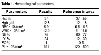
The dog presented a normal attitude during the clinical examination and a normal body condition (previously considered obese). No fever was present, but severe periodontal disease was detected. A large abdominal mass was palpated in the cranial abdomen, and excess of fluid and gas were palpated in some areas of the intestines. The palpation of the mass caused evident discomfort in the patient. The auscultation of the abdomen revealed excessive noises related to increased peristaltic movements. Blood was collected for a CBC and chemistry panel. Hematological and biochemical parameters were all within established reference ranges (<a href="#t1">see Table 1</a>, <a href="#t2">Table 2</a>).</P >     <P    ><img src="/img/revistas/rccp/v18n1/v18n1a08t01.jpg"><a name="t1"></a></P >     <P    ><img src="/img/revistas/rccp/v18n1/v18n1a08t02.jpg"><a name="t2"></a></P >     ]]></body>
<body><![CDATA[<P    ><img src="/img/revistas/rccp/v18n1/v18n1a08t03.jpg"><a name="t3"></a></P >     <P    >&nbsp;</P >     <P    ><I>Radiographic interpretation
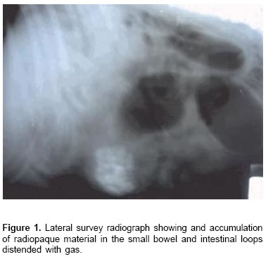
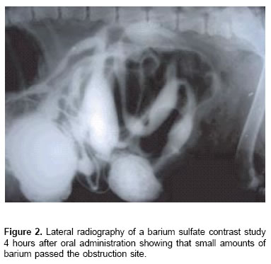
 </I></P >    <P    >Lateral (<a href="#f1">see Figure 1</a>) and ventrodorsal abdominal radiographs were obtained and a barium sulfate contrast study was performed. The abdominal radiographs clearly showed an accumulation of a radio opaque foreign material in the small intestine (between 10 and 15 cm in diameter) in the cranial region of the abdomen and a great dilatation of the intestinal loop at this point. These findings are called the graveling sign and are due to the accumulation of indigestible material anterior to the obstruction. Small intestine showed excessive distension due to accumulation of gas (<a href="#f1">see Figure 1</a>). In the contrast study the barium sulfate reached the site of the suspected obstruction about one hour after oral administration and showed very little contrast medium had passed this point in the next four hours (<a href="#f2">see Figure 2</a>). </P > <b>Surgical treatment and evolution </b>     <P    >A standard midline laparotomy procedure, using isofluorane as the anesthetic agent was performed in order to explore the suspected intestinal obstruction. A segment of the jejunum presented a severe distended loop in the site of the obstruction and also a large segment anterior to this point showed distension (<a href="#f3">see Figure 3</a>). The aspect of the mucosa was edematized with no signs of wall necrosis. An initial enterotomy was performed and a large amount of foreign material including stones and grass, was extracted from the site of the obstruction. Once the material was completely retired, a circular mass inside the lumen of the intestinal segment was palpated. The suspicion of a tumoral mass followed to a bowel resection of a 15 cm segment. And end-to-end anastomosis was performed. Since the mass was suspected as tumoral, an exploration of the abdominal organs was done before the abdominal closure, showing no macroscopic abnormal findings. </P >     <P    >The patient recovered normally from the anesthesia and any oral food or water was restricted during 36 hours. After this period, water was given and well-tolerated, so liquated food was allowed 12 hours later. In the next 5 days the patient was gradually introduced to solid food. </P >     <P    >
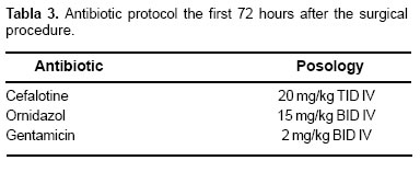
During the first 72 hours after the surgical procedure the dog was maintained to the protocol showed in <a href="#t3">Table 3</a>. After this time, 1 gram of cefalexin (40 mg/kg) BID PO and 500 miligrams of Metronidazol (20 mg/kg) BID PO, were maintained during seven more days. The patient finished its recuperation at home without any complications. </P >     <P    ><I>Macroscopic and Histological findings </I></P >    <P    >
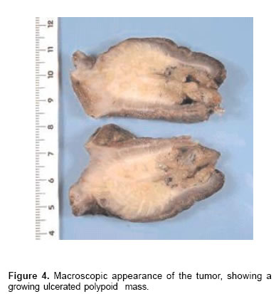
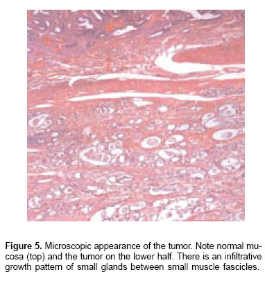
The tumoral mass was polypoid and ulcerative, as a bulky mass with an elevated surface centrally and occluding the greater part of the luminal circumference    <br>   (<a href="#f4">see Figure 4</a>). Microscopically, the intestinal mucosa   was ulcerated and lined by fibrin and neutrophils. The   submucosa was invaded by a well-differentiated    ]]></body>
<body><![CDATA[<br>   adenocarcinoma that secreted variable amounts of   mucina. The tumor infiltrated all the layers of the bowel   and extended into the serosa (<a href="#f5">see Figure 5</a>).</P >     <P    ><img src="/img/revistas/rccp/v18n1/v18n1a08f01.jpg"><a name="f1"></a></P >     <P    ><img src="/img/revistas/rccp/v18n1/v18n1a08f02.jpg"><a name="f2"></a></P >     <P    ><img src="/img/revistas/rccp/v18n1/v18n1a08f03.jpg"><a name="f3"></a></P >     <P    ><img src="/img/revistas/rccp/v18n1/v18n1a08f04.jpg"><a name="f4"></a></P >     <P    ><img src="/img/revistas/rccp/v18n1/v18n1a08f05.jpg"><a name="f5"></a></P >     <P    >&nbsp;</P >     <P    ><b>Discussion </b></P >     <P    >Clinical signs associated with small intestinal carcinomas may include chronic vomiting, diarrhea, weight loss, and anorexia (4,5,8). This patient presented a history of recurrent vomiting and occasional diarrhea, which resulted from the partial obstruction caused by the growing mass in the jejunum. Finally, in the last 48 hours, the patient suffered a complete obstruction due to the chronic ingestion of indigestible material that could not pass through the reduced intestinal lumen. Diarrhea was present occasionally in this case and is common in animals with partial obstruction of the small bowel, usually due to bacterial overgrowth and deranged intestinal motility (3). </P >     <P    >Plain abdominal radiographs demonstrated in this patient the presence of the palpated abdominal mass and an intestinal obstructive pattern (<a href="#f1">see Figure 1</a>). When the adenocarcinoma results in a concentric narrowing of the intestinal lumen, contrast radiography in the gastrointestinal tract can highlight areas of stenosis and aid in the diagnosis of the condition and planning of surgery (8). When the obstruction is still partial, the intestinal radiographs do not show a markedly dilated bowel loops but occasionally will be revealed by the &ldquo;graveling sign&rdquo;. The graveling sign refers to the accumulation of indigestible material anterior to the partial obstruction (3). In this case, was obvious the accumulation of the radiopaque not digested material and the excessive accumulation of gas resulting from the complete obstruction. </P >     ]]></body>
<body><![CDATA[<P    >Laboratory findings in this patient were normal. In animals with small intestinal neoplasia the laboratory parameters are usually unspecific. Hematological parameters in affected dogs can show a degenerative left shift in cases of obstructions and severely affected by the inflammatory or the toxemic process. Anemia and/or melena may be observed if any blood loss occurs (3,4). Serum chemistry is usually normal in patients without complete obstructions. Electrolyte and blood gas estimations are a valuable aid in selecting the appropriate fluid therapy in obstructed animals (3,4). </P >    <P    >Although ultrasonography has been proved to be an important tool in the diagnosis of an intestinal neoplasm and endoscopy occasionally helps to identify masses in the lumen of the proximal duodenum or distal ileum (1,6), the definitive diagnosis of a small bowel intramural neoplasm requires an exploratory laparotomy. Besides this, it allows a complete visualization of the primary tumor and the explorations of the entire abdominal cavity to locate metastasis. In the present case, no macroscopic evidence of metastasis was detected in any organ of the abdominal cavity, and no samples of the mesenteric lymph nodes were obtained. However, biopsy of the lymph nodes may yield additional prognostic information (8). Other sites of metastasis besides the lymph nodes have been reported, including the liver, lungs, peritoneum, prostate, testicles and rarer in the lumbar spinal cord and skin. (5) </P >    <P    >Histopathological examination confirmed the presence of a well-differentiated adenocarcinoma. Adenocarcinoma of the small intestine tends to be annular in the dog (4). The gross appearance of adenocarcinomas is variable. They may appear as expansive, plaque-like, ulcerated masses; they may invade adjacent bowel wall producing an annular or fusiform obstruction; they may cause discrete ulcers or polypoid growths; or may widely infiltrate the submucosa causing diffuse thickening without causing obstruction (4). In the present case, the mass caused an ulcerated polypoid growth that caused a partial obstruction of the small intestine segment. </P >     <P    >It was decided that the patient will not be treated with chemotherapeutic agents; in general, no benefic effects are seen in dogs treated with chemotherapy (2). The prognosis is guarded but long-term survival is common if patients with well-differentiated adenocarcinomas survive the immediate postoperative clinical background, patient history, and results of period (as in this case) (4). No signs of metastasis or physical examination and laboratory tests. Conventional other diseases have been found nine months after the radiography and barium sulfate contrast studies remain surgical procedure and the Pit-bull dog regained weight the initial methods of imaging in patientswith suspected and actually eats a normal commercial food. small bowel obstruction. The definitive diagnosis of a small bowel intramural neoplasm causing the intestinal Conclusions obstruction often requires an exploratory laparotomy in veterinary patients. Surgery may provide unique The diagnosis of small bowel obstruction in dogs is information about tumorextension and local or distant based on a comprehensive approach that includes metastasis. </P >     <P    >&nbsp;</P > <I>Resumen </I>     <P    ><I>Adenocarcinoma en el yeyuno de un perro: reporte de un caso </I></P >    <P    ><I>Un perro macho de raza Pitbull de 10 a&ntilde;os de edad fue atendido por presentar v&oacute;mito espor&aacute;dico en los &uacute;ltimos 2 meses y diarrea ocasional. En las &uacute;ltimas 48 horas el paciente hab&iacute;a presentado m&uacute;ltiples v&oacute;mitos sin tolerar alimentaci&oacute;n oral. Al examen cl&iacute;nico se palp&oacute; una masa abdominal. Las radiograf&iacute;as abdominales simples y contrastadas con sulfato de bario mostraron un ac&uacute;mulo de material radiopaco causando un patr&oacute;n caracter&iacute;stico de obstrucci&oacute;n intestinal. En la exploraci&oacute;n quir&uacute;rgica el material causante de la obstrucci&oacute;n que se encontraba en el yeyuno fue retirado mediante enterotom&iacute;a pero se detect&oacute; una masa intraluminal intestinal, por lo que se extrajo el segmento completo y se realiz&oacute; una enteroanastomosis termino-terminal. El an&aacute;lisis histol&oacute;gico de la masa extra&iacute;da evidenci&oacute; un adenocarcinoma envolviendo todo el espesor de la pared intestinal. El paciente se recuper&oacute; normalmente y hasta los nueve meses posteriores a la cirug&iacute;a, no ha aparecido ning&uacute;n signo de met&aacute;stasis. </I></P >    <P    >Palabras clave: <I>enteroanastomosis, intestino, neoplasia, obstrucci&oacute;n. </I></P >     <P    >&nbsp;</P > <b>References </b>     <!-- ref --><P    >1. Burk RL, Ackerman N. The abdomen. In: Burk RL, Ackerman   N, editors. Small animal radiology and ultrasonography, a   diagnostic atlas and text. 2nd ed. Saunders, Philadelphia; 1996.   p. 215-426.</P >    &nbsp;&nbsp;&nbsp;&nbsp;&nbsp;&nbsp;&nbsp;&nbsp;[&#160;<a href="javascript:void(0);" onclick="javascript: window.open('/scielo.php?script=sci_nlinks&ref=000052&pid=S0120-0690200500010000800001&lng=','','width=640,height=500,resizable=yes,scrollbars=1,menubar=yes,');">Links</a>&#160;]<!-- end-ref --><!-- ref --><P    > 2. Couto CG. Neoplasia gastrointestinal en perros y gatos. En:   Bonagura JD, Kirk R (eds). Terap&eacute;utica Veterinaria de   Peque&ntilde;os Animales, XI ed. Madrid, McGraw-Hill   Interamericana; 1994. p. 659-66.</P >    &nbsp;&nbsp;&nbsp;&nbsp;&nbsp;&nbsp;&nbsp;&nbsp;[&#160;<a href="javascript:void(0);" onclick="javascript: window.open('/scielo.php?script=sci_nlinks&ref=000053&pid=S0120-0690200500010000800002&lng=','','width=640,height=500,resizable=yes,scrollbars=1,menubar=yes,');">Links</a>&#160;]<!-- end-ref --><!-- ref --><P    > 3. Guilford WG, Strombeck DR. Intestinal obstruction, pseudoobstruction, and    foreign bodies. En: Guilford WG, Center SA, Strombeck DR (Eds). Strombeks' Small    Animal Gastroenterology. 3ed Philadelphia, W.B. Saunders Company; 1996:487-502.</P >    &nbsp;&nbsp;&nbsp;&nbsp;&nbsp;&nbsp;&nbsp;&nbsp;[&#160;<a href="javascript:void(0);" onclick="javascript: window.open('/scielo.php?script=sci_nlinks&ref=000054&pid=S0120-0690200500010000800003&lng=','','width=640,height=500,resizable=yes,scrollbars=1,menubar=yes,');">Links</a>&#160;]<!-- end-ref --><!-- ref --><P    > 4. Guilford WG, Strombeck DR. Neoplasm of the gastrointestinal tract, APUD tumors,    endocrinopathies and the gastrointestinal tract. En: Guilford WG, Center SA,    Strombeck DR (Eds). Strombeks' Small Animal Gastroenterology. 3ed Philadelphia,    W.B. Saunders Company; 1996:519-531.</P >    &nbsp;&nbsp;&nbsp;&nbsp;&nbsp;&nbsp;&nbsp;&nbsp;[&#160;<a href="javascript:void(0);" onclick="javascript: window.open('/scielo.php?script=sci_nlinks&ref=000055&pid=S0120-0690200500010000800004&lng=','','width=640,height=500,resizable=yes,scrollbars=1,menubar=yes,');">Links</a>&#160;]<!-- end-ref --><!-- ref --><P    >5. Juopperi TA, Cesta M, Tomlinson L, et al. Extensive   cutaneous metastases in a dog with duodenal adenocarcinoma. Vet Clin Path 2003; 32:88-91</P >    &nbsp;&nbsp;&nbsp;&nbsp;&nbsp;&nbsp;&nbsp;&nbsp;[&#160;<a href="javascript:void(0);" onclick="javascript: window.open('/scielo.php?script=sci_nlinks&ref=000056&pid=S0120-0690200500010000800005&lng=','','width=640,height=500,resizable=yes,scrollbars=1,menubar=yes,');">Links</a>&#160;]<!-- end-ref --><!-- ref --><P    > 6. Lecoindre P. Tumors of the Gastrointestinal Tract:   Endoscopic Diagnosis. Presented in the 27th World Small   Animal Veterinary Association Congress - Granada, Spain.   2002.</P >    &nbsp;&nbsp;&nbsp;&nbsp;&nbsp;&nbsp;&nbsp;&nbsp;[&#160;<a href="javascript:void(0);" onclick="javascript: window.open('/scielo.php?script=sci_nlinks&ref=000057&pid=S0120-0690200500010000800006&lng=','','width=640,height=500,resizable=yes,scrollbars=1,menubar=yes,');">Links</a>&#160;]<!-- end-ref --><!-- ref --><P    > 7. Neugut AI, Marvin MR, Rella VA, Chabot JA. An overview   of adenocarcinoma of the small intestine. Oncology 1997; 11:   529-536.</P >    &nbsp;&nbsp;&nbsp;&nbsp;&nbsp;&nbsp;&nbsp;&nbsp;[&#160;<a href="javascript:void(0);" onclick="javascript: window.open('/scielo.php?script=sci_nlinks&ref=000058&pid=S0120-0690200500010000800007&lng=','','width=640,height=500,resizable=yes,scrollbars=1,menubar=yes,');">Links</a>&#160;]<!-- end-ref --><!-- ref --><P    > 8. O&otilde;brien CR, Wong WT. Intermittent vomiting and weight   loss in an old dog. Aust Vet J 2001; 79:251-261</P >    &nbsp;&nbsp;&nbsp;&nbsp;&nbsp;&nbsp;&nbsp;&nbsp;[&#160;<a href="javascript:void(0);" onclick="javascript: window.open('/scielo.php?script=sci_nlinks&ref=000059&pid=S0120-0690200500010000800008&lng=','','width=640,height=500,resizable=yes,scrollbars=1,menubar=yes,');">Links</a>&#160;]<!-- end-ref --><!-- ref --><P    > 9. Wheeler J M, Warren B F, McC Mortensen N J, et all. An   insight into the genetic pathway of adenocarcinoma of the   small intestine Gut 2002;50:218-223.</P >    &nbsp;&nbsp;&nbsp;&nbsp;&nbsp;&nbsp;&nbsp;&nbsp;[&#160;<a href="javascript:void(0);" onclick="javascript: window.open('/scielo.php?script=sci_nlinks&ref=000060&pid=S0120-0690200500010000800009&lng=','','width=640,height=500,resizable=yes,scrollbars=1,menubar=yes,');">Links</a>&#160;]<!-- end-ref --><P    >&nbsp;</P >      ]]></body><back>
<ref-list>
<ref id="B1">
<label>1</label><nlm-citation citation-type="book">
<person-group person-group-type="author">
<name>
<surname><![CDATA[Burk]]></surname>
<given-names><![CDATA[RL]]></given-names>
</name>
<name>
<surname><![CDATA[Ackerman]]></surname>
<given-names><![CDATA[N]]></given-names>
</name>
</person-group>
<article-title xml:lang="en"><![CDATA[The abdomen]]></article-title>
<person-group person-group-type="editor">
<name>
<surname><![CDATA[Burk]]></surname>
<given-names><![CDATA[RL]]></given-names>
</name>
<name>
<surname><![CDATA[Ackerman]]></surname>
<given-names><![CDATA[N]]></given-names>
</name>
</person-group>
<source><![CDATA[Small animal radiology and ultrasonography, a diagnostic atlas and text]]></source>
<year>1996</year>
<edition>2</edition>
<page-range>215-426</page-range><publisher-loc><![CDATA[Philadelphia ]]></publisher-loc>
<publisher-name><![CDATA[Saunders]]></publisher-name>
</nlm-citation>
</ref>
<ref id="B2">
<label>2</label><nlm-citation citation-type="book">
<person-group person-group-type="author">
<name>
<surname><![CDATA[Couto]]></surname>
<given-names><![CDATA[CG]]></given-names>
</name>
</person-group>
<article-title xml:lang="es"><![CDATA[Neoplasia gastrointestinal en perros y gatos]]></article-title>
<person-group person-group-type="editor">
<name>
<surname><![CDATA[Bonagura]]></surname>
<given-names><![CDATA[JD]]></given-names>
</name>
<name>
<surname><![CDATA[Kirk]]></surname>
<given-names><![CDATA[R]]></given-names>
</name>
</person-group>
<source><![CDATA[Terapéutica Veterinaria de Pequeños Animales]]></source>
<year>1994</year>
<edition>XI</edition>
<page-range>659-66</page-range><publisher-loc><![CDATA[Madrid ]]></publisher-loc>
<publisher-name><![CDATA[McGraw-Hill Interamericana]]></publisher-name>
</nlm-citation>
</ref>
<ref id="B3">
<label>3</label><nlm-citation citation-type="book">
<person-group person-group-type="author">
<name>
<surname><![CDATA[Guilford]]></surname>
<given-names><![CDATA[WG]]></given-names>
</name>
<name>
<surname><![CDATA[Strombeck]]></surname>
<given-names><![CDATA[DR]]></given-names>
</name>
</person-group>
<article-title xml:lang="en"><![CDATA[Intestinal obstruction, pseudoobstruction, and foreign bodies.]]></article-title>
<person-group person-group-type="editor">
<name>
<surname><![CDATA[Guilford]]></surname>
<given-names><![CDATA[WG]]></given-names>
</name>
<name>
<surname><![CDATA[Center]]></surname>
<given-names><![CDATA[SA]]></given-names>
</name>
<name>
<surname><![CDATA[Strombeck]]></surname>
<given-names><![CDATA[DR]]></given-names>
</name>
</person-group>
<source><![CDATA[Strombeks' Small Animal Gastroenterology]]></source>
<year>1996</year>
<edition>3</edition>
<page-range>487-502</page-range><publisher-loc><![CDATA[Philadelphia ]]></publisher-loc>
<publisher-name><![CDATA[W.B. Saunders Company]]></publisher-name>
</nlm-citation>
</ref>
<ref id="B4">
<label>4</label><nlm-citation citation-type="book">
<person-group person-group-type="author">
<name>
<surname><![CDATA[Guilford]]></surname>
<given-names><![CDATA[WG]]></given-names>
</name>
<name>
<surname><![CDATA[Strombeck]]></surname>
<given-names><![CDATA[DR]]></given-names>
</name>
</person-group>
<article-title xml:lang="en"><![CDATA[Neoplasm of the gastrointestinal tract, APUD tumors, endocrinopathies and the gastrointestinal tract]]></article-title>
<person-group person-group-type="editor">
<name>
<surname><![CDATA[Guilford]]></surname>
<given-names><![CDATA[WG]]></given-names>
</name>
<name>
<surname><![CDATA[Center]]></surname>
<given-names><![CDATA[SA]]></given-names>
</name>
<name>
<surname><![CDATA[Strombeck]]></surname>
<given-names><![CDATA[DR]]></given-names>
</name>
</person-group>
<source><![CDATA[Strombeks' Small Animal Gastroenterology]]></source>
<year>1996</year>
<edition>3</edition>
<page-range>519-531</page-range><publisher-loc><![CDATA[Philadelphia ]]></publisher-loc>
<publisher-name><![CDATA[W.B. Saunders Company]]></publisher-name>
</nlm-citation>
</ref>
<ref id="B5">
<label>5</label><nlm-citation citation-type="journal">
<person-group person-group-type="author">
<name>
<surname><![CDATA[Juopperi]]></surname>
<given-names><![CDATA[TA]]></given-names>
</name>
<name>
<surname><![CDATA[Cesta]]></surname>
<given-names><![CDATA[M]]></given-names>
</name>
<name>
<surname><![CDATA[Tomlinson]]></surname>
<given-names><![CDATA[L]]></given-names>
</name>
</person-group>
<article-title xml:lang="en"><![CDATA[Extensive cutaneous metastases in a dog with duodenal adenocarcinoma]]></article-title>
<source><![CDATA[Vet Clin Path]]></source>
<year>2003</year>
<volume>32</volume>
<page-range>88-91</page-range></nlm-citation>
</ref>
<ref id="B6">
<label>6</label><nlm-citation citation-type="confpro">
<person-group person-group-type="author">
<name>
<surname><![CDATA[Lecoindre]]></surname>
<given-names><![CDATA[P]]></given-names>
</name>
</person-group>
<source><![CDATA[Tumors of the Gastrointestinal Tract: Endoscopic Diagnosis]]></source>
<year></year>
<conf-name><![CDATA[27 World Small Animal Veterinary Association Congress]]></conf-name>
<conf-date>2002</conf-date>
<conf-loc>Granada </conf-loc>
</nlm-citation>
</ref>
<ref id="B7">
<label>7</label><nlm-citation citation-type="journal">
<person-group person-group-type="author">
<name>
<surname><![CDATA[Neugut]]></surname>
<given-names><![CDATA[AI]]></given-names>
</name>
<name>
<surname><![CDATA[Marvin]]></surname>
<given-names><![CDATA[MR]]></given-names>
</name>
<name>
<surname><![CDATA[Rella]]></surname>
<given-names><![CDATA[VA]]></given-names>
</name>
<name>
<surname><![CDATA[Chabot]]></surname>
<given-names><![CDATA[JA]]></given-names>
</name>
</person-group>
<article-title xml:lang="en"><![CDATA[An overview of adenocarcinoma of the small intestine]]></article-title>
<source><![CDATA[Oncology]]></source>
<year>1997</year>
<volume>11</volume>
<page-range>529-536</page-range></nlm-citation>
</ref>
<ref id="B8">
<label>8</label><nlm-citation citation-type="journal">
<person-group person-group-type="author">
<name>
<surname><![CDATA[Oõbrien]]></surname>
<given-names><![CDATA[CR]]></given-names>
</name>
<name>
<surname><![CDATA[Wong]]></surname>
<given-names><![CDATA[WT]]></given-names>
</name>
</person-group>
<article-title xml:lang="en"><![CDATA[Intermittent vomiting and weight loss in an old dog]]></article-title>
<source><![CDATA[Aust Vet J]]></source>
<year>2001</year>
<volume>79</volume>
<page-range>251-261</page-range></nlm-citation>
</ref>
<ref id="B9">
<label>9</label><nlm-citation citation-type="journal">
<person-group person-group-type="author">
<name>
<surname><![CDATA[Wheeler]]></surname>
<given-names><![CDATA[J M]]></given-names>
</name>
<name>
<surname><![CDATA[Warren]]></surname>
<given-names><![CDATA[B F]]></given-names>
</name>
<name>
<surname><![CDATA[McC Mortensen]]></surname>
<given-names><![CDATA[N J]]></given-names>
</name>
</person-group>
<article-title xml:lang="en"><![CDATA[An insight into the genetic pathway of adenocarcinoma of the small intestine]]></article-title>
<source><![CDATA[Gut]]></source>
<year>2002</year>
<volume>50</volume>
<page-range>218-223</page-range></nlm-citation>
</ref>
</ref-list>
</back>
</article>
