<?xml version="1.0" encoding="ISO-8859-1"?><article xmlns:mml="http://www.w3.org/1998/Math/MathML" xmlns:xlink="http://www.w3.org/1999/xlink" xmlns:xsi="http://www.w3.org/2001/XMLSchema-instance">
<front>
<journal-meta>
<journal-id>0120-0690</journal-id>
<journal-title><![CDATA[Revista Colombiana de Ciencias Pecuarias]]></journal-title>
<abbrev-journal-title><![CDATA[Rev Colom Cienc Pecua]]></abbrev-journal-title>
<issn>0120-0690</issn>
<publisher>
<publisher-name><![CDATA[Facultad de Ciencias Agrarias, Universidad de Antioquia]]></publisher-name>
</publisher>
</journal-meta>
<article-meta>
<article-id>S0120-06902006000400009</article-id>
<title-group>
<article-title xml:lang="en"><![CDATA[Pituitary tumor diagnosis using gadolinium-enhanced magnetic resonance imaging in a dog with hyperadrenocorticism: A case report]]></article-title>
<article-title xml:lang="es"><![CDATA[Diagnóstico de tumor pituitario utilizando resonancia magnética contrastada con gadolinio en un perro con hiperadrenocorticismo: Reporte de un caso]]></article-title>
</title-group>
<contrib-group>
<contrib contrib-type="author">
<name>
<surname><![CDATA[Hernández]]></surname>
<given-names><![CDATA[Carlos A]]></given-names>
</name>
<xref ref-type="aff" rid="A01"/>
</contrib>
<contrib contrib-type="author">
<name>
<surname><![CDATA[Alzate]]></surname>
<given-names><![CDATA[Gildardo]]></given-names>
</name>
<xref ref-type="aff" rid="A02"/>
</contrib>
</contrib-group>
<aff id="A01">
<institution><![CDATA[,Universidad de Antioquia Facultad de Ciencias Agrarias Grupo de Investigación CENTAURO]]></institution>
<addr-line><![CDATA[Medellín ]]></addr-line>
<country>Colombia</country>
</aff>
<aff id="A02">
<institution><![CDATA[,Universidad de Antioquia Facultad de Ciencias Agrarias ]]></institution>
<addr-line><![CDATA[Medellín ]]></addr-line>
<country>Colombia</country>
</aff>
<pub-date pub-type="pub">
<day>00</day>
<month>12</month>
<year>2006</year>
</pub-date>
<pub-date pub-type="epub">
<day>00</day>
<month>12</month>
<year>2006</year>
</pub-date>
<volume>19</volume>
<numero>4</numero>
<fpage>437</fpage>
<lpage>441</lpage>
<copyright-statement/>
<copyright-year/>
<self-uri xlink:href="http://www.scielo.org.co/scielo.php?script=sci_arttext&amp;pid=S0120-06902006000400009&amp;lng=en&amp;nrm=iso"></self-uri><self-uri xlink:href="http://www.scielo.org.co/scielo.php?script=sci_abstract&amp;pid=S0120-06902006000400009&amp;lng=en&amp;nrm=iso"></self-uri><self-uri xlink:href="http://www.scielo.org.co/scielo.php?script=sci_pdf&amp;pid=S0120-06902006000400009&amp;lng=en&amp;nrm=iso"></self-uri><abstract abstract-type="short" xml:lang="en"><p><![CDATA[Cushing’s disease is a common endocrine disorder of dogs, caused by persistent high concentration of cortisol in circulating blood. Almost eighty five percent of the cases of spontaneous hyperadrenocorticism in dogs are the result of a functional pituitary tumor or hyperplasia. Advanced imaging techniques like computed tomography (CT) or magnetic resonance imaging (MRI) are the best means to obtain a diagnosis of a pituitary tumor in dogs with pituitary-dependent hyperadrenocorticism (PDH). Moreover these imaging techniques are required to asses the size and location of the pituitary tumor when planning surgical removal of the tumor mass or of the complete pituitary gland. In Colombia, the use of these imaging techniques is very limited in the small animal practice, but it has recently become available for veterinary patients thanks to agreements with human clinical institutions. This report describes the case of an eleven year-old golden retriever with PDH as a result of a functional pituitary tumor, visualized by magnetic resonance imaging using gadolinium as contrast agent.]]></p></abstract>
<abstract abstract-type="short" xml:lang="es"><p><![CDATA[La enfermedad de Cushing es un trastorno endocrino común en perros causado por altas y persistentes concentraciones de cortisol en sangre. Casi el 85% de los casos de hiperadrenocorticismo espontáneo en perros son el resultado de un tumor funcional o una hiperplasia pituitaria. Las técnicas avanzadas de diagnóstico por imagen como la tomografía axial computarizada (TAC) o la resonancia magnética (RM) son los mejores métodos para obtener el diagnóstico de un tumor pituitario en pacientes con hiperadrenocorticismo dependiente de la hipófisis (PDH). Además estas técnicas son necesarias para determinar el tamaño y localización del tumor pituitario cuando se planea la extracción quirúrgica de la masa o la hipófisis completa. En nuestro país, el uso de estas técnicas por imagen está muy restringido en la práctica de pequeñas especies, sin embargo recientemente han estado disponibles para los pacientes veterinarios gracias al uso de instituciones médicas humanas. En este reporte se describe el caso de un Golden retriever de 11 años con PDH como resultado de un tumor funcional pituitario, que fue visualizado usando RM utilizando gadolinio como medio de contraste.]]></p></abstract>
<kwd-group>
<kwd lng="en"><![CDATA[canine]]></kwd>
<kwd lng="en"><![CDATA[Cushing]]></kwd>
<kwd lng="en"><![CDATA[imaging]]></kwd>
<kwd lng="en"><![CDATA[pituitary]]></kwd>
<kwd lng="en"><![CDATA[tumor]]></kwd>
<kwd lng="es"><![CDATA[canino]]></kwd>
<kwd lng="es"><![CDATA[Cushing]]></kwd>
<kwd lng="es"><![CDATA[hipófisis]]></kwd>
<kwd lng="es"><![CDATA[imágenes]]></kwd>
<kwd lng="es"><![CDATA[tumor]]></kwd>
</kwd-group>
</article-meta>
</front><body><![CDATA[ <p><b>CASOS CL&Iacute;NICOS</b>    <br>       <br>   <b>Pituitary tumor diagnosis using gadolinium-enhanced magnetic resonance imaging in a dog with hyperadrenocorticism. A case report.</b>    <br>     <br> Carlos A Hern&aacute;ndez<sup>1</sup> MV, Gildardo Alzate<sup>2</sup> MV esp    <br>     <br>   <sup>1</sup>Grupo de Investigaci&oacute;n CENTAURO, Facultad de Ciencias Agrarias, Universidad de Antioquia, A.A. 1226, Medell&iacute;n, Colombia    <br> <sup>2</sup>Docente Facultad de Ciencias Agrarias, Universidad de Antioquia, Medell&iacute;n, Colombia     <br> carlitosevi@yahoo.com    <br>       ]]></body>
<body><![CDATA[<br>   (Recibido: 10 mayo, 2006; aceptado: 20 Octubre, 2006)    <br>       <br>   <b>Summary</b>    <br>       <br>   <i><b>Cushing&rsquo;s disease is a common endocrine disorder of dogs, caused by persistent high concentration of cortisol in circulating blood. Almost eighty five percent of the cases of spontaneous hyperadrenocorticism in dogs are the result of a functional pituitary tumor or hyperplasia. Advanced imaging techniques like computed tomography (CT) or magnetic resonance imaging (MRI) are the best means to obtain a diagnosis of a pituitary tumor in dogs with pituitary-dependent hyperadrenocorticism (PDH). Moreover these imaging techniques are required to asses the size and location of the pituitary tumor when planning surgical removal of the tumor mass or of the complete pituitary gland. In Colombia, the use of these imaging techniques is very limited in the small animal practice, but it has recently become available for veterinary patients thanks to agreements with human clinical institutions. This report describes the case of an eleven year-old golden retriever with PDH as a result of a functional pituitary tumor, visualized by magnetic resonance imaging using gadolinium as contrast agent.   Key words: canine, Cushing, imaging, pituitary, tumor.</b></i>    <br>       <br>   <b>Introduction</b>    <br>       <br>   Hyperadrenocorticism is a common clinical disorder, and is one of the most common endocrinophaties in dogs (1, 2). Clinical signs in naturally occurring hyperadrenocorticism are the result of cortisol overproduction that may result from excess secretion of ACTH (2). A functional pituitary tumor causing pituitary dependant hyperadrenocorticism (PDH) is the most common cause of canine hyperadrenocorticism (2, 7).    <br>       ]]></body>
<body><![CDATA[<br>   In Medell&iacute;n, in regular clinical practice, the clinical diagnosis of hyperadrenocorticism based usually in systemic and dermatologic signs is common. The etiologic determination is unusual due to the cost implicated in laboratory tests and the restrictions in the availability of the advanced imaging techniques that are not yet available for veterinary diagnosis.    <br>       <br>   The recent cost-based accessibility to laboratory tests and the availability of advanced imaging techniques due to human clinical institutions that allow the use in small animals in the city permited   the diagnostic of a PDH in a golden retriever   caused by a pituitary tumor visualized using gadolinium-enhanced magnetic resonance imaging. This imaging technique is the first choice in the diagnostic of this kind of tumors in human clinical practice.    <br>       <br>   <b>Patient evaluation</b>    <br>       <br>   <i>Anamnesis</i>    <br>       <br>   An 11-year-old golden retriever was examined for a two months history of polyuria &ndash; polydipsia, polyphagia, muscle weakness and abdominal enlargement. The dog had endocrine alopecia and a previous diagnostic of hypothyroidism. The dog received 800 mcg of levotiroxin twice a day (BID) without any clinical improvement.    <br>       ]]></body>
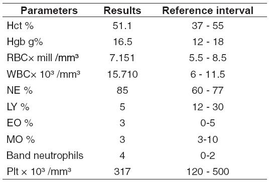
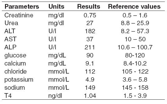
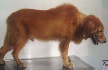
<body><![CDATA[<br>   <i>Physical examination and laboratory findings</i>    <br>       <br>   The dog was overweight (43 kg) and its attitude was normal at the time of clinical examination.   Previous laboratory test revealed T4 values   between 0.6 and 1.05 pc/dL, and the dog had been receiving 800 mcg of levotiroxin BID during one   year. However, endocrine alopecia had become worst and polyuria- polydipsia and polyphagia   were developed during the last two months (see Figure 1). During the neurological examination no deficits   were detected and no abdominal masses were palpated.     <br>       <br>   Hyperadrenocorticism was suspected on the basis of the clinical examination and the   laboratory findings that included CBC (complete blood count) (see Table 1), serum biochemical analysis (see Table 2), and urinalysis, and low-dose dexamethasone suppression test were made. Administering 0.01 mg/kg of dexamethasone in polyethylene glycol intravenously after a blood sample had been obtained to determine a plasma cortisol concentration baseline (see Table 3). Blood samples were obtained for determination   of plasma cortisol concentration at 4 and 8 hours   after administration of dexamethasone. An abdominal ultrasonography was performed with   no evident changes in the adrenal glands. A solitary calicial calculus measuring 7 mm of diameter was detected in the left kidney with no hidronephrotic signs. The urine analysis found no abnormal   changes with a density in the isostenuric range.    <br>       <br>   <b>Table 1</b>. Hematology values.    <br>   <img src="/img/revistas/rccp/v19n4/4a09t1.JPG" width="542" height="373" />    <br>       <br>   <b>Table 2</b>. Serum biochemical profile.    ]]></body>
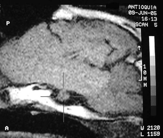
<body><![CDATA[<br>   <img src="/img/revistas/rccp/v19n4/4a09t2.JPG" width="536" height="335" />    <br>       <br>   <b>Table 3</b>. Low-dose dexamethasone suppression test    <br>   <img src="/img/revistas/rccp/v19n4/4a09t3.JPG" width="546" height="105" />    <br>       <br>   <i>Magnetic resonance imaging</i>    <br>       <br>   Evaluation of the low-dose dexamethasone suppression test and the abdominal ultrasonography suggested a diagnosis of PDH, and then a dynamic magnetic resonance imaging (MRI) examination   was performed. Food was withheld 24 hours prior to MRI. After administration of atropine (0.022 mg/kg, IM) and acepromazine (0.05 mg/kg), the dog was anesthetized by IV administration of a mixture of ketamine HCL (5 mg/kg) and diazepam (0.4 mg/kg) and placed in sternal recumbence. Anesthesia was maintained by repeated doses of IV ketamine (2 mg/kg).    <br>       <br>   Gadolinium-diethylenetriaminepentaacetic acid (Gd-DTPA) was administered as an intravenous   bolus injection to increase the diagnostic sensivity   and the delineation of the lesion suspected.   According to the evaluation, it is noticeable the presence of a 6 mm diameter heterogeneous nodule with an hemorrhage component in a late subacute   phase which was located on the sella turcica overextending it and slightly overlapping the   chiasm and optic nerve (see Figure 2).    ]]></body>
<body><![CDATA[<br>       <br>   <img src="/img/revistas/rccp/v19n4/4a09f1.jpg" width="378" height="246" />      <br>   <b>Figure 1</b>. Fat redistributed to the abdomen and symmetrical alopecia noticed in the patient.    <br>       <br>   <img src="/img/revistas/rccp/v19n4/4a09f2.jpg" width="573" height="484" />    <br>   <b>Figure 2</b>. In the MRI the presence of one heterogeneous nodule (red lines) located on the sella turcica was noticed.    <br>       <br>   <b>Discussion</b>    <br>       <br>   International data shows that 80% to 85% of the dogs with spontaneous hyperadrenocorticism have pituitary-dependent hyperadrenocorticism (PDH), and the remaining dogs have an autonomously functioning adrenocortical tumor (AT) (2,7). There are no dates known by the authors about the   incidence of hyperadrenocorticism in dogs in Colombia.    ]]></body>
<body><![CDATA[<br>       <br>   Spontaneous PDH in dog is the result of a   functional pituitary tumor in 90% of the cases,   either in the anterior pituitary (85% of PDH cases caused by tumors) or the intermediate lobe (15% of PDH cases caused by tumors). The 10% remaining dogs with PDH have pituitary hyperplasia (1, 4).    <br>       <br>   Clinical signs, basic laboratory tests (CBC, serum biochemical analysis) and abdominal ultrasonography aid in the diagnostic of hyperadrenocorticism but did not confirm the   PDH diagnosis, however, there are other useful tests to evaluate adrenal and pituitary function. A low-dose dexamethasone suppression test was   performed to confirm the presumptive   diagnosis and it is the most commonly used   screening test (2). Other test like High-dose dexamethasone tests and ACTH stimulation test may help to differentiate between PDH and AT   in dogs with hyperadrenocorticism that do not   have adrenal suppression in response to the   low-dose test (5). Other tests including the corticotrophin-releasing hormone response test have been reported but are not frequently used (7). In the present case, the low-dose   dexamethasone suppression test helped   discriminate between PDH and AT because the plasma cortisol concentration at 4 hours was less than 50% of the baseline value (7).    <br>       <br>   Adrenal ultrasonography is a powerful tool   for the evaluation of adrenal size, tissue changes   in the gland and the presence of unilateral or bilateral masses causing adrenocortical tumor hyperadrenocorticism (7). In this case, no abnormalities were found using this screening test.    <br>       <br>   There are several techniques for visualizing the pituitary gland including conventional radiography and linear tomography following the injection of radiographic contrast medium in the subarachnoid space (cisternography), although the most   informative techniques to obtain an antemorten diagnosis of a pituitary tumor in dogs with PDH   are computed tomography (CT) and magnetic   resonance imaging (MRI) (3, 12).    <br>       <br>   Magnetic resonance imaging (MRI) is a noninvasive procedure that does not involve ionizing radiation and is the imaging modality of choice for the detection of pituitary adenomas in humans combined with the use of the paramagnetic contrast agent, gadolinium-diethylenetriaminepentaacetic acid (Gd-DTPA) to enhance the capacity to visualize the pituitary gland and to screen patients for pituitary   adenomas (8, 11, 12). Moreover, the use of   Gd-DTPA increases lesion detectability and delineation by enhancing the border between certain brain tumors and normal tissue. This allows accurate localization of the pituitary gland   relative to surgical landmarks, which is essential   when pituitary surgery is considered (6, 10, 12). The gadolinium is not visualized directly but indirectly by the influence of paramagnetic gadolinium on the tissue protons (6, 10).    ]]></body>
<body><![CDATA[<br>       <br>   MRI is based on mapping the density and magnetic properties of hydrogen atoms when placed in a strong magnetic field. Hydrogen nuclei or protons (H<sup>+</sup>) are present in all tissues and fluids of the body. The H<sup>+</sup> binding degree in the different tissues or organs being examined,   can affect the sensitivity to the applied magnetic field and thus their appearance which is   interpreted and transformed into images by magnetic resonance (MR) instruments (9, 10).    <br>       <br>   MR images are based primarily on proton density and proton relaxation dynamics. There are various excitation and detection sequences available. The various sequences provide images of the proton   density and the two basic forms of relaxation, MR instruments are sensitive to these two different relaxation processes, the T1 (spin-lattice or   longitudinal relaxation time) and T2 (spin-spin or transverse relaxation time). The most common sequence used has been a spin-echo sequence   yielding a proton density and T2-weighted sequence before and after the administrations of a   paramagnetic contrast agent (11).    <br>       <br>   Paramagnetic agents contain one or more   unpaired electrons that enhance the T1 and T2   relaxation rates of protons in their molecular environment. The most common used contrast   agent is the form of gadolinium as mentioned earlier. When placed in a magnetic field, Gd-DTPA   shortens the T1 and T2 relaxation times in tissues where it accumulates, making possible that two tissues that would otherwise have similar relaxation characteristics appear more distinct (9). Gd-DTPA does not pass the intact blood-brain barrier, and then enhances visualization of normal tissues that lack a blood-brain barrier, such as the pituitary gland and   the choroid plexus. Lesions that disrupt de   blood-brain barrier allows accumulation of   Gd-DTPA in lesions such as neoplasm, abscesses,   and sub acute infarcts, resulting in a high signal   intensity influencing the relaxation of tissue   protons (11).    <br>       <br>   <b>Conclusions</b>    <br>       <br>   The high cost and limited availability of MR scanners has restricted its use in veterinary medicine in Colombia, and it is available only in veterinary teaching institutions in other countries. We occasionally utilize human intended facilities   and institutions, but they do not have the   appropriate infrastructure to work with animals, however, it is important to know that we now have the access to the advance imaging techniques, and when the cost it is not an issue for the dog owner, we have the capacity to improve our diagnostic approach.    ]]></body>
<body><![CDATA[<br>       <br>   <b>Resumen</b>    <br>       <br>   <b><i>Diagn&oacute;stico de tumor pituitario utilizando resonancia magn&eacute;tica contrastada con gadolinio en un perro con hiperadrenocorticismo. Reporte de un caso.</i></b>    <br>       <br>   <b><i> La enfermedad de Cushing es un trastorno endocrino com&uacute;n en perros causado por altas y persistentes concentraciones de cortisol en sangre. Casi el 85% de los casos de hiperadrenocorticismo espont&aacute;neo en perros son el resultado de un tumor funcional o una hiperplasia pituitaria. Las t&eacute;cnicas avanzadas de diagn&oacute;stico por imagen como la tomograf&iacute;a axial computarizada (TAC) o la resonancia magn&eacute;tica (RM) son los mejores m&eacute;todos para obtener el diagn&oacute;stico de un tumor pituitario en pacientes con hiperadrenocorticismo dependiente de la hip&oacute;fisis (PDH). Adem&aacute;s estas t&eacute;cnicas son necesarias para determinar el tama&ntilde;o y localizaci&oacute;n del tumor pituitario cuando se planea la extracci&oacute;n quir&uacute;rgica de la masa o la hip&oacute;fisis completa. En nuestro pa&iacute;s, el uso de estas t&eacute;cnicas por imagen est&aacute; muy restringido en la pr&aacute;ctica de peque&ntilde;as especies, sin embargo recientemente han estado disponibles para los pacientes veterinarios gracias al uso de instituciones m&eacute;dicas humanas. En este reporte se describe el caso de un Golden retriever de 11 a&ntilde;os con PDH como resultado de un tumor funcional pituitario, que fue visualizado usando RM utilizando gadolinio como medio de contraste.</i></b>    <br>       <br>   <b>Palabras clave</b>: <i>canino, Cushing, hip&oacute;fisis, im&aacute;genes, tumor.    <br>   </i>    <br>   <b>References</b>    ]]></body>
<body><![CDATA[<br>       <!-- ref --><br>   1. Axlund TW, Behrend EN. Surgical treatment of canine  Hyperadrenocorticism. Compendium 2003; 25: 334-347.    &nbsp;&nbsp;&nbsp;&nbsp;&nbsp;&nbsp;&nbsp;&nbsp;[&#160;<a href="javascript:void(0);" onclick="javascript: window.open('/scielo.php?script=sci_nlinks&ref=000094&pid=S0120-0690200600040000900001&lng=','','width=640,height=500,resizable=yes,scrollbars=1,menubar=yes,');">Links</a>&#160;]<!-- end-ref --><br>       <!-- ref --><br>   2. Behrend E, Kemppainen RJ, Clark TP. Diagnosis of  hyperadrenocorticism in dogs: a survey of internists and dermatologists. JAVMA  2002; 220: 1643-1649.    &nbsp;&nbsp;&nbsp;&nbsp;&nbsp;&nbsp;&nbsp;&nbsp;[&#160;<a href="javascript:void(0);" onclick="javascript: window.open('/scielo.php?script=sci_nlinks&ref=000096&pid=S0120-0690200600040000900002&lng=','','width=640,height=500,resizable=yes,scrollbars=1,menubar=yes,');">Links</a>&#160;]<!-- end-ref --><br>       <!-- ref --><br>   3. Duesburg  C, Feldman E, Richard W <i>et. al</i>. Magnetic resonance imaging for diagnosis of  pituitary macrotumors in dogs. JAVMA 1995; 206: 657-662.    &nbsp;&nbsp;&nbsp;&nbsp;&nbsp;&nbsp;&nbsp;&nbsp;[&#160;<a href="javascript:void(0);" onclick="javascript: window.open('/scielo.php?script=sci_nlinks&ref=000098&pid=S0120-0690200600040000900003&lng=','','width=640,height=500,resizable=yes,scrollbars=1,menubar=yes,');">Links</a>&#160;]<!-- end-ref --><br>       <br>   4. Feldman EC, Nelson RW. Hyperadrenocorticism (Cushing&rsquo;s  syndrome). In: Feldman BF, Nelson RW (editors): Canine and feline endocrinology  and Reproduction, 2nd ed. Philadelphia: WB Saunders; 1996. p 187-265.    <br>       <!-- ref --><br>   5. Feldman  EC, Nelson RW, Feldman MS. Use of low- and high-dose dexamethasone tests for  distinguishing pituitary-dependent from adrenal tumor hyperadrenocorticism &nbsp;in dogs. JAVMA 1996; 209:772-775.    &nbsp;&nbsp;&nbsp;&nbsp;&nbsp;&nbsp;&nbsp;&nbsp;[&#160;<a href="javascript:void(0);" onclick="javascript: window.open('/scielo.php?script=sci_nlinks&ref=000102&pid=S0120-0690200600040000900005&lng=','','width=640,height=500,resizable=yes,scrollbars=1,menubar=yes,');">Links</a>&#160;]<!-- end-ref --><br>       <!-- ref --><br>   6.  Goldstein EJ, Burnett KR, Wolf GL, <i>et. al</i>. Contrast enhancement of spontaneous  animal CNS tumors with gadolinium DTPA: a correlation of MRI with x-ray CT). Physiol  Chem Phys Med NMR 1985; 17: 113-122.    &nbsp;&nbsp;&nbsp;&nbsp;&nbsp;&nbsp;&nbsp;&nbsp;[&#160;<a href="javascript:void(0);" onclick="javascript: window.open('/scielo.php?script=sci_nlinks&ref=000104&pid=S0120-0690200600040000900006&lng=','','width=640,height=500,resizable=yes,scrollbars=1,menubar=yes,');">Links</a>&#160;]<!-- end-ref --><br>       <!-- ref --><br>   7. Guptill  L, Scott-Moncrieff JC, Widmer W. Diagnosis of canine hyperadrenocorticism. Vet  Clin North Am Small Anim Prac 1997; 27:215-225.    &nbsp;&nbsp;&nbsp;&nbsp;&nbsp;&nbsp;&nbsp;&nbsp;[&#160;<a href="javascript:void(0);" onclick="javascript: window.open('/scielo.php?script=sci_nlinks&ref=000106&pid=S0120-0690200600040000900007&lng=','','width=640,height=500,resizable=yes,scrollbars=1,menubar=yes,');">Links</a>&#160;]<!-- end-ref --><br>       <!-- ref --><br>   8.&nbsp;Hall WA, Luciano M,  Doppman Jl, Patrpmas NJ, <i>et. al</i>. Pituitary Magnetic Resonance Imaging  in Normal Human Volunteers Occult Adenomas in  the General Population. Ann Intern Med 1994; 120: 817-820.    &nbsp;&nbsp;&nbsp;&nbsp;&nbsp;&nbsp;&nbsp;&nbsp;[&#160;<a href="javascript:void(0);" onclick="javascript: window.open('/scielo.php?script=sci_nlinks&ref=000108&pid=S0120-0690200600040000900008&lng=','','width=640,height=500,resizable=yes,scrollbars=1,menubar=yes,');">Links</a>&#160;]<!-- end-ref --><br>       <!-- ref --><br>   9. Pooya  HA. Magnetic Resonance Imaging in Small Animal Medicine: Clinical Applications.  Compendium 2004; 26: 292-301.    &nbsp;&nbsp;&nbsp;&nbsp;&nbsp;&nbsp;&nbsp;&nbsp;[&#160;<a href="javascript:void(0);" onclick="javascript: window.open('/scielo.php?script=sci_nlinks&ref=000110&pid=S0120-0690200600040000900009&lng=','','width=640,height=500,resizable=yes,scrollbars=1,menubar=yes,');">Links</a>&#160;]<!-- end-ref --><br>       <!-- ref --><br>   10. Snellman M. Magnetic resonance Imaging in canine  spontaneous neurological disorders: an evaluation of equipment and methods. Academic  Dissertation    presented in the Auditorium Maximum, Helsinki. 2000.    &nbsp;&nbsp;&nbsp;&nbsp;&nbsp;&nbsp;&nbsp;&nbsp;[&#160;<a href="javascript:void(0);" onclick="javascript: window.open('/scielo.php?script=sci_nlinks&ref=000112&pid=S0120-0690200600040000900010&lng=','','width=640,height=500,resizable=yes,scrollbars=1,menubar=yes,');">Links</a>&#160;]<!-- end-ref --><br>       <!-- ref --><br>   11.&nbsp;Tucker RL, Gavin PR.&nbsp;Brain imaging. Vet Clin North Am Small Anim  Pract&nbsp;1996; 26: 735-758.    &nbsp;&nbsp;&nbsp;&nbsp;&nbsp;&nbsp;&nbsp;&nbsp;[&#160;<a href="javascript:void(0);" onclick="javascript: window.open('/scielo.php?script=sci_nlinks&ref=000114&pid=S0120-0690200600040000900011&lng=','','width=640,height=500,resizable=yes,scrollbars=1,menubar=yes,');">Links</a>&#160;]<!-- end-ref --><br>       <!-- ref --><br>   12. Van der Vlugt-Meijer RH, Voorhout G, P. Meij B. Imaging  of the pituitary gland in dogs with pituitary-dependent hyperadrenocorticism. Mol  Cel endocrinal 2002.   197: 81-87. &nbsp;&nbsp;&nbsp;&nbsp;&nbsp;&nbsp;&nbsp;&nbsp;[&#160;<a href="javascript:void(0);" onclick="javascript: window.open('/scielo.php?script=sci_nlinks&ref=000116&pid=S0120-0690200600040000900012&lng=','','width=640,height=500,resizable=yes,scrollbars=1,menubar=yes,');">Links</a>&#160;]<!-- end-ref --> ]]></body><back>
<ref-list>
<ref id="B1">
<label>1</label><nlm-citation citation-type="journal">
<person-group person-group-type="author">
<name>
<surname><![CDATA[Axlund]]></surname>
<given-names><![CDATA[TW]]></given-names>
</name>
<name>
<surname><![CDATA[Behrend]]></surname>
<given-names><![CDATA[EN]]></given-names>
</name>
</person-group>
<article-title xml:lang="en"><![CDATA[Surgical treatment of canine Hyperadrenocorticism]]></article-title>
<source><![CDATA[Compendium]]></source>
<year>2003</year>
<volume>25</volume>
<page-range>334-347</page-range></nlm-citation>
</ref>
<ref id="B2">
<label>2</label><nlm-citation citation-type="journal">
<person-group person-group-type="author">
<name>
<surname><![CDATA[Behrend]]></surname>
<given-names><![CDATA[E]]></given-names>
</name>
<name>
<surname><![CDATA[Kemppainen]]></surname>
<given-names><![CDATA[RJ]]></given-names>
</name>
<name>
<surname><![CDATA[Clark]]></surname>
<given-names><![CDATA[TP]]></given-names>
</name>
</person-group>
<article-title xml:lang="en"><![CDATA[Diagnosis of hyperadrenocorticism in dogs: a survey of internists and dermatologists]]></article-title>
<source><![CDATA[JAVMA]]></source>
<year>2002</year>
<volume>220</volume>
<page-range>1643-1649</page-range></nlm-citation>
</ref>
<ref id="B3">
<label>3</label><nlm-citation citation-type="journal">
<person-group person-group-type="author">
<name>
<surname><![CDATA[Duesburg]]></surname>
<given-names><![CDATA[C]]></given-names>
</name>
<name>
<surname><![CDATA[Feldman]]></surname>
<given-names><![CDATA[E]]></given-names>
</name>
<name>
<surname><![CDATA[Richard]]></surname>
<given-names><![CDATA[W]]></given-names>
</name>
</person-group>
<article-title xml:lang="en"><![CDATA[Magnetic resonance imaging for diagnosis of pituitary macrotumors in dogs]]></article-title>
<source><![CDATA[JAVMA]]></source>
<year>1995</year>
<volume>206</volume>
<page-range>657-662</page-range></nlm-citation>
</ref>
<ref id="B4">
<label>4</label><nlm-citation citation-type="book">
<person-group person-group-type="author">
<name>
<surname><![CDATA[Feldman]]></surname>
<given-names><![CDATA[EC]]></given-names>
</name>
<name>
<surname><![CDATA[Nelson]]></surname>
<given-names><![CDATA[RW]]></given-names>
</name>
</person-group>
<article-title xml:lang="en"><![CDATA[Hyperadrenocorticism: (Cushing’s syndrome)]]></article-title>
<person-group person-group-type="editor">
<name>
<surname><![CDATA[Feldman]]></surname>
<given-names><![CDATA[BF]]></given-names>
</name>
<name>
<surname><![CDATA[Nelson]]></surname>
<given-names><![CDATA[RW]]></given-names>
</name>
</person-group>
<source><![CDATA[Canine and feline endocrinology and Reproduction]]></source>
<year>1996</year>
<edition>2</edition>
<page-range>187-265</page-range><publisher-loc><![CDATA[Philadelphia ]]></publisher-loc>
<publisher-name><![CDATA[WB Saunders]]></publisher-name>
</nlm-citation>
</ref>
<ref id="B5">
<label>5</label><nlm-citation citation-type="journal">
<person-group person-group-type="author">
<name>
<surname><![CDATA[Feldman]]></surname>
<given-names><![CDATA[EC]]></given-names>
</name>
<name>
<surname><![CDATA[Nelson]]></surname>
<given-names><![CDATA[RW]]></given-names>
</name>
<name>
<surname><![CDATA[Feldman]]></surname>
<given-names><![CDATA[MS]]></given-names>
</name>
</person-group>
<article-title xml:lang="en"><![CDATA[Use of low- and high-dose dexamethasone tests for distinguishing pituitary-dependent from adrenal tumor hyperadrenocorticism in dogs]]></article-title>
<source><![CDATA[JAVMA]]></source>
<year>1996</year>
<volume>209</volume>
<page-range>772-775</page-range></nlm-citation>
</ref>
<ref id="B6">
<label>6</label><nlm-citation citation-type="journal">
<person-group person-group-type="author">
<name>
<surname><![CDATA[Goldstein]]></surname>
<given-names><![CDATA[EJ]]></given-names>
</name>
<name>
<surname><![CDATA[Burnett]]></surname>
<given-names><![CDATA[KR]]></given-names>
</name>
<name>
<surname><![CDATA[Wolf]]></surname>
<given-names><![CDATA[GL]]></given-names>
</name>
</person-group>
<article-title xml:lang="en"><![CDATA[Contrast enhancement of spontaneous animal CNS tumors with gadolinium DTPA: a correlation of MRI with x-ray CT)]]></article-title>
<source><![CDATA[Physiol Chem Phys Med NMR]]></source>
<year>1985</year>
<volume>17</volume>
<page-range>113-122</page-range></nlm-citation>
</ref>
<ref id="B7">
<label>7</label><nlm-citation citation-type="journal">
<person-group person-group-type="author">
<name>
<surname><![CDATA[Guptill]]></surname>
<given-names><![CDATA[L]]></given-names>
</name>
<name>
<surname><![CDATA[Scott-Moncrieff]]></surname>
<given-names><![CDATA[JC]]></given-names>
</name>
<name>
<surname><![CDATA[Widmer]]></surname>
<given-names><![CDATA[W]]></given-names>
</name>
</person-group>
<article-title xml:lang="en"><![CDATA[Diagnosis of canine hyperadrenocorticism]]></article-title>
<source><![CDATA[Vet Clin North Am Small Anim Prac]]></source>
<year>1997</year>
<volume>27</volume>
<page-range>215-225</page-range></nlm-citation>
</ref>
<ref id="B8">
<label>8</label><nlm-citation citation-type="journal">
<person-group person-group-type="author">
<name>
<surname><![CDATA[Hall]]></surname>
<given-names><![CDATA[WA]]></given-names>
</name>
<name>
<surname><![CDATA[Luciano]]></surname>
<given-names><![CDATA[M]]></given-names>
</name>
<name>
<surname><![CDATA[Doppman]]></surname>
<given-names><![CDATA[Jl]]></given-names>
</name>
<name>
<surname><![CDATA[Patrpmas]]></surname>
<given-names><![CDATA[NJ]]></given-names>
</name>
</person-group>
<article-title xml:lang="en"><![CDATA[Pituitary Magnetic Resonance Imaging in Normal Human Volunteers Occult Adenomas in the General Population]]></article-title>
<source><![CDATA[Ann Intern Med]]></source>
<year>1994</year>
<volume>120</volume>
<page-range>817-820</page-range></nlm-citation>
</ref>
<ref id="B9">
<label>9</label><nlm-citation citation-type="journal">
<person-group person-group-type="author">
<name>
<surname><![CDATA[Pooya]]></surname>
<given-names><![CDATA[HA]]></given-names>
</name>
</person-group>
<article-title xml:lang="en"><![CDATA[Magnetic Resonance Imaging in Small Animal Medicine: Clinical Applications]]></article-title>
<source><![CDATA[Compendium]]></source>
<year>2004</year>
<volume>26</volume>
<page-range>292-301</page-range></nlm-citation>
</ref>
<ref id="B10">
<label>10</label><nlm-citation citation-type="">
<person-group person-group-type="author">
<name>
<surname><![CDATA[Snellman]]></surname>
<given-names><![CDATA[M]]></given-names>
</name>
</person-group>
<source><![CDATA[Magnetic resonance Imaging in canine spontaneous neurological disorders: an evaluation of equipment and methods]]></source>
<year>2000</year>
<publisher-loc><![CDATA[Helsinki ]]></publisher-loc>
</nlm-citation>
</ref>
<ref id="B11">
<label>11</label><nlm-citation citation-type="journal">
<person-group person-group-type="author">
<name>
<surname><![CDATA[Tucker]]></surname>
<given-names><![CDATA[RL]]></given-names>
</name>
<name>
<surname><![CDATA[Gavin]]></surname>
<given-names><![CDATA[PR]]></given-names>
</name>
</person-group>
<article-title xml:lang="en"><![CDATA[Brain imaging]]></article-title>
<source><![CDATA[Vet Clin North Am Small Anim Pract]]></source>
<year>1996</year>
<volume>26</volume>
<page-range>735-758</page-range></nlm-citation>
</ref>
<ref id="B12">
<label>12</label><nlm-citation citation-type="journal">
<person-group person-group-type="author">
<name>
<surname><![CDATA[Van der Vlugt-Meijer]]></surname>
<given-names><![CDATA[RH]]></given-names>
</name>
<name>
<surname><![CDATA[Voorhout]]></surname>
<given-names><![CDATA[G]]></given-names>
</name>
<name>
<surname><![CDATA[P. Meij]]></surname>
<given-names><![CDATA[B]]></given-names>
</name>
</person-group>
<article-title xml:lang="en"><![CDATA[Imaging of the pituitary gland in dogs with pituitary-dependent hyperadrenocorticism]]></article-title>
<source><![CDATA[Mol Cel endocrinal]]></source>
<year>2002</year>
<volume>197</volume>
<page-range>81-87</page-range></nlm-citation>
</ref>
</ref-list>
</back>
</article>