<?xml version="1.0" encoding="ISO-8859-1"?><article xmlns:mml="http://www.w3.org/1998/Math/MathML" xmlns:xlink="http://www.w3.org/1999/xlink" xmlns:xsi="http://www.w3.org/2001/XMLSchema-instance">
<front>
<journal-meta>
<journal-id>0120-0690</journal-id>
<journal-title><![CDATA[Revista Colombiana de Ciencias Pecuarias]]></journal-title>
<abbrev-journal-title><![CDATA[Rev Colom Cienc Pecua]]></abbrev-journal-title>
<issn>0120-0690</issn>
<publisher>
<publisher-name><![CDATA[Facultad de Ciencias Agrarias, Universidad de Antioquia]]></publisher-name>
</publisher>
</journal-meta>
<article-meta>
<article-id>S0120-06902010000100004</article-id>
<title-group>
<article-title xml:lang="en"><![CDATA[Survival of vitrified mouse blastocysts loaded into glass micro-capillaries&curren;]]></article-title>
<article-title xml:lang="es"><![CDATA[Sobrevivencia embrionaria de blastocistos murinos vitrificados en microcapilares de vidrio]]></article-title>
<article-title xml:lang="pt"><![CDATA[Sobrevivência embrionária de blastocistos murinos vitrificados em micro capilares de vidro]]></article-title>
</title-group>
<contrib-group>
<contrib contrib-type="author">
<name>
<surname><![CDATA[Rodríguez V]]></surname>
<given-names><![CDATA[Paula]]></given-names>
</name>
<xref ref-type="aff" rid="A01"/>
</contrib>
<contrib contrib-type="author">
<name>
<surname><![CDATA[Ongaratto]]></surname>
<given-names><![CDATA[Felipe]]></given-names>
</name>
<xref ref-type="aff" rid="A01"/>
</contrib>
<contrib contrib-type="author">
<name>
<surname><![CDATA[Scherer]]></surname>
<given-names><![CDATA[Daniela]]></given-names>
</name>
<xref ref-type="aff" rid="A01"/>
</contrib>
<contrib contrib-type="author">
<name>
<surname><![CDATA[de Ávila Rodrigues]]></surname>
<given-names><![CDATA[Berenice]]></given-names>
</name>
<xref ref-type="aff" rid="A01"/>
</contrib>
<contrib contrib-type="author">
<name>
<surname><![CDATA[Rodrigues]]></surname>
<given-names><![CDATA[José L]]></given-names>
</name>
<xref ref-type="aff" rid="A01"/>
</contrib>
</contrib-group>
<aff id="A01">
<institution><![CDATA[,Universidade Federal do Rio Grande do Sul Faculdade de Medicina Veterinária Laboratório de Embriologia e biotecnicas da reprodução]]></institution>
<addr-line><![CDATA[Porto Alegre ]]></addr-line>
<country>Brasil</country>
</aff>
<pub-date pub-type="pub">
<day>00</day>
<month>03</month>
<year>2010</year>
</pub-date>
<pub-date pub-type="epub">
<day>00</day>
<month>03</month>
<year>2010</year>
</pub-date>
<volume>23</volume>
<numero>1</numero>
<fpage>28</fpage>
<lpage>34</lpage>
<copyright-statement/>
<copyright-year/>
<self-uri xlink:href="http://www.scielo.org.co/scielo.php?script=sci_arttext&amp;pid=S0120-06902010000100004&amp;lng=en&amp;nrm=iso"></self-uri><self-uri xlink:href="http://www.scielo.org.co/scielo.php?script=sci_abstract&amp;pid=S0120-06902010000100004&amp;lng=en&amp;nrm=iso"></self-uri><self-uri xlink:href="http://www.scielo.org.co/scielo.php?script=sci_pdf&amp;pid=S0120-06902010000100004&amp;lng=en&amp;nrm=iso"></self-uri><abstract abstract-type="short" xml:lang="en"><p><![CDATA[The purpose of this study was to determine the in vitro expansion and hatching rates of vitrified mouse blastocysts loaded into glass micro-capillaries (Brand® - 5 &mu;L). Early morning on day 4 of pregnancy, blastocysts were collected from donors, morphologically evaluated, and then allocated in three groups: Group 1 (Control): embryos were transferred into 100 &mu;L of KSOM medium drops and in vitro cultured during 72 h; Groups 2 and 3: embryos initially exposed to the equilibration solution (PBSm + 10% EG + 10% PROH and 0.5% PVA) for 1 min, and then to vitrification solution (PBSm + 20% EG + 20% PROH + 0.5% PVA) for 30 sec. After that, blastocysts were loaded into glass micropipettes (GMP) or glass microcapillaries (GMC) and plunged into super-cooled liquid nitrogen (-200 &deg;C). Embryo warming and cryoprotectant dilution were carried out into 500 &mu;L droplets of PBSm supplemented with 0.25 M sucrose maintained at 37 &deg;C. After 5 min embryos were transferred to 100 mL droplets of KSOM medium and cultured in vitro for 72 h. Blastocyst expansion rates after in vitro culture were 77% (138/177) and 74% (131/175), for blastocysts vitrified in GMP and GMC, respectively. Blastocyst hatching rate (control group) was 91% (134/146), which was higher than for embryos loaded in GMP 61% (108/177) and GMC 53% (93/175). ICM number in control group embryos contained 25.7 &plusmn; 2.5 cells and did not differ from the mean cell number observed in vitrified embryos loaded in GMP (24.2&plusmn;2.3) or GMC (22.5&plusmn;2.59). Regarding the trophoectoderm cell number, Group 1 embryos displayed 63.1&plusmn;3.0 cells, and also not differ from the cell numbers of the embryos loaded into GMP (58.0&plusmn;1.8) or GMC (58.0&plusmn;.3.7). In conclusion, manufactured GMC (Brand®) tested in this study showed same efficiency as GMP for vitrification of mouse blastocysts.]]></p></abstract>
<abstract abstract-type="short" xml:lang="es"><p><![CDATA[El objetivo de este estudio fue determinar las tazas de expansión y eclosión in vitro de los blastocistos murinos vitrificados en micro-capilares de vidrio (Brand® - 5 &mu;L). En el día 4 de preñez, los blastocistos eran colectados de las donantes, evaluados morfológicamente y localizados en tres diferentes grupos: Grupo 1 (Control): compuesto por los embriones que eran transferidos a gotas de 100 &mu;L de medio KSOM y cultivados in vitro por un periodo de 72 h; Grupos 2 y 3: compuesto por los embriones que eran expuestos inicialmente a la solución de equilibrio (PBSm + 10% EG + 10% PROH and 0.5% PVA) por 1 min, y posteriormente a la solución de vitrificación (PBSm + 20% EG + 20% PROH + 0.5% PVA) por un periodo de 30 seg. Posteriormente, los blastocistos, eran almacenados dentro de micro-pipetas de vidrio (GMP) o micro-capilares de vidrio (GMC) y sumergidos en nitrógeno líquido (-200 &deg;C). La dilución de los crioprotectores y desvitrificación de los embriones fue realizada al colocarlos en gotas de 500 &mu;L de PBSm suplementado con 0.25 M de sacarosa a una temperatura de 37 &deg;C. Después de 5 minutos los embriones fueron transferidos a gotas de 100 &mu;L de medio KSOM y cultivados in vitro por 72 h. Las tazas de expansión de los blastocistos, posteriores al cultivo fueron de 77% (138/177) y 74% (131/175), para los blastocistos vitrificados en GMP y GMC, respectivamente. Las tazas de eclosión fueron de 91% (134/146) para el grupo control, siendo mayores que para los embriones vitrificados en GMP 61% (108/177) y GMC 53% (93/175). El número del índice de masa celular interna (ICM) para los embriones del grupo control fue de 25.7 &plusmn; 2.5 células, no teniendo diferencia significativa con el número de células observado en los embriones vitrificados en GMP (24.2&plusmn;2.3) ó GMC (22.5&plusmn;2.59). Además, las células del trofoectodermo, en el grupo control presentaron 63.1&plusmn;3.0 células, no siendo tampoco diferentes de las células de los embriones vitrificados en GMP (58.0&plusmn;1.8) ó GMC (58.0&plusmn;.3.7). En conclusión, las GMC comerciales (Brand®) probadas en este estudio muestran la misma eficiencia que las GMP para la vitrificación de embriones murinos.]]></p></abstract>
<abstract abstract-type="short" xml:lang="pt"><p><![CDATA[O objectivo de este estudo foi determinar as taxas de expansão e eclosão in vitro dos blastocitos murinos vitrificados em micro capilares de vidro (Brand® - 5 &mu;L). No quarto dia de prenhes, os blastocitos foram colectados das doadoras, avaliados morfologicamente e localizados em três diferentes grupos: Grupo 1 (Controle): composto por os embriões que foram transferidos a gotas de 100 &mu;L de KSOM e cultivados in vitro por um período de 72 h; Grupos 2 e 3: composto por os embriões que foram expostos inicialmente á solução de equilíbrio (PBSm + 10% EG + 10% PROH e 0.5% PVA) por 1 min, e posteriormente á solução de vitrificação (PBSm + 20% EG + 20% PROH + 0.5% PVA) por um período de 30 seg. Posteriormente, os blastocistos, foram armazenados dentro de micro pipetas de vidro (GMP) ou micro capilares de vidro (GMC) e submergidos em nitrogénio líquido super-resfriado (-200&deg;C). A diluição dos crioprotetores e desvitrificação dos embriões foi realizada ao colocar-lhos em gotas de 500 &mu;L de PBSm suplementado com 0.25 M de sacarose a uma temperatura de 37 &deg;C. Depois de 5 minutos, os embriões foram transferidos a gotas de 100 &mu;L de KSOM e cultivados in vitro por 72 h. As taxas de expansão dos blastocistos, posteriores ao cultivo foram de 77% (138/177) e 74% (131/175), para os blastocistos vitrificados em GMP e GMC, respectivamente. As taxas de eclosão foram de 91% (134/146) para o grupo controle, e foram maiores os embriões vitrificados em GMP 61% (108/177) e GMC 53% (93/175). O número do índice de massa celular interna (ICM) para os embriões do grupo controle foi de 25.7 &plusmn; 2.5 células, não havendo diferencia significativa com o número de células observado em embriões vitrificados em GMP (24.2&plusmn;2.3) ou GMC (22.5&plusmn;2.59). Alem do mais, as células do trofoectodermo, no grupo controle apresentaram 63.1&plusmn;3.0 células, no sendo diferente às células dos embriões vitrificados em GMP (58.0&plusmn;1.8) ou GMC (58.0&plusmn;.3.7). Em conclusão, as GMC comerciais (Brand®) provadas neste estudo indicam a mesma eficiência que as GMP para a vitrificação de embriões murinos.]]></p></abstract>
<kwd-group>
<kwd lng="en"><![CDATA[blastocyst]]></kwd>
<kwd lng="en"><![CDATA[glass micropipettes]]></kwd>
<kwd lng="en"><![CDATA[micro capillaries]]></kwd>
<kwd lng="en"><![CDATA[Mouse]]></kwd>
<kwd lng="en"><![CDATA[vitrification]]></kwd>
<kwd lng="es"><![CDATA[blastocisto]]></kwd>
<kwd lng="es"><![CDATA[micro-capilar]]></kwd>
<kwd lng="es"><![CDATA[micro-pipeta de vidrio]]></kwd>
<kwd lng="es"><![CDATA[murino]]></kwd>
<kwd lng="es"><![CDATA[vitrificación]]></kwd>
<kwd lng="pt"><![CDATA[blastocisto]]></kwd>
<kwd lng="pt"><![CDATA[micro capilar]]></kwd>
<kwd lng="pt"><![CDATA[micro pipeta]]></kwd>
<kwd lng="pt"><![CDATA[murino]]></kwd>
<kwd lng="pt"><![CDATA[vitrificação]]></kwd>
</kwd-group>
</article-meta>
</front><body><![CDATA[ <p><font face="Verdana, Arial, Helvetica, sans-serif" size="4"><b>Survival of vitrified mouse blastocysts loaded into glass micro-capillaries<Sup>&curren; </Sup> </b></font></p>     <p><font face="Verdana, Arial, Helvetica, sans-serif" size="3"><b><I>Sobrevivencia embrionaria de blastocistos murinos vitrificados en microcapilares de vidrio </I></b></font></p>     <p><font face="Verdana, Arial, Helvetica, sans-serif" size="3"><b><I>Sobreviv&ecirc;ncia embrion&aacute;ria de blastocistos murinos vitrificados em micro capilares de vidro</I></b></font></p>     <p>&nbsp;</p>     <p><font face="Verdana, Arial, Helvetica, sans-serif" size="2">Paula Rodr&iacute;guez V<Sup><I>1*</I></Sup>, MV; Felipe Ongaratto<Sup><I>1</I></Sup>; Daniela Scherer <Sup><I>1</I></Sup>, MV, MSc; Berenice de &Aacute;vila Rodrigues<Sup><I>1</I></Sup>, MV,  PhD and Jos&eacute; L Rodrigues<Sup><I>1</I></Sup>, MV, PhD.  </font></p>    <p><font face="Verdana, Arial, Helvetica, sans-serif" size="2"><Sup><I>1 </I></Sup><I>Laborat&oacute;rio de Embriologia e biotecnicas da reprodu&ccedil;&atilde;o, Faculdade de Medicina Veterin&aacute;ria, Universidade Federal do Rio Grande do Sul, Porto Alegre, Brasil. </I></font></p>    <p><font face="Verdana, Arial, Helvetica, sans-serif" size="2"><I>(Recibido: 31 julio, 2009; aceptado: 26 enero, 2010) </I></font></p>     <p>&nbsp;</p><hr size="1">     <p><font face="Verdana, Arial, Helvetica, sans-serif" size="2"><I><b>Summary</b></I></font></p>    <p><font face="Verdana, Arial, Helvetica, sans-serif" size="2"><I>The purpose of this study was to determine the in vitro expansion and hatching rates of vitrified mouse blastocysts loaded into glass micro-capillaries (Brand&reg; - 5 </I><I>&mu;</I><I>L). Early morning on day 4 of pregnancy, blastocysts were collected from donors, morphologically evaluated, and then allocated in three groups: Group 1 (Control): embryos were transferred into 100 </I><I>&mu;</I><I>L of KSOM medium drops and in vitro cultured during 72 h; Groups 2 and 3: embryos initially exposed to the equilibration solution (PBSm + 10% EG </I><I>+</I><I> 10% PROH and 0.5% PVA) for 1 min, and then to vitrification solution (PBSm + 20% EG + 20% PROH + 0.5% PVA) for 30 sec. After that, blastocysts were loaded into glass micropipettes (GMP) or glass microcapillaries (GMC) and plunged into super-cooled liquid nitrogen (-200 &deg;C). Embryo warming and cryoprotectant dilution were carried out into 500 </I><I>&mu;</I><I>L droplets of PBSm supplemented with 0.25 M sucrose maintained at 37 &deg;C. After 5 min embryos were transferred to 100 mL droplets of KSOM medium and cultured in vitro for 72 h. Blastocyst expansion rates after in vitro culture were 77% (138/177) and 74% (131/175), for blastocysts vitrified in GMP and GMC, respectively. Blastocyst hatching rate (control group) was 91% (134/146), which was higher than for embryos loaded in GMP 61% (108/177) and GMC 53% (93/175). ICM number in control group embryos contained 25.7 &plusmn; 2.5 cells and did not differ from the mean cell number observed in vitrified embryos loaded in GMP (24.2&plusmn;2.3) or GMC (22.5&plusmn;2.59). Regarding the trophoectoderm cell number, Group 1 embryos displayed 63.1&plusmn;3.0 cells, and also not differ from the cell numbers of the embryos loaded into GMP (58.0&plusmn;1.8) or GMC (58.0&plusmn;.3.7). In conclusion, manufactured GMC (Brand&reg;) tested in this study showed same efficiency as GMP for vitrification of mouse blastocysts.  </I></font></p>     ]]></body>
<body><![CDATA[<p><font face="Verdana, Arial, Helvetica, sans-serif" size="2"><b>Key words</b>: <I>blastocyst, glass micropipettes, micro capillaries, Mouse, vitrification. </I></font></p>    <p><font face="Verdana, Arial, Helvetica, sans-serif" size="2">&curren; 	Para citar este art&iacute;culo: Rodr&iacute;guez P, Ongaratto F, Scherer D, De &Aacute;vila B, Rodrigues JL. Survival of vitrified mouse blastocysts loaded into glass microcapillaries. . </font></p>     <p><font face="Verdana, Arial, Helvetica, sans-serif" size="2">* 	Autor para correspondencia: Laborat&oacute;rio de Embriologia e biotecnicas da reprodu&ccedil;&atilde;o. Faculdade de Medicina Veterinaria, UFRGS. 91501-970, Porto Alegre, RS- Brasil. Correo electr&oacute;nico: <a href="mailto:prodriguezv@unal.edu.co">prodriguezv@unal.edu.co</a></font></p>     <p>&nbsp;</p><hr size="1">     <p><font face="Verdana, Arial, Helvetica, sans-serif" size="2"><I><b>Resumen</b></I></font></p>    <p><font face="Verdana, Arial, Helvetica, sans-serif" size="2"><I>El objetivo de este estudio fue determinar las tazas de expansi&oacute;n y eclosi&oacute;n in vitro de los blastocistos murinos vitrificados en micro-capilares de vidrio (Brand&reg; - 5 </I><I>&mu;</I><I>L). En el d&iacute;a 4 de pre&ntilde;ez, los blastocistos eran colectados de las donantes, evaluados morfol&oacute;gicamente y localizados en tres diferentes grupos: Grupo 1 (Control): compuesto por los embriones que eran transferidos a gotas de 100 </I><I>&mu;</I><I>L de medio KSOM y cultivados in vitro por un periodo de 72 h; Grupos 2 y 3: compuesto por los embriones que eran expuestos inicialmente a la soluci&oacute;n de equilibrio (PBSm + 10% EG + 10% PROH and 0.5% PVA) por 1 min, y posteriormente a la soluci&oacute;n de vitrificaci&oacute;n (PBSm + 20% EG + 20% PROH + 0.5% PVA) por un periodo de 30 seg. Posteriormente, los blastocistos, eran almacenados dentro de micro-pipetas de vidrio (GMP) o micro-capilares de vidrio (GMC) y sumergidos en nitr&oacute;geno l&iacute;quido (-200 &deg;C). La diluci&oacute;n de los crioprotectores y desvitrificaci&oacute;n de los embriones fue realizada al colocarlos en gotas de 500 </I><I>&mu;</I><I>L de PBSm suplementado con 0.25 M de sacarosa a una temperatura de 37 &deg;C. Despu&eacute;s de 5 minutos los embriones fueron transferidos a gotas de 100 </I><I>&mu;</I><I>L de medio KSOM y cultivados in vitro por 72 h. Las tazas de expansi&oacute;n de los blastocistos, posteriores al cultivo fueron de 77% (138/177) y 74% (131/175), para los blastocistos vitrificados en GMP y GMC, respectivamente. Las tazas de eclosi&oacute;n fueron de 91% (134/146) para el grupo control, siendo mayores que para los embriones vitrificados en GMP 61% (108/177) y GMC 53% (93/175). El n&uacute;mero del &iacute;ndice de masa celular interna (ICM) para los embriones del grupo control fue de 25.7 &plusmn; 2.5 c&eacute;lulas, no teniendo diferencia significativa con el n&uacute;mero de c&eacute;lulas observado en los embriones vitrificados en GMP (24.2&plusmn;2.3) &oacute; GMC (22.5&plusmn;2.59). Adem&aacute;s, las c&eacute;lulas del trofoectodermo, en el grupo control presentaron 63.1&plusmn;3.0 c&eacute;lulas, no siendo tampoco diferentes de las c&eacute;lulas de los embriones vitrificados en GMP (58.0&plusmn;1.8) &oacute; GMC (58.0&plusmn;.3.7). En conclusi&oacute;n, las GMC comerciales (Brand&reg;) probadas en este estudio muestran la misma eficiencia que las GMP para la vitrificaci&oacute;n de embriones murinos. </I></font></p>    <p><font face="Verdana, Arial, Helvetica, sans-serif" size="2"><b>Palabras clave</b>: <I>blastocisto, micro-capilar, micro-pipeta de vidrio, murino, vitrificaci&oacute;n. </I></font></p>     <p>&nbsp;</p><hr size="1">     <p><font face="Verdana, Arial, Helvetica, sans-serif" size="2"><I><b>Resumo</b></I></font></p>    <p><font face="Verdana, Arial, Helvetica, sans-serif" size="2"><I>O objectivo de este estudo foi determinar as taxas de expans&atilde;o e eclos&atilde;o in vitro dos blastocitos murinos vitrificados em micro capilares de vidro (Brand&reg; - 5 </I><I>&mu;</I><I>L). No quarto dia de prenhes, os blastocitos foram colectados das doadoras, avaliados morfologicamente e localizados em tr&ecirc;s diferentes grupos: Grupo 1 (Controle): composto por os embri&otilde;es que foram transferidos a gotas de 100 </I><I>&mu;</I><I>L de KSOM e cultivados in vitro por um per&iacute;odo de 72 h; Grupos 2 e 3: composto por os embri&otilde;es que foram expostos inicialmente &aacute; solu&ccedil;&atilde;o de equil&iacute;brio (PBSm + 10% EG + 10% PROH e 0.5% PVA) por 1 min, e posteriormente &aacute; solu&ccedil;&atilde;o de vitrifica&ccedil;&atilde;o (PBSm + 20% EG + 20% PROH + 0.5% PVA) por um per&iacute;odo de 30 seg. Posteriormente, os blastocistos, foram armazenados dentro de micro pipetas de vidro (GMP) ou micro capilares de vidro (GMC) e submergidos em nitrog&eacute;nio l&iacute;quido super-resfriado (-200&deg;C). A dilui&ccedil;&atilde;o dos crioprotetores e desvitrifica&ccedil;&atilde;o dos embri&otilde;es foi realizada ao colocar-lhos em gotas de 500 </I><I>&mu;</I><I>L de PBSm suplementado com 0.25 M de sacarose a uma temperatura de 37 &deg;C. Depois de 5 minutos, os embri&otilde;es foram transferidos a gotas de 100 </I><I>&mu;</I><I>L de KSOM e cultivados in vitro por 72 h. As taxas de expans&atilde;o dos blastocistos, posteriores ao cultivo foram de 77% (138/177) e 74% (131/175), para os blastocistos vitrificados em GMP e GMC, respectivamente. As taxas de eclos&atilde;o foram de 91% (134/146) para o grupo controle, e foram maiores os embri&otilde;es vitrificados em GMP 61% (108/177) e GMC 53% (93/175). O n&uacute;mero do &iacute;ndice de massa celular interna (ICM) para os embri&otilde;es do grupo controle foi de 25.7 &plusmn; 2.5 c&eacute;lulas, n&atilde;o havendo diferencia significativa com o n&uacute;mero de c&eacute;lulas observado em embri&otilde;es vitrificados em GMP (24.2&plusmn;2.3) ou GMC (22.5&plusmn;2.59). Alem do mais, as c&eacute;lulas do trofoectodermo, no grupo controle apresentaram 63.1&plusmn;3.0 c&eacute;lulas, no sendo diferente &agrave;s c&eacute;lulas dos embri&otilde;es vitrificados em GMP (58.0&plusmn;1.8) ou GMC (58.0&plusmn;.3.7). Em conclus&atilde;o, as GMC comerciais (Brand&reg;) provadas neste estudo indicam a mesma efici&ecirc;ncia que as GMP para a vitrifica&ccedil;&atilde;o de embri&otilde;es murinos. </I></font></p>    ]]></body>
<body><![CDATA[<p><font face="Verdana, Arial, Helvetica, sans-serif" size="2"><b>Palavras chave</b>: <I>blastocisto, micro capilar, micro pipeta, murino, vitrifica&ccedil;&atilde;o. </I></font></p>    <p> <hr size="1">     <p><font face="Verdana, Arial, Helvetica, sans-serif" size="3"><b>Introduction</b></font>   </p> </p>     <p><font face="Verdana, Arial, Helvetica, sans-serif" size="2">The first container successfully used for vitrification of mouse oocytes and embryos was the French insemination straw (Rall y Fahy, 1985), which uses large volume samples (&gt;20 &mu;L) and achieves &asymp;2500 &deg;C/min cooling and warming rates (Palasz y Mapletof, 1996). </font>   </p> </p>     <p><font face="Verdana, Arial, Helvetica, sans-serif" size="2">According to He <I>et al.</I> (2008) containers have significantly been improved since then, leading to the development of more effi cient devices. Almost all devices attempt to use minimum volumes to provide low or non-toxic cryoprotectants concentration and higher cooling rates in order to minimize cell injury and toxicity damage caused by vitrification process. These devices include minimum drop size (MDS) (Arav, 1992), electron microscopic grids (Martino <I>et al</I>., 1996; Arav y Zeron, 1997), cryoloops (Lane <I>et al.</I>, 1999), hemistraw system (Vanderzwalmen <I>et al.</I>, 2000), gel loading tips (Tominaga y Hanada, 2001), nylon mesh (Matsumoto <I>et al</I>., 2001), fine diameter plastic micropipette (Cremades <I>et al.</I>, 2004), and 100 &mu;L pipetting tip (Hredzak <I>et al.</I>, 2005). The most commonly used is the open pulled straw (OPS) method, which raises cooling and warming rates (over 20,000 &deg;C/min), decreasing toxic and osmotic cell damage (Vajta, 1997). </font></p>    <p><font face="Verdana, Arial, Helvetica, sans-serif" size="2">However, plastic materials have lower heat conductivity (0.2 WmK), which limits cooling rates. Using other materials with higher heat conductivity such as glass (0.8 WmK), could enhance heat transfer and achieve faster cooling rates, to attain more than 20.000 &deg;C/min (He <I>et al.</I>, 2008). Kong <I>et al.</I>, 2000 demonstrated that glass micropipettes (GMP) allowed higher cooling and warming rates when compared to OPS, due to higher heat conductivity of glass and lower volume of cryoprotectant solution containing embryos. Also, GMP size can be reduced more than OPS (i.d 0.3 mm vs 0.8 mm), avoiding embryo damage and increasing viability rates (Kong <I>et al.</I>, 2000; Cho <I>et al.</I>, 2002). </font></p>    <p><font face="Verdana, Arial, Helvetica, sans-serif" size="2">Nevertheless, GMPs are handmade containers, and do not allow to control loaded volumes and standard parameters among pipettes. Glass microcapillaries (GMC) of 5 &mu;L produced by Brand&reg; provide standard volumes (2 &mu;L), avoiding handmade GMP; and a convenient manipulation either during stepwise procedure or under liquid nitrogen, indicated that it can be considered as a tool for murine embryo vitrifi cation. The aim of this study was to determine in vitro survival of mouse blastocysts loaded into standard micro-capillaries (Brand&reg;, Germany) and plunged directly into super-cooled LN2. </font></p>     <p><font face="Verdana, Arial, Helvetica, sans-serif" size="2">&nbsp;</font></p>     <p><font face="Verdana, Arial, Helvetica, sans-serif" size="3"><b>Materials and methods </b></font></p> <font face="Verdana, Arial, Helvetica, sans-serif" size="2">The study followed the guidelines for animal care and animal use introduced by Sociedade Brasileira de Ci&ecirc;ncias em Animais de Laborat&oacute;rio (SBCAL). </font></p>    <p><font face="Verdana, Arial, Helvetica, sans-serif" size="2">Except where otherwise indicated, all chemicals were obtained from Sigma Chemical Co. (St. Louis, MO, USA) Culture media and solutions were prepared using water purified by Milli-Q synthesis system (Millipore, Bedford, MA, USA). </font></p>    ]]></body>
<body><![CDATA[<p><font face="Verdana, Arial, Helvetica, sans-serif" size="2"><I>Production of Mouse Embryos </I></font></p>    <p><font face="Verdana, Arial, Helvetica, sans-serif" size="2">Fertile male mice, aged between two and ten months and fertile females aged between six and eight weeks of <I>Mus dosmesticus domesticus, </I>swiss albino strain, were used in the experiments. Animals were kept under controlled temperature (22 &plusmn; 2 &ordm;C) and light (14h light/10h dark cycle) conditions. Food and water were provided <I>ad libitum</I>. Females were superovulated by intraperitoneal 10 IU injection of equine chorionic gonadotropin (eCG - Folligon&reg;, Intervet) followed 46h later by intraperitoneal 10 IU injection of human chorionic gonadotropin (hCG-Chorulon&reg;, Intervet). Afterwards, donors were mated overnight with males. Early next morning those females with a vaginal plug were selected for embryo collection. Ninety-six hours after hCG injection, females were subjected to cervical dislocation and uterine horns were flushed individually with 0.5 mL PBSm medium. Only embryos at blastocyst and expanded blastocyst stages of excellent morphological quality were randomly allocated into experimental groups. </font></p>    <p><font face="Verdana, Arial, Helvetica, sans-serif" size="2"><I>Preparation of GMP and GMC characteristics </I></font></p>    <p><font face="Verdana, Arial, Helvetica, sans-serif" size="2">GMC (5 &mu;L) manufacturated by Brand&reg; (Germany), has 1.0 mm outer and 0.6 mm inner diameter. GMP were made according Vajta <I>et al.</I>, 1998, using capillary tubes with outer/ inner diameter of 1.5/1.0 mm, respectively (microhematocrit tubes, Perfecta&reg;). They were softened by heat, and pulled manually until outer central part diameter decreased from 1.0 mm to approximately 0.6 mm. Then the hand pulled GMPs were cooled in air, broken at the narrowest point after being scribed with a diamond tip and sterilized by heat as described by Viera <I>et al.</I> (2008). </font></p>    <p><font face="Verdana, Arial, Helvetica, sans-serif" size="2"><I>Vitri</I><I>fi</I><I>cation procedure </I></font></p>    <p><font face="Verdana, Arial, Helvetica, sans-serif" size="2">Vitrification procedure was based on OPS technology, originally described by Vajta <I>et al</I>. (2008), with modifications. Equilibration solution (VS<Sub>1</Sub>) was PBSm added with 10% EG + 10% propanediol (PROH) + 0.5% polyvinyl alcohol (PVA). Embryos were kept at 37 &ordm;C into VS<Sub>1</Sub> for 1 min before being transferred and maintained during 30 s into vitrification solution (VS<Sub>2</Sub>) made of PBSm supplemented with 20% EG + 20% PROH + 0.5% PVA. Five embryos were loaded into each GMP or GMC by capillarity, placing the narrowest end of capillary tubes into 2&mu;l VS droplets, which were subsequently plunged into super-cooled nitrogen (LN<Sub>2</Sub>). </font></p>    <p><font face="Verdana, Arial, Helvetica, sans-serif" size="2"><I>Embryo warming and cryoprotectant dilution </I></font></p>    <p><font face="Verdana, Arial, Helvetica, sans-serif" size="2">Embryo warming and cryoprotectant dilution were carried out by plunging the narrowest end of the capillaries into 300 &mu;l droplets of PBSm supplemented with 0.25 M sucrose at 37 &ordm;C, and after 5 min embryos were transferred into 100&mu;l KSOM medium droplets for <I>in vitro</I> culture.   </font></p>     <p><font face="Verdana, Arial, Helvetica, sans-serif" size="2"><img src="/img/revistas/rccp/v23n1/v23n1a04f01.jpg"></font></p>     <p><font face="Verdana, Arial, Helvetica, sans-serif" size="2"><I>Assessment of in vitro development </I></font></p>    ]]></body>
<body><![CDATA[<p><font face="Verdana, Arial, Helvetica, sans-serif" size="2">Control group (non-vitrified) and vitrified embryos were <I>in vitro</I> cultured into 100 &mu;L droplets of KSOM medium supplemented with 0.4% of BSA, under oil, at 37&ordm;C, 5% CO<Sub>2, </Sub>5% O<Sub>2</Sub> and 90% N<Sub>2</Sub> under saturated humidity. The re-expansion and hatching status of embryos were examined at 24 h intervals during 72 h. </font></p>    <p><font face="Verdana, Arial, Helvetica, sans-serif" size="2"><I>Differential ICM and TE cell counts </I></font></p>    <p><font face="Verdana, Arial, Helvetica, sans-serif" size="2">
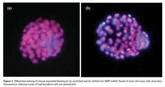
All embryos that re-expanded and hatched were considered viable and were differentially stained to count trophectoderm (TE) and inner cell mass (ICM) nucleus. Blastocysts were differentially stained using ionic detergent, Triton-X 100 and fluorochromes, Hoechst 33342 and propidium iodide, as previously described (Thouas <I>et al</I>., 2001). Stained blastocysts were mounted on a glass slide containing a 10uL drop of glycerol. Counts were performed on blastocysts which were gently flattened with a cover slip and examined under fl uorescence microscopy. </font></p>    <p><font face="Verdana, Arial, Helvetica, sans-serif" size="2"><I>Statistical analysis </I></font></p>    <p><font face="Verdana, Arial, Helvetica, sans-serif" size="2">Re-expansion and hatching rates were compared by Chi-square test, for a significance level of (p&lt;0.05). ICM and trophectoderm mean cells numbers were compared by ANOVA (p&lt;0.05). </font></p>     <p><font face="Verdana, Arial, Helvetica, sans-serif" size="2">&nbsp;</font></p>     <p><font face="Verdana, Arial, Helvetica, sans-serif" size="3"><b>Results</b></font></p> <font face="Verdana, Arial, Helvetica, sans-serif" size="2">
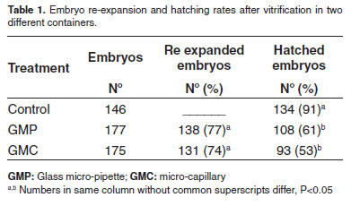
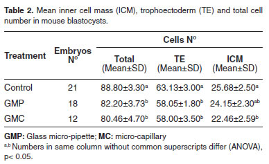
The embryo recovery rate after warming was 95.67% (177/185) of those loaded into GMP, and 94.5% (175/185) into GMC, showing that procedure efficiency was similar in both containers. Embryo viability after warming, observed at 24 and 72 h after <I>in vitro </I>culture is summarized in <a href="#t1">Table 1</a>. After 24 h of <I>in vitro</I> culture, re- expanded mouse blastocysts rates did not differ whether loading embryos into GMP (77%) or GMC (74%). <a href="#t1">Table 1</a> shows the same results on embryo viability after 72 h of <I>in vitro</I> culture. Hatching rates were similar among two experimental groups, 53% and 61%, achieved by embryos loaded into GMC and GMP, respectively. </font></p>    <p><font face="Verdana, Arial, Helvetica, sans-serif" size="2"><img src="/img/revistas/rccp/v23n1/v23n1a04t01.jpg"><a name="t1"></a></font></p>     <p><font face="Verdana, Arial, Helvetica, sans-serif" size="2">Hatched blastocysts from control group showed significantly more cells when compared to vitrified, warmed and <I>in vitro</I> cultured hatched blastocysts (<a href="#t2">see Table 2</a>). However, embryos loaded into GMP showed similar ICM cell number when compared to control group. Cell numbers (total number, ICM and TE) did not differ among vitrified embryos loaded into GMP or GMC. </font></p>    <p><font face="Verdana, Arial, Helvetica, sans-serif" size="2"><img src="/img/revistas/rccp/v23n1/v23n1a04t02.jpg"><a name="t2"></a></font></p>     ]]></body>
<body><![CDATA[<p><font face="Verdana, Arial, Helvetica, sans-serif" size="2">&nbsp;</font></p>     <p><font face="Verdana, Arial, Helvetica, sans-serif" size="3"><b>Discussion</b></font></p> <font face="Verdana, Arial, Helvetica, sans-serif" size="2">It is known that increasing cooling rates results in higher embryo survival (Vajta <I>et al</I>., 1998; He <I>et al</I>., 2008). The increase of cooling rates is an important parameter of vitrification process, because passage through critical low temperature is more rapid when compared with conventional vitrification methods (Vajta, 1997). </font></p>    <p><font face="Verdana, Arial, Helvetica, sans-serif" size="2">The cooling rate is directly influenced by temperature, thermal conductivity of containers, and related holding volume of the medium surrounding the embryos (Bunn <I>et al</I>., 2006; Sun <I>et al</I>., 2008). Minimum vitrification volumes increment cooling and warming rates and decrease chances of crystal nucleation formation in small samples (Rall, 1987). Procedures like loading embryos into OPS could diminish vitrification solution volumes and lead to higher embryo survival rates. OPS allows to obtain faster cooling rates (&gt;20,000 &deg;C/min) than French conventional straws (2,500 &deg;C/min), decreasing cell chilling and toxicity. However, it has been shown in previously experiments that GMP improved embryo survival rates when compared to OPS (Kong <I>et al</I>., 2000; Cho <I>et al</I>., 2002). GMP and GMC also allow an increased cooling rate due to higher heat conductivity of glass and a smaller volume of the frozen sample. These two vessels can be reduced more than OPS, for example the inner diameter of GMP can be reduced to 0.3 mm, GMC has 0,6 mm of inner diameter, compared to 0.8 mm of OPS straws, minimizing dimension and loaded volumes inside the container. </font></p>    <p><font face="Verdana, Arial, Helvetica, sans-serif" size="2">In this study, the results confirm these observations showing that the two glass capillary systems are efficient for vitrification of murine embryos. The pos warming embryo recovery rates of 95.67% (177/185) observed with the GMP and 94.5% (175/185) obtained with GMC did not differ, showing easy embryo manipulation and similar efficiency as embryo containers. The <I>in vitro</I> survival rates after vitrification observed with embryos loaded into GMP or GMC did not differ. The re-expanded showed similar results using the GMP (77%) or the GMC (74%) to load the embryos, and the hatching rates (61% and 53%, respectively) were also similar among the embryos loaded into the two different containers. </font></p>    <p><font face="Verdana, Arial, Helvetica, sans-serif" size="2">Nevertheless, differences between GMP and GMC survival rates are probably due to dimension differences. GMP and GMC contain the same loading volumes, but wall thickness causes different heat conductivity. However, these two containers had an inner diameter of less than 0.4 mm and used glass material, which allowed to achieve higher cooling rates (&gt; 20.000 &deg;C/min) than other usual vessels (Palasz y Mapletof, 1996; Vajta, 1997; He <I>et al</I>., 2008; Sun <I>et al</I>., 2008). </font></p>    <p><font face="Verdana, Arial, Helvetica, sans-serif" size="2">Harmful effects of vitrification solutions or vitrification procedure <I>per se </I>may impair development kinetics, causing viable embryos to delay development (Bertolini <I>et al.</I>, 2005). Taking this information into account, we used differential staining to obtain more accurate information about embryo quality and embryo cell differentiation. Results show that vitrified embryos regardless of the embryo container have lower cell numbers than embryos in control group. This cell number reduction shown in vitrified embryos, might be related to the fact that these embryos were exposed to cryoprotectant solutions and cooling rates that have proven to produce changes in the cell membranes of both the inner cell mass (ICM) and the trophectoderm (TE) (Mazur, 1970; Tominaga y Hanada, 2001; Kaidi <i>et al.</i>, 2001; Bertolini <i>et al.</i>, 2005) diminishing development rates of embryos when compared with unvitrified embryos. ICM cell number of embryos loaded into GMP or GMC survival rate after warming did not differ from ICM cell number in the control group embryos. However, vitrified embryo TE cells were not as well protected as the ICM cells, when we looked at the control group TE cell numbers, which could impair further embryo development (Kaidi <i>et al.</i>, 2001). At the same time we observed that the cell number (total number, ICM and TE) did not differ among vitrified embryos loaded into GMP or GMC. </font></p>     <p><font face="Verdana, Arial, Helvetica, sans-serif" size="2">Manufacturated GMC (Brand&reg;) tested showed the same efficiency as GMP to load mouse blastocysts for vitrification. </font></p>     <p><font face="Verdana, Arial, Helvetica, sans-serif" size="2">&nbsp;</font></p>     <p><font face="Verdana, Arial, Helvetica, sans-serif" size="3"><b>Acknowledgement</b> </font></p>     <p><font face="Verdana, Arial, Helvetica, sans-serif" size="2">This study was supported by a grant from the Brazilian National Science and Technology Development Council (CNPq). </font></p>     ]]></body>
<body><![CDATA[<p><font face="Verdana, Arial, Helvetica, sans-serif" size="2">&nbsp; </font></p>     <!-- ref --><p><font face="Verdana, Arial, Helvetica, sans-serif" size="3"><b>References</b></font>&nbsp;&nbsp;&nbsp;&nbsp;&nbsp;&nbsp;&nbsp;&nbsp;[&#160;<a href="javascript:void(0);" onclick="javascript: window.open('/scielo.php?script=sci_nlinks&ref=000064&pid=S0120-0690201000010000400001&lng=','','width=640,height=500,resizable=yes,scrollbars=1,menubar=yes,');">Links</a>&#160;]<!-- end-ref --><!-- ref --><p><font face="Verdana, Arial, Helvetica, sans-serif" size="2">Arav A, Zeron Y. Vitrification of bovine oocytes using modified minimum drop size technique (MDS) is effected by the composition and the concentration of the vitrifi cation solution and by the cooling conditions. Theriogenology, 47;341. 1997. </font>&nbsp;&nbsp;&nbsp;&nbsp;&nbsp;&nbsp;&nbsp;&nbsp;[&#160;<a href="javascript:void(0);" onclick="javascript: window.open('/scielo.php?script=sci_nlinks&ref=000065&pid=S0120-0690201000010000400002&lng=','','width=640,height=500,resizable=yes,scrollbars=1,menubar=yes,');">Links</a>&#160;]<!-- end-ref --><!-- ref --><p><font face="Verdana, Arial, Helvetica, sans-serif" size="2">Cho SK, Cho SG, Bae IH, Park CS, Kong IK<I>.</I> Improvement in post-thaw viability of in vitro-produced bovine blastocysts vitrified by glass micropipette (GMP). Anim Reprod Sci 2002; 73:151-158. </font>&nbsp;&nbsp;&nbsp;&nbsp;&nbsp;&nbsp;&nbsp;&nbsp;[&#160;<a href="javascript:void(0);" onclick="javascript: window.open('/scielo.php?script=sci_nlinks&ref=000066&pid=S0120-0690201000010000400003&lng=','','width=640,height=500,resizable=yes,scrollbars=1,menubar=yes,');">Links</a>&#160;]<!-- end-ref --><!-- ref --><p><font face="Verdana, Arial, Helvetica, sans-serif" size="2">Bertolini M, Lange MC, Rodrigues JL. <I>In vitro</I> and <I>in vivo</I> survival of mouse morulas and blastocysts following vitrification in 45% glycerol. Acta Scient Vet 2005; 33:245-251. </font>&nbsp;&nbsp;&nbsp;&nbsp;&nbsp;&nbsp;&nbsp;&nbsp;[&#160;<a href="javascript:void(0);" onclick="javascript: window.open('/scielo.php?script=sci_nlinks&ref=000067&pid=S0120-0690201000010000400004&lng=','','width=640,height=500,resizable=yes,scrollbars=1,menubar=yes,');">Links</a>&#160;]<!-- end-ref --><!-- ref --><p><font face="Verdana, Arial, Helvetica, sans-serif" size="2">Bunn S, Bertolini M, Cruz FB, Vieira AD, Pedrazzi C, Cesaro MP, Ortigari I, Ribeiro ES, Mezzalira JC, Mezzalira A. Vitrification of immature bovine oocytes loaded in containers with distinct heat conductivities. Acta Scient Vet 2006; 34:322. </font>&nbsp;&nbsp;&nbsp;&nbsp;&nbsp;&nbsp;&nbsp;&nbsp;[&#160;<a href="javascript:void(0);" onclick="javascript: window.open('/scielo.php?script=sci_nlinks&ref=000068&pid=S0120-0690201000010000400005&lng=','','width=640,height=500,resizable=yes,scrollbars=1,menubar=yes,');">Links</a>&#160;]<!-- end-ref --><!-- ref --><p><font face="Verdana, Arial, Helvetica, sans-serif" size="2">Cremades N, Sousa M, Silva J, Viana P, Sousa S, Oliveira C, Texeira JS, Barros A<I>.</I> Experimental vitrification of human compacted morulae and early blastocysts using fi ne diameter plastic micropipettes. Human Reprod 2004; 19:300-305. </font>&nbsp;&nbsp;&nbsp;&nbsp;&nbsp;&nbsp;&nbsp;&nbsp;[&#160;<a href="javascript:void(0);" onclick="javascript: window.open('/scielo.php?script=sci_nlinks&ref=000069&pid=S0120-0690201000010000400006&lng=','','width=640,height=500,resizable=yes,scrollbars=1,menubar=yes,');">Links</a>&#160;]<!-- end-ref --><!-- ref --><p><font face="Verdana, Arial, Helvetica, sans-serif" size="2">He X, Park EHY, Fowler A, Yarmush ML, Toner M, Vitrification by ultra-fast cooling at a low concentration of cryoprotectants in a quartz micro-capillary: A study using murine embryonic stem cells. Cryobiology 2008; 56:223-232. </font>&nbsp;&nbsp;&nbsp;&nbsp;&nbsp;&nbsp;&nbsp;&nbsp;[&#160;<a href="javascript:void(0);" onclick="javascript: window.open('/scielo.php?script=sci_nlinks&ref=000070&pid=S0120-0690201000010000400007&lng=','','width=640,height=500,resizable=yes,scrollbars=1,menubar=yes,');">Links</a>&#160;]<!-- end-ref --><!-- ref --><p><font face="Verdana, Arial, Helvetica, sans-serif" size="2">Hredz&aacute;k R, Ostro A, Maraaek I, Kaamarik J, Idilova V, Vesela </font><font face="Verdana, Arial, Helvetica, sans-serif" size="2">J. Influence of slow-rate freezing and vitrification on mouse embryos. Acta Vet. Brno 2005; 74:23-27. </font>&nbsp;&nbsp;&nbsp;&nbsp;&nbsp;&nbsp;&nbsp;&nbsp;[&#160;<a href="javascript:void(0);" onclick="javascript: window.open('/scielo.php?script=sci_nlinks&ref=000071&pid=S0120-0690201000010000400008&lng=','','width=640,height=500,resizable=yes,scrollbars=1,menubar=yes,');">Links</a>&#160;]<!-- end-ref --><!-- ref --><p><font face="Verdana, Arial, Helvetica, sans-serif" size="2">Kaidi S, Bernard S, Lambert P, Massip A, Dessy F, Donnay I, Effect of conventional controlled-rate freezing and vitrification on morphology and metabolism of bovine blastocysts produced in vitro. Biol Reprod 2001; 65:1127-1134. </font>&nbsp;&nbsp;&nbsp;&nbsp;&nbsp;&nbsp;&nbsp;&nbsp;[&#160;<a href="javascript:void(0);" onclick="javascript: window.open('/scielo.php?script=sci_nlinks&ref=000072&pid=S0120-0690201000010000400009&lng=','','width=640,height=500,resizable=yes,scrollbars=1,menubar=yes,');">Links</a>&#160;]<!-- end-ref --><!-- ref --><p><font face="Verdana, Arial, Helvetica, sans-serif" size="2">Kong IK, Lee SI, Cho SG, Cho SK, Park CS. Comparison of open pulled straw (OPS) vs glass micropipette (GMP) vitrification in mouse blastocysts. Theriogenology 2000; 53:1817-1826. </font>&nbsp;&nbsp;&nbsp;&nbsp;&nbsp;&nbsp;&nbsp;&nbsp;[&#160;<a href="javascript:void(0);" onclick="javascript: window.open('/scielo.php?script=sci_nlinks&ref=000073&pid=S0120-0690201000010000400010&lng=','','width=640,height=500,resizable=yes,scrollbars=1,menubar=yes,');">Links</a>&#160;]<!-- end-ref --><!-- ref --><p><font face="Verdana, Arial, Helvetica, sans-serif" size="2">Martino A, Songsasen N, Leibo SP. Development into blastocysts of bovine oocytes cryopreserved by ultra-rapid cooling. Biol Reprod 1996; 54:1059-1069. </font>&nbsp;&nbsp;&nbsp;&nbsp;&nbsp;&nbsp;&nbsp;&nbsp;[&#160;<a href="javascript:void(0);" onclick="javascript: window.open('/scielo.php?script=sci_nlinks&ref=000074&pid=S0120-0690201000010000400011&lng=','','width=640,height=500,resizable=yes,scrollbars=1,menubar=yes,');">Links</a>&#160;]<!-- end-ref --><!-- ref --><p><font face="Verdana, Arial, Helvetica, sans-serif" size="2">Matsumoto, H, Jiang JY, Tanaka T, Sasada H, Sato E. Vitrification of large quantities of immature bovine oocytes using nylon mesh. Cryobiology 2001; 42:139-144. </font>&nbsp;&nbsp;&nbsp;&nbsp;&nbsp;&nbsp;&nbsp;&nbsp;[&#160;<a href="javascript:void(0);" onclick="javascript: window.open('/scielo.php?script=sci_nlinks&ref=000075&pid=S0120-0690201000010000400012&lng=','','width=640,height=500,resizable=yes,scrollbars=1,menubar=yes,');">Links</a>&#160;]<!-- end-ref --><!-- ref --><p><font face="Verdana, Arial, Helvetica, sans-serif" size="2">Mazur, P. Cryobiology: the freezing of biological systems. Science 1970; 168:939-949. </font>&nbsp;&nbsp;&nbsp;&nbsp;&nbsp;&nbsp;&nbsp;&nbsp;[&#160;<a href="javascript:void(0);" onclick="javascript: window.open('/scielo.php?script=sci_nlinks&ref=000076&pid=S0120-0690201000010000400013&lng=','','width=640,height=500,resizable=yes,scrollbars=1,menubar=yes,');">Links</a>&#160;]<!-- end-ref --><!-- ref --><p><font face="Verdana, Arial, Helvetica, sans-serif" size="2">Palasz, AT.; Mapletof, R. J. Cryopreservation of mammalian embryos and oocytes: Recent advances. Biotech Adv 1996; 14:127-149. </font>&nbsp;&nbsp;&nbsp;&nbsp;&nbsp;&nbsp;&nbsp;&nbsp;[&#160;<a href="javascript:void(0);" onclick="javascript: window.open('/scielo.php?script=sci_nlinks&ref=000077&pid=S0120-0690201000010000400014&lng=','','width=640,height=500,resizable=yes,scrollbars=1,menubar=yes,');">Links</a>&#160;]<!-- end-ref --><!-- ref --><p><font face="Verdana, Arial, Helvetica, sans-serif" size="2">Rall WF, Fahy GM. Ice-free cryopreservation of mouse embryos at -196 &deg;C by vitrification. Nature 1985; 313:573-575. </font>&nbsp;&nbsp;&nbsp;&nbsp;&nbsp;&nbsp;&nbsp;&nbsp;[&#160;<a href="javascript:void(0);" onclick="javascript: window.open('/scielo.php?script=sci_nlinks&ref=000078&pid=S0120-0690201000010000400015&lng=','','width=640,height=500,resizable=yes,scrollbars=1,menubar=yes,');">Links</a>&#160;]<!-- end-ref --><!-- ref --><p><font face="Verdana, Arial, Helvetica, sans-serif" size="2">Rall, WF. Factors affecting the survival of mouse embryos cryopreserved by vitrification. Cryobiology 1987; 24:387-402. </font>&nbsp;&nbsp;&nbsp;&nbsp;&nbsp;&nbsp;&nbsp;&nbsp;[&#160;<a href="javascript:void(0);" onclick="javascript: window.open('/scielo.php?script=sci_nlinks&ref=000079&pid=S0120-0690201000010000400016&lng=','','width=640,height=500,resizable=yes,scrollbars=1,menubar=yes,');">Links</a>&#160;]<!-- end-ref --><!-- ref --><p><font face="Verdana, Arial, Helvetica, sans-serif" size="2">Sun X, Li Z, Yi Y, Chen J, Leno GH, Engelhardt JF. Efficient term development of vitrified ferret embryos using a novel pipette chamber technique. Biol Reprod 2008; doi:10.1095/ biolreprod.107.<I>067371</I>. </font>&nbsp;&nbsp;&nbsp;&nbsp;&nbsp;&nbsp;&nbsp;&nbsp;[&#160;<a href="javascript:void(0);" onclick="javascript: window.open('/scielo.php?script=sci_nlinks&ref=000080&pid=S0120-0690201000010000400017&lng=','','width=640,height=500,resizable=yes,scrollbars=1,menubar=yes,');">Links</a>&#160;]<!-- end-ref --><!-- ref --><p><font face="Verdana, Arial, Helvetica, sans-serif" size="2">Thouas GA, Korfiatis NA, French AJ, Jones JM, Trounson AO. Simplified technique for differential staining of inner cell mass and tropectoderm cells of mouse and bovine blastocyst. Reprod BioMed Online 2001; 3:25-29. </font>&nbsp;&nbsp;&nbsp;&nbsp;&nbsp;&nbsp;&nbsp;&nbsp;[&#160;<a href="javascript:void(0);" onclick="javascript: window.open('/scielo.php?script=sci_nlinks&ref=000081&pid=S0120-0690201000010000400018&lng=','','width=640,height=500,resizable=yes,scrollbars=1,menubar=yes,');">Links</a>&#160;]<!-- end-ref --><!-- ref --><p><font face="Verdana, Arial, Helvetica, sans-serif" size="2">Tominaga K, Hanada Y. Gel Loading tips as containers for vitrifi cation of <I>in vitro</I> produced bovine embryos. J Reprod Develop 2001;47: 267-273. </font>&nbsp;&nbsp;&nbsp;&nbsp;&nbsp;&nbsp;&nbsp;&nbsp;[&#160;<a href="javascript:void(0);" onclick="javascript: window.open('/scielo.php?script=sci_nlinks&ref=000082&pid=S0120-0690201000010000400019&lng=','','width=640,height=500,resizable=yes,scrollbars=1,menubar=yes,');">Links</a>&#160;]<!-- end-ref --><!-- ref --><p><font face="Verdana, Arial, Helvetica, sans-serif" size="2">Vanderzwalmen P, Bertin G, Debauche CH, Standaart V, Schoysman E. In vitro survival of metaphase II oocytes (MII) and blastocysts after vitrification in a hemi-straw (HS) system. Fertility and Sterility 2000; 74:215-216. </font>&nbsp;&nbsp;&nbsp;&nbsp;&nbsp;&nbsp;&nbsp;&nbsp;[&#160;<a href="javascript:void(0);" onclick="javascript: window.open('/scielo.php?script=sci_nlinks&ref=000083&pid=S0120-0690201000010000400020&lng=','','width=640,height=500,resizable=yes,scrollbars=1,menubar=yes,');">Links</a>&#160;]<!-- end-ref --><!-- ref --><p><font face="Verdana, Arial, Helvetica, sans-serif" size="2">Vajta, G. Vitrification of bovine oocytes and embryos. Embryo Transfer Newsletter  1997; 15:12-8. </font>&nbsp;&nbsp;&nbsp;&nbsp;&nbsp;&nbsp;&nbsp;&nbsp;[&#160;<a href="javascript:void(0);" onclick="javascript: window.open('/scielo.php?script=sci_nlinks&ref=000084&pid=S0120-0690201000010000400021&lng=','','width=640,height=500,resizable=yes,scrollbars=1,menubar=yes,');">Links</a>&#160;]<!-- end-ref --><!-- ref --><p><font face="Verdana, Arial, Helvetica, sans-serif" size="2">Vajta G, Holm P, Kuwayama M, Booth PJ, Jacobsen H, Greve T, Callesen H. Open pulled straw (OPS) vitrification: a new way to reduce cryoinjuries of bovine ova and embryos. Mol Reprod Dev 1998; 5:53-58. </font>&nbsp;&nbsp;&nbsp;&nbsp;&nbsp;&nbsp;&nbsp;&nbsp;[&#160;<a href="javascript:void(0);" onclick="javascript: window.open('/scielo.php?script=sci_nlinks&ref=000085&pid=S0120-0690201000010000400022&lng=','','width=640,height=500,resizable=yes,scrollbars=1,menubar=yes,');">Links</a>&#160;]<!-- end-ref --><!-- ref --><p><font face="Verdana, Arial, Helvetica, sans-serif" size="2">Vieira AD, Forell F, Feltrin C, Rodrigues JL. Calves Born after Direct Transfer of Vitrifi ed Bovine <I>In Vitro</I>-produced Blastocysts Derived from Vitrified Immature Oocytes. Reprod Dom Anim 2008; 43: 314-318. </font>&nbsp;&nbsp;&nbsp;&nbsp;&nbsp;&nbsp;&nbsp;&nbsp;[&#160;<a href="javascript:void(0);" onclick="javascript: window.open('/scielo.php?script=sci_nlinks&ref=000086&pid=S0120-0690201000010000400023&lng=','','width=640,height=500,resizable=yes,scrollbars=1,menubar=yes,');">Links</a>&#160;]<!-- end-ref --> ]]></body><back>
<ref-list>
<ref id="B1">
<nlm-citation citation-type="book">
<person-group person-group-type="author">
<name>
<surname><![CDATA[Arav]]></surname>
<given-names><![CDATA[A]]></given-names>
</name>
</person-group>
<article-title xml:lang="en"><![CDATA[Vitrification of oocytes and embryos]]></article-title>
<person-group person-group-type="editor">
<name>
<surname><![CDATA[Lauria]]></surname>
<given-names><![CDATA[A]]></given-names>
</name>
<name>
<surname><![CDATA[Gandolfi]]></surname>
<given-names><![CDATA[F]]></given-names>
</name>
</person-group>
<source><![CDATA[Embryonic Development and Manipulation in Animal Production]]></source>
<year>1992</year>
<page-range>255-264</page-range><publisher-name><![CDATA[Portland Press, London and Chapel Hill]]></publisher-name>
</nlm-citation>
</ref>
<ref id="B2">
<nlm-citation citation-type="journal">
<person-group person-group-type="author">
<name>
<surname><![CDATA[Arav]]></surname>
<given-names><![CDATA[A]]></given-names>
</name>
<name>
<surname><![CDATA[Zeron]]></surname>
<given-names><![CDATA[Y]]></given-names>
</name>
</person-group>
<article-title xml:lang="en"><![CDATA[Vitrification of bovine oocytes using modified minimum drop size technique (MDS) is effected by the composition and the concentration of the vitrifi cation solution and by the cooling conditions]]></article-title>
<source><![CDATA[Theriogenology]]></source>
<year>1997</year>
<volume>47</volume>
<page-range>341</page-range></nlm-citation>
</ref>
<ref id="B3">
<nlm-citation citation-type="journal">
<person-group person-group-type="author">
<name>
<surname><![CDATA[Cho]]></surname>
<given-names><![CDATA[SK]]></given-names>
</name>
<name>
<surname><![CDATA[Cho]]></surname>
<given-names><![CDATA[SG]]></given-names>
</name>
<name>
<surname><![CDATA[Bae]]></surname>
<given-names><![CDATA[IH]]></given-names>
</name>
<name>
<surname><![CDATA[Park]]></surname>
<given-names><![CDATA[CS]]></given-names>
</name>
<name>
<surname><![CDATA[Kong]]></surname>
<given-names><![CDATA[IK]]></given-names>
</name>
</person-group>
<article-title xml:lang="en"><![CDATA[Improvement in post-thaw viability of in vitro-produced bovine blastocysts vitrified by glass micropipette (GMP)]]></article-title>
<source><![CDATA[Anim Reprod Sci]]></source>
<year>2002</year>
<volume>73</volume>
<page-range>151-158</page-range></nlm-citation>
</ref>
<ref id="B4">
<nlm-citation citation-type="journal">
<person-group person-group-type="author">
<name>
<surname><![CDATA[Bertolini]]></surname>
<given-names><![CDATA[M]]></given-names>
</name>
<name>
<surname><![CDATA[Lange]]></surname>
<given-names><![CDATA[MC]]></given-names>
</name>
<name>
<surname><![CDATA[Rodrigues]]></surname>
<given-names><![CDATA[JL]]></given-names>
</name>
</person-group>
<article-title xml:lang="en"><![CDATA[In vitro and in vivo survival of mouse morulas and blastocysts following vitrification in 45% glycerol]]></article-title>
<source><![CDATA[Acta Scient Vet]]></source>
<year>2005</year>
<volume>33</volume>
<page-range>245-251</page-range></nlm-citation>
</ref>
<ref id="B5">
<nlm-citation citation-type="journal">
<person-group person-group-type="author">
<name>
<surname><![CDATA[Bunn]]></surname>
<given-names><![CDATA[S]]></given-names>
</name>
<name>
<surname><![CDATA[Bertolini]]></surname>
<given-names><![CDATA[M]]></given-names>
</name>
<name>
<surname><![CDATA[Cruz]]></surname>
<given-names><![CDATA[FB]]></given-names>
</name>
<name>
<surname><![CDATA[Vieira]]></surname>
<given-names><![CDATA[AD]]></given-names>
</name>
<name>
<surname><![CDATA[Pedrazzi]]></surname>
<given-names><![CDATA[C]]></given-names>
</name>
<name>
<surname><![CDATA[Cesaro]]></surname>
<given-names><![CDATA[MP]]></given-names>
</name>
<name>
<surname><![CDATA[Ortigari]]></surname>
<given-names><![CDATA[I]]></given-names>
</name>
<name>
<surname><![CDATA[Ribeiro]]></surname>
<given-names><![CDATA[ES]]></given-names>
</name>
<name>
<surname><![CDATA[Mezzalira]]></surname>
<given-names><![CDATA[JC]]></given-names>
</name>
<name>
<surname><![CDATA[Mezzalira]]></surname>
<given-names><![CDATA[A]]></given-names>
</name>
</person-group>
<article-title xml:lang="en"><![CDATA[Vitrification of immature bovine oocytes loaded in containers with distinct heat conductivities]]></article-title>
<source><![CDATA[Acta Scient Vet]]></source>
<year>2006</year>
<volume>34</volume>
<page-range>322</page-range></nlm-citation>
</ref>
<ref id="B6">
<nlm-citation citation-type="journal">
<person-group person-group-type="author">
<name>
<surname><![CDATA[Cremades]]></surname>
<given-names><![CDATA[N]]></given-names>
</name>
<name>
<surname><![CDATA[Sousa]]></surname>
<given-names><![CDATA[M]]></given-names>
</name>
<name>
<surname><![CDATA[Silva]]></surname>
<given-names><![CDATA[J]]></given-names>
</name>
<name>
<surname><![CDATA[Viana]]></surname>
<given-names><![CDATA[P]]></given-names>
</name>
<name>
<surname><![CDATA[Sousa]]></surname>
<given-names><![CDATA[S]]></given-names>
</name>
<name>
<surname><![CDATA[Oliveira]]></surname>
<given-names><![CDATA[C]]></given-names>
</name>
<name>
<surname><![CDATA[Texeira]]></surname>
<given-names><![CDATA[JS]]></given-names>
</name>
<name>
<surname><![CDATA[Barros]]></surname>
<given-names><![CDATA[A]]></given-names>
</name>
</person-group>
<article-title xml:lang="en"><![CDATA[Experimental vitrification of human compacted morulae and early blastocysts using fi ne diameter plastic micropipettes]]></article-title>
<source><![CDATA[Human Reprod]]></source>
<year>2004</year>
<volume>19</volume>
<page-range>300-305</page-range></nlm-citation>
</ref>
<ref id="B7">
<nlm-citation citation-type="journal">
<person-group person-group-type="author">
<name>
<surname><![CDATA[He]]></surname>
<given-names><![CDATA[X]]></given-names>
</name>
<name>
<surname><![CDATA[Park]]></surname>
<given-names><![CDATA[EHY]]></given-names>
</name>
<name>
<surname><![CDATA[Fowler]]></surname>
<given-names><![CDATA[A]]></given-names>
</name>
<name>
<surname><![CDATA[Yarmush]]></surname>
<given-names><![CDATA[ML]]></given-names>
</name>
<name>
<surname><![CDATA[Toner]]></surname>
<given-names><![CDATA[M]]></given-names>
</name>
</person-group>
<article-title xml:lang="en"><![CDATA[Vitrification by ultra-fast cooling at a low concentration of cryoprotectants in a quartz micro-capillary: A study using murine embryonic stem cells]]></article-title>
<source><![CDATA[Cryobiology]]></source>
<year>2008</year>
<volume>56</volume>
<page-range>223-232</page-range></nlm-citation>
</ref>
<ref id="B8">
<nlm-citation citation-type="journal">
<person-group person-group-type="author">
<name>
<surname><![CDATA[Hredzák]]></surname>
<given-names><![CDATA[R]]></given-names>
</name>
<name>
<surname><![CDATA[Ostro]]></surname>
<given-names><![CDATA[A]]></given-names>
</name>
<name>
<surname><![CDATA[Maraaek]]></surname>
<given-names><![CDATA[I]]></given-names>
</name>
<name>
<surname><![CDATA[Kaamarik]]></surname>
<given-names><![CDATA[J]]></given-names>
</name>
<name>
<surname><![CDATA[Idilova]]></surname>
<given-names><![CDATA[V]]></given-names>
</name>
<name>
<surname><![CDATA[Vesela]]></surname>
<given-names><![CDATA[J]]></given-names>
</name>
</person-group>
<article-title xml:lang="en"><![CDATA[Influence of slow-rate freezing and vitrification on mouse embryos]]></article-title>
<source><![CDATA[Acta Vet. Brno]]></source>
<year>2005</year>
<volume>74</volume>
<page-range>23-27</page-range></nlm-citation>
</ref>
<ref id="B9">
<nlm-citation citation-type="journal">
<person-group person-group-type="author">
<name>
<surname><![CDATA[Kaidi]]></surname>
<given-names><![CDATA[S]]></given-names>
</name>
<name>
<surname><![CDATA[Bernard]]></surname>
<given-names><![CDATA[S]]></given-names>
</name>
<name>
<surname><![CDATA[Lambert]]></surname>
<given-names><![CDATA[P]]></given-names>
</name>
<name>
<surname><![CDATA[Massip]]></surname>
<given-names><![CDATA[A]]></given-names>
</name>
<name>
<surname><![CDATA[Dessy]]></surname>
<given-names><![CDATA[F]]></given-names>
</name>
<name>
<surname><![CDATA[Donnay]]></surname>
<given-names><![CDATA[I]]></given-names>
</name>
</person-group>
<article-title xml:lang="en"><![CDATA[Effect of conventional controlled-rate freezing and vitrification on morphology and metabolism of bovine blastocysts produced in vitro]]></article-title>
<source><![CDATA[Biol Reprod]]></source>
<year>2001</year>
<volume>65</volume>
<page-range>1127-1134</page-range></nlm-citation>
</ref>
<ref id="B10">
<nlm-citation citation-type="journal">
<person-group person-group-type="author">
<name>
<surname><![CDATA[Kong]]></surname>
<given-names><![CDATA[IK]]></given-names>
</name>
<name>
<surname><![CDATA[Lee]]></surname>
<given-names><![CDATA[SI]]></given-names>
</name>
<name>
<surname><![CDATA[Cho]]></surname>
<given-names><![CDATA[SG]]></given-names>
</name>
<name>
<surname><![CDATA[Cho]]></surname>
<given-names><![CDATA[SK]]></given-names>
</name>
<name>
<surname><![CDATA[Park]]></surname>
<given-names><![CDATA[CS]]></given-names>
</name>
</person-group>
<article-title xml:lang="en"><![CDATA[Comparison of open pulled straw (OPS) vs glass micropipette (GMP) vitrification in mouse blastocysts]]></article-title>
<source><![CDATA[Theriogenology]]></source>
<year>2000</year>
<volume>53</volume>
<page-range>1817-1826</page-range></nlm-citation>
</ref>
<ref id="B11">
<nlm-citation citation-type="journal">
<person-group person-group-type="author">
<name>
<surname><![CDATA[Martino]]></surname>
<given-names><![CDATA[A]]></given-names>
</name>
<name>
<surname><![CDATA[Songsasen]]></surname>
<given-names><![CDATA[N]]></given-names>
</name>
<name>
<surname><![CDATA[Leibo]]></surname>
<given-names><![CDATA[SP]]></given-names>
</name>
</person-group>
<article-title xml:lang="en"><![CDATA[Development into blastocysts of bovine oocytes cryopreserved by ultra-rapid cooling]]></article-title>
<source><![CDATA[Biol Reprod]]></source>
<year>1996</year>
<volume>54</volume>
<page-range>1059-1069</page-range></nlm-citation>
</ref>
<ref id="B12">
<nlm-citation citation-type="journal">
<person-group person-group-type="author">
<name>
<surname><![CDATA[Matsumoto]]></surname>
<given-names><![CDATA[H]]></given-names>
</name>
<name>
<surname><![CDATA[Jiang]]></surname>
<given-names><![CDATA[JY]]></given-names>
</name>
<name>
<surname><![CDATA[Tanaka]]></surname>
<given-names><![CDATA[T]]></given-names>
</name>
<name>
<surname><![CDATA[Sasada]]></surname>
<given-names><![CDATA[H]]></given-names>
</name>
<name>
<surname><![CDATA[Sato]]></surname>
<given-names><![CDATA[E]]></given-names>
</name>
</person-group>
<article-title xml:lang="en"><![CDATA[Vitrification of large quantities of immature bovine oocytes using nylon mesh]]></article-title>
<source><![CDATA[Cryobiology]]></source>
<year>2001</year>
<volume>42</volume>
<page-range>139-144</page-range></nlm-citation>
</ref>
<ref id="B13">
<nlm-citation citation-type="journal">
<person-group person-group-type="author">
<name>
<surname><![CDATA[Mazur]]></surname>
<given-names><![CDATA[P]]></given-names>
</name>
</person-group>
<article-title xml:lang="en"><![CDATA[Cryobiology: the freezing of biological systems]]></article-title>
<source><![CDATA[Science]]></source>
<year>1970</year>
<volume>168</volume>
<page-range>939-949</page-range></nlm-citation>
</ref>
<ref id="B14">
<nlm-citation citation-type="journal">
<person-group person-group-type="author">
<name>
<surname><![CDATA[Palasz]]></surname>
<given-names><![CDATA[AT]]></given-names>
</name>
<name>
<surname><![CDATA[Mapletof]]></surname>
<given-names><![CDATA[R. J]]></given-names>
</name>
</person-group>
<article-title xml:lang="en"><![CDATA[Cryopreservation of mammalian embryos and oocytes: Recent advances]]></article-title>
<source><![CDATA[Biotech Adv]]></source>
<year>1996</year>
<volume>14</volume>
<page-range>127-149</page-range></nlm-citation>
</ref>
<ref id="B15">
<nlm-citation citation-type="journal">
<person-group person-group-type="author">
<name>
<surname><![CDATA[Rall]]></surname>
<given-names><![CDATA[WF]]></given-names>
</name>
<name>
<surname><![CDATA[Fahy]]></surname>
<given-names><![CDATA[GM]]></given-names>
</name>
</person-group>
<article-title xml:lang="en"><![CDATA[Ice-free cryopreservation of mouse embryos at -196 &deg;C by vitrification]]></article-title>
<source><![CDATA[Nature]]></source>
<year>1985</year>
<volume>313</volume>
<page-range>573-575</page-range></nlm-citation>
</ref>
<ref id="B16">
<nlm-citation citation-type="journal">
<person-group person-group-type="author">
<name>
<surname><![CDATA[Rall]]></surname>
<given-names><![CDATA[WF]]></given-names>
</name>
</person-group>
<article-title xml:lang="en"><![CDATA[Factors affecting the survival of mouse embryos cryopreserved by vitrification]]></article-title>
<source><![CDATA[Cryobiology]]></source>
<year>1987</year>
<volume>24</volume>
<page-range>387-402</page-range></nlm-citation>
</ref>
<ref id="B17">
<nlm-citation citation-type="journal">
<person-group person-group-type="author">
<name>
<surname><![CDATA[Sun]]></surname>
<given-names><![CDATA[X]]></given-names>
</name>
<name>
<surname><![CDATA[Li]]></surname>
<given-names><![CDATA[Z]]></given-names>
</name>
<name>
<surname><![CDATA[Yi]]></surname>
<given-names><![CDATA[Y]]></given-names>
</name>
<name>
<surname><![CDATA[Chen]]></surname>
<given-names><![CDATA[J]]></given-names>
</name>
<name>
<surname><![CDATA[Leno]]></surname>
<given-names><![CDATA[GH]]></given-names>
</name>
<name>
<surname><![CDATA[Engelhardt]]></surname>
<given-names><![CDATA[JF]]></given-names>
</name>
</person-group>
<article-title xml:lang="en"><![CDATA[Efficient term development of vitrified ferret embryos using a novel pipette chamber technique]]></article-title>
<source><![CDATA[Biol Reprod]]></source>
<year>2008</year>
</nlm-citation>
</ref>
<ref id="B18">
<nlm-citation citation-type="journal">
<person-group person-group-type="author">
<name>
<surname><![CDATA[Thouas]]></surname>
<given-names><![CDATA[GA]]></given-names>
</name>
<name>
<surname><![CDATA[Korfiatis]]></surname>
<given-names><![CDATA[NA]]></given-names>
</name>
<name>
<surname><![CDATA[French]]></surname>
<given-names><![CDATA[AJ]]></given-names>
</name>
<name>
<surname><![CDATA[Jones]]></surname>
<given-names><![CDATA[JM]]></given-names>
</name>
<name>
<surname><![CDATA[Trounson]]></surname>
<given-names><![CDATA[AO]]></given-names>
</name>
</person-group>
<article-title xml:lang="en"><![CDATA[Simplified technique for differential staining of inner cell mass and tropectoderm cells of mouse and bovine blastocyst]]></article-title>
<source><![CDATA[Reprod BioMed Online]]></source>
<year>2001</year>
<volume>3</volume>
<page-range>25-29</page-range></nlm-citation>
</ref>
<ref id="B19">
<nlm-citation citation-type="journal">
<person-group person-group-type="author">
<name>
<surname><![CDATA[Tominaga]]></surname>
<given-names><![CDATA[K]]></given-names>
</name>
<name>
<surname><![CDATA[Hanada]]></surname>
<given-names><![CDATA[Y]]></given-names>
</name>
</person-group>
<article-title xml:lang="en"><![CDATA[Gel Loading tips as containers for vitrifi cation of in vitro produced bovine embryos]]></article-title>
<source><![CDATA[J Reprod Develop]]></source>
<year>2001</year>
<volume>47</volume>
<page-range>267-273</page-range></nlm-citation>
</ref>
<ref id="B20">
<nlm-citation citation-type="journal">
<person-group person-group-type="author">
<name>
<surname><![CDATA[Vanderzwalmen]]></surname>
<given-names><![CDATA[P]]></given-names>
</name>
<name>
<surname><![CDATA[Bertin]]></surname>
<given-names><![CDATA[G]]></given-names>
</name>
<name>
<surname><![CDATA[Debauche]]></surname>
<given-names><![CDATA[CH]]></given-names>
</name>
<name>
<surname><![CDATA[Standaart]]></surname>
<given-names><![CDATA[V]]></given-names>
</name>
<name>
<surname><![CDATA[Schoysman]]></surname>
<given-names><![CDATA[E]]></given-names>
</name>
</person-group>
<article-title xml:lang="en"><![CDATA[In vitro survival of metaphase II oocytes (MII) and blastocysts after vitrification in a hemi-straw (HS) system]]></article-title>
<source><![CDATA[Fertility and Sterility]]></source>
<year>2000</year>
<volume>74</volume>
<page-range>215-216</page-range></nlm-citation>
</ref>
<ref id="B21">
<nlm-citation citation-type="journal">
<person-group person-group-type="author">
<name>
<surname><![CDATA[Vajta]]></surname>
<given-names><![CDATA[G]]></given-names>
</name>
</person-group>
<article-title xml:lang="en"><![CDATA[Vitrification of bovine oocytes and embryos]]></article-title>
<source><![CDATA[Embryo Transfer Newsletter]]></source>
<year>1997</year>
<volume>15</volume>
<page-range>12-8</page-range></nlm-citation>
</ref>
<ref id="B22">
<nlm-citation citation-type="journal">
<person-group person-group-type="author">
<name>
<surname><![CDATA[Vajta]]></surname>
<given-names><![CDATA[G]]></given-names>
</name>
<name>
<surname><![CDATA[Holm]]></surname>
<given-names><![CDATA[P]]></given-names>
</name>
<name>
<surname><![CDATA[Kuwayama]]></surname>
<given-names><![CDATA[M]]></given-names>
</name>
<name>
<surname><![CDATA[Booth]]></surname>
<given-names><![CDATA[PJ]]></given-names>
</name>
<name>
<surname><![CDATA[Jacobsen]]></surname>
<given-names><![CDATA[H]]></given-names>
</name>
<name>
<surname><![CDATA[Greve]]></surname>
<given-names><![CDATA[T]]></given-names>
</name>
<name>
<surname><![CDATA[Callesen]]></surname>
<given-names><![CDATA[H]]></given-names>
</name>
</person-group>
<article-title xml:lang="en"><![CDATA[Open pulled straw (OPS) vitrification: a new way to reduce cryoinjuries of bovine ova and embryos]]></article-title>
<source><![CDATA[Mol Reprod Dev]]></source>
<year>1998</year>
<volume>5</volume>
<page-range>53-58</page-range></nlm-citation>
</ref>
<ref id="B23">
<nlm-citation citation-type="journal">
<person-group person-group-type="author">
<name>
<surname><![CDATA[Vieira]]></surname>
<given-names><![CDATA[AD]]></given-names>
</name>
<name>
<surname><![CDATA[Forell]]></surname>
<given-names><![CDATA[F]]></given-names>
</name>
<name>
<surname><![CDATA[Feltrin]]></surname>
<given-names><![CDATA[C]]></given-names>
</name>
<name>
<surname><![CDATA[Rodrigues]]></surname>
<given-names><![CDATA[JL]]></given-names>
</name>
</person-group>
<article-title xml:lang="en"><![CDATA[Calves Born after Direct Transfer of Vitrifi ed Bovine In Vitro-produced Blastocysts Derived from Vitrified Immature Oocytes]]></article-title>
<source><![CDATA[Reprod Dom Anim]]></source>
<year>2008</year>
<volume>43</volume>
<page-range>314-318</page-range></nlm-citation>
</ref>
</ref-list>
</back>
</article>
