<?xml version="1.0" encoding="ISO-8859-1"?><article xmlns:mml="http://www.w3.org/1998/Math/MathML" xmlns:xlink="http://www.w3.org/1999/xlink" xmlns:xsi="http://www.w3.org/2001/XMLSchema-instance">
<front>
<journal-meta>
<journal-id>0120-0690</journal-id>
<journal-title><![CDATA[Revista Colombiana de Ciencias Pecuarias]]></journal-title>
<abbrev-journal-title><![CDATA[Rev Colom Cienc Pecua]]></abbrev-journal-title>
<issn>0120-0690</issn>
<publisher>
<publisher-name><![CDATA[Facultad de Ciencias Agrarias, Universidad de Antioquia]]></publisher-name>
</publisher>
</journal-meta>
<article-meta>
<article-id>S0120-06902010000400009</article-id>
<title-group>
<article-title xml:lang="en"><![CDATA[Uveitis and glaucoma associated with Hepatozoon canis infection: a case report]]></article-title>
<article-title xml:lang="es"><![CDATA[Uveítis y glaucoma asociados a infección por Hepatozoon canis: reporte de un caso]]></article-title>
<article-title xml:lang="pt"><![CDATA[Uveitis e glaucoma em um canino compatível com infecção por Hepatozoon canis: Reporte de um caso]]></article-title>
</title-group>
<contrib-group>
<contrib contrib-type="author">
<name>
<surname><![CDATA[Acevedo T]]></surname>
<given-names><![CDATA[Sandra P]]></given-names>
</name>
<xref ref-type="aff" rid="A01"/>
</contrib>
<contrib contrib-type="author">
<name>
<surname><![CDATA[Ramírez L]]></surname>
<given-names><![CDATA[Mauricio]]></given-names>
</name>
<xref ref-type="aff" rid="A02"/>
</contrib>
<contrib contrib-type="author">
<name>
<surname><![CDATA[Restrepo R]]></surname>
<given-names><![CDATA[Luis G]]></given-names>
</name>
<xref ref-type="aff" rid="A03"/>
</contrib>
</contrib-group>
<aff id="A01">
<institution><![CDATA[,University of Antioquia Faculty of Agricultural Sciences CENTAURO Research Group]]></institution>
<addr-line><![CDATA[Medellín ]]></addr-line>
<country>Colombia</country>
</aff>
<aff id="A02">
<institution><![CDATA[,University of Antioquia Faculty of Agricultural Sciences CENTAURO Research Group]]></institution>
<addr-line><![CDATA[Medellin ]]></addr-line>
<country>Colombia</country>
</aff>
<aff id="A03">
<institution><![CDATA[,University of Antioquia Faculty of Agricultural Sciences CENTAURO Research Group]]></institution>
<addr-line><![CDATA[Medellin ]]></addr-line>
<country>Colombia</country>
</aff>
<pub-date pub-type="pub">
<day>00</day>
<month>12</month>
<year>2010</year>
</pub-date>
<pub-date pub-type="epub">
<day>00</day>
<month>12</month>
<year>2010</year>
</pub-date>
<volume>23</volume>
<numero>4</numero>
<fpage>485</fpage>
<lpage>491</lpage>
<copyright-statement/>
<copyright-year/>
<self-uri xlink:href="http://www.scielo.org.co/scielo.php?script=sci_arttext&amp;pid=S0120-06902010000400009&amp;lng=en&amp;nrm=iso"></self-uri><self-uri xlink:href="http://www.scielo.org.co/scielo.php?script=sci_abstract&amp;pid=S0120-06902010000400009&amp;lng=en&amp;nrm=iso"></self-uri><self-uri xlink:href="http://www.scielo.org.co/scielo.php?script=sci_pdf&amp;pid=S0120-06902010000400009&amp;lng=en&amp;nrm=iso"></self-uri><abstract abstract-type="short" xml:lang="en"><p><![CDATA[We report the case of a five years old German Shepherd dog, who presented anterior uveitis in his left eye which then led him to glaucoma and corneal ulcer. Clinical examination found a patient with low body condition, muscle weakness, dull and brittle coat, and unspecific symptomatology. During both the cell blood count (CBC) and blood smears, gamonts of Hepatozoon canis were found in blood and aqueous humor. Diagnosis, treatment and progress are reported.]]></p></abstract>
<abstract abstract-type="short" xml:lang="es"><p><![CDATA[Se expone el caso de un perro Pastor Alemán de cinco años y medio de edad, el cual presentó uveítis anterior en ojo izquierdo que luego le condujo a glaucoma y úlcera corneal. En el examen clínico se encontró un paciente con baja condición corporal, debilidad muscular, además de un pelaje opaco y quebradizo, con sintomatología inespecífica. Tanto en el hemograma como en los extendidos sanguíneos se encontraron gamontes de Hepatozoon canis en sangre y humor acuoso. Se discute su diagnóstico, tratamiento y evolución.]]></p></abstract>
<abstract abstract-type="short" xml:lang="pt"><p><![CDATA[Expõe-se um caso de um cão Pastor Alemão de cinco anos de idade, o qual apresentou uveitis anterior no olho esquerdo e que levou à presencia de um glaucoma e uma úlcera de córnea. No exame clínico encontrou-se um paciente com baixa condição corporal, debilidade muscular e pelagem opaca e quebradiça, com sintomatologia inespecífica. No hemograma e nos estendidos sanguíneos encontraramse gamontes de Hepatozoon canis no sangue e humor aquoso. Discute-se o seu diagnóstico, tratamento e evolução.]]></p></abstract>
<kwd-group>
<kwd lng="en"><![CDATA[glaucoma]]></kwd>
<kwd lng="en"><![CDATA[Hepatozoon canis]]></kwd>
<kwd lng="en"><![CDATA[Rhipicephalus sanguineus]]></kwd>
<kwd lng="en"><![CDATA[uveítis]]></kwd>
<kwd lng="es"><![CDATA[glaucoma]]></kwd>
<kwd lng="es"><![CDATA[Hepatozoon canis]]></kwd>
<kwd lng="es"><![CDATA[Rhipicephalus sanguineus]]></kwd>
<kwd lng="es"><![CDATA[uveítis]]></kwd>
<kwd lng="pt"><![CDATA[glaucoma]]></kwd>
<kwd lng="pt"><![CDATA[Hepatozoon canis]]></kwd>
<kwd lng="pt"><![CDATA[Rhipicephalus sanguineus]]></kwd>
<kwd lng="pt"><![CDATA[uveitis]]></kwd>
</kwd-group>
</article-meta>
</front><body><![CDATA[ <p align="right"><font face="Verdana, Arial, Helvetica, sans-serif" size="2"><b>Casos cl&iacute;nicos  </b></font></p>     <p align="center"><b><font face="Verdana, Arial, Helvetica, sans-serif" size="4">Uveitis and glaucoma associated with <I>Hepatozoon canis</I> infection: a case report<Sup><Sup>&curren; </Sup></Sup></font></b></p>     <p align="center"><b><font face="Verdana, Arial, Helvetica, sans-serif" size="3"><I>Uve&iacute;tis y glaucoma asociados a infecci&oacute;n por </I><U><I>Hepatozoon canis</I></U><I>: reporte de un caso </I></font></b></p>     <p align="center"><b><font face="Verdana, Arial, Helvetica, sans-serif" size="3"><I>Uveitis e glaucoma em um canino compat&iacute;vel com infec&ccedil;&atilde;o por </I><U><I>Hepatozoon canis</I></U><I>: Reporte de um caso. </I></font></b></p>     <p>&nbsp;</p>     <p><font face="Verdana, Arial, Helvetica, sans-serif" size="2">Sandra P Acevedo T<Sup><I>1*</I></Sup>, MV, Esp Medic; Mauricio Ram&iacute;rez L<Sup><I>2</I></Sup>, MV, cEsp.Clin;  Luis G Restrepo R<Sup><I>3</I></Sup>, MV, cEsp Medic;  </font></p>    <p><font face="Verdana, Arial, Helvetica, sans-serif" size="2"><Sup><I>1</I></Sup><I>CENTAURO Research Group, Faculty of Agricultural Sciences, University of Antioquia, AA 1226, Medell&iacute;n, Colombia. </I></font></p>     <p><font face="Verdana, Arial, Helvetica, sans-serif" size="2"><Sup><I>2</I></Sup><I>Student of Clinical specialization in small animal species, CENTAURO Research Group, Faculty of Agricultural Sciences</I><I>,  </I><I>University of Antioquia, A.A. 1226, Medellin, Colombia</I><I>.  </I><Sup><I>3</I></Sup><I>Student of Medical specialization in small animal species, CENTAURO Research Group, Faculty of Agricultural Sciences</I><I>,  </I><I>University of Antioquia, A.A. 1226, Medellin, Colombia.</I></font></p>     <p><font face="Verdana, Arial, Helvetica, sans-serif" size="2"><I>(Received: 3 march, 2009; accepted: 20 august, 2009)     <br>   Published in spanish: Rev Colomb Cienc Pecu 2009; 22:287-295</I></font></p>     ]]></body>
<body><![CDATA[<p>&nbsp;</p><hr size="1">     <p><font face="Verdana, Arial, Helvetica, sans-serif" size="2"><I><b>Summary </b></I></font></p>    <p><font face="Verdana, Arial, Helvetica, sans-serif" size="2"><i>We report the case of a five years old German Shepherd dog, who presented anterior uveitis in his left eye which then led him to glaucoma and corneal ulcer. Clinical examination found a patient with low body condition, muscle weakness, dull and brittle coat, and unspecific symptomatology. During both the   cell blood count (CBC) and blood smears, gamonts of <u>Hepatozoon canis</u> were found in blood and aqueous   humor. Diagnosis, treatment and progress are reported.</i></font></p>     <p><font face="Verdana, Arial, Helvetica, sans-serif" size="2"><b>Key words:</b><I> glaucoma, </I><U><I>Hepatozoon canis</I></U><I>, </I><U><I>Rhipicephalus sanguineus</I></U><I>, uve&iacute;tis. </I></font></p>     <p>&nbsp;</p><hr size="1">     <p><font face="Verdana, Arial, Helvetica, sans-serif" size="2"><I><b>Resumen </b></I></font></p>    <p><font face="Verdana, Arial, Helvetica, sans-serif" size="2"><I>Se expone el caso de un perro Pastor Alem&aacute;n de cinco a&ntilde;os y medio de edad, el cual present&oacute; uve&iacute;tis anterior en ojo izquierdo que luego le condujo a glaucoma y &uacute;lcera corneal. En el examen cl&iacute;nico se encontr&oacute; un paciente con baja condici&oacute;n corporal, debilidad muscular, adem&aacute;s de un pelaje opaco y </I><I>quebradizo, con sintomatolog&iacute;a inespec&iacute;fica. Tanto en el hemograma como en los extendidos sangu&iacute;neos se encontraron gamontes de </I><U><I>Hepatozoon canis </I></U><I>en sangre y humor acuoso. Se discute su diagn&oacute;stico, tratamiento y evoluci&oacute;n. </I></font></p>     <p><font face="Verdana, Arial, Helvetica, sans-serif" size="2"><b>Palabras clave:</b> <I>glaucoma, </I><U><I>Hepatozoon canis</I></U><I>, </I><U><I>Rhipicephalus sanguineus</I></U><I>, uve&iacute;tis. </I></font></p>     <p>&nbsp;</p><hr size="1">     <p><font face="Verdana, Arial, Helvetica, sans-serif" size="2"><I><b>Resumo </b></I></font></p>    ]]></body>
<body><![CDATA[<p><font face="Verdana, Arial, Helvetica, sans-serif" size="2"><I>Exp&otilde;e-se um caso de um c&atilde;o Pastor Alem&atilde;o de cinco anos de idade, o qual apresentou uveitis anterior no olho esquerdo e que levou &agrave; presencia de um glaucoma e uma &uacute;lcera de c&oacute;rnea. No exame cl&iacute;nico encontrou-se um paciente com baixa condi&ccedil;&atilde;o corporal, debilidade muscular e pelagem opaca e quebradi&ccedil;a, com sintomatologia inespec&iacute;fica. No hemograma e nos estendidos sangu&iacute;neos encontraramse gamontes de </I><U><I>Hepatozoon canis </I></U><I>no sangue e humor aquoso. Discute-se o seu diagn&oacute;stico, tratamento e evolu&ccedil;&atilde;o. </I></font></p>    <p><font face="Verdana, Arial, Helvetica, sans-serif" size="2"><b>Palavras chave: </b><I>glaucoma, </I><U><I>Hepatozoon canis</I></U><I>, </I><U><I>Rhipicephalus sanguineus</I></U><I>, uveitis. </I></font></p>    <p><font face="Verdana, Arial, Helvetica, sans-serif" size="2">&curren; 	To cite this paper: Acevedo SP, Ram&iacute;rez M, Restrepo LG. Uveitis and glaucoma associated with <I>Hepatozoon canis </I>infection: a case report. Rev Colom Cienc Pecu 2010; 23:485-491. </font></p>     <p><font face="Verdana, Arial, Helvetica, sans-serif" size="2">* 	Corresponding Author: Sandra Patricia Acevedo. Facultad de Ciencias Agrarias. Universidad de Antioquia. Carrera 75 No. 65-87. Ciudadela de Robledo. Medell&iacute;n, Colombia. Tel: (574) 219 91 18. E-mail: <a href="mailto:crisalida18@hotmail.com">crisalida18@hotmail.com</a></font></p>     <p><font face="Verdana, Arial, Helvetica, sans-serif" size="2">&nbsp;</font></p><hr size="1">     <p><font face="Verdana, Arial, Helvetica, sans-serif" size="3"><b>Introduction </b></font></p>     <p><font face="Verdana, Arial, Helvetica, sans-serif" size="2">Hepatozoonosis in dogs is a worldwide disease, mostly prevalent in rural areas. The condition is caused by a protozoan of the phylum <I>Apicomplexa, </I>genus <I>Hepatozoon</I>, species <I>canis </I>(Aguiar <I>et al., </I>2004; Baneth et al., 2003; Stades <I>et al., </I>1999). Besides the dog, this protozoan infects other mammals such as fox, coyote, jackal, hyena, and leopard (Mateus <I>et al.</I>, 2007; Stades <I>et al.</I>, 1999). </font></p>    <p><font face="Verdana, Arial, Helvetica, sans-serif" size="2"><I>Hepatozoon canis </I>is transmitted through ingestion of ticks of the genus <I>Rhipicephalus sanguineus </I>contaminated with mature oocysts of <I>Hepatozoon canis</I>. There is no reported evidence regarding the transmission through the saliva of the tick (Aguiar <I>et al.</I>, 2004, Allen <I>et al.</I>, 2008). In dogs, the disease is often asymptomatic but can cause varying degrees of granulomatous inflammation in various organs, mainly in the muscles, which leads to anorexia, weight loss, weakness, diarrhea and eye discharge (Allen <I>et al.</I>, 2008, O'Dwyer <I>et al.</I>, 2006; Eiras <I>et al.</I>, 2007; Mundim <I>et al.</I>, 2008). </font></p>    <p><font face="Verdana, Arial, Helvetica, sans-serif" size="2">In this paper we describe the case of a canine patient who presented anterior uveitis in the left eye, and later developed glaucoma. The dog had clinical signs of infection with symptoms associated with Hepatozoon canis, a hemoparasite which was isolated from the blood and aqueous humor of the patient. </font></p>     <p><font face="Verdana, Arial, Helvetica, sans-serif" size="2">&nbsp;</font></p>     ]]></body>
<body><![CDATA[<p><font face="Verdana, Arial, Helvetica, sans-serif" size="3"><b>Patient assessment </b></font></p>    <p><font face="Verdana, Arial, Helvetica, sans-serif" size="2"><I>Review </I></font></p>    <p><font face="Verdana, Arial, Helvetica, sans-serif" size="2">Male German Shepherd dog, five years-old, weighing 31 kg, dewormed and vaccinated. During four years living in a farm located in the eastern region of Antioquia, Colombia. </font></p>    <p><font face="Verdana, Arial, Helvetica, sans-serif" size="2"><I>Anamnesis </I></font></p>    <p><font face="Verdana, Arial, Helvetica, sans-serif" size="2">The owners took the patient for consultation when they noticed the dog had progressive vision loss in his left eye for about a week, along with purulent discharge and great pain. Gentamicin, dexamethasone and lidocaine-based eye drops were administered, but failed to improve the condition. </font></p>    <p><font face="Verdana, Arial, Helvetica, sans-serif" size="2">For several months he had lost weight, had a poor appetite, malaise and severe hair loss. A year ago, the patient had lost vision in his right eye after a trauma. </font></p>    <p><font face="Verdana, Arial, Helvetica, sans-serif" size="2">Findings on physical examination </font></p>    <p><font face="Verdana, Arial, Helvetica, sans-serif" size="2">
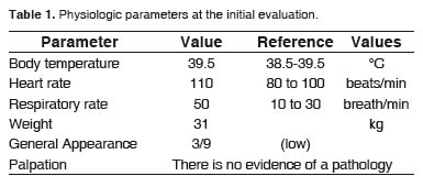
Clinical evaluation found a fearful patient, with marked anxiety, hyperemic ocular and oral mucous membranes, panting (50 breaths per minute, bpm), strong pulse (110 beats per minute, bpm), 39.5 &ordm;C body temperature, 2 seconds capillary refill time, dull coat, low body condition (3/9), and difficulty to walk and to recognize objects and his environment (<a href="#t1">Table 1</a>). </font></p>    <p align="center"><font face="Verdana, Arial, Helvetica, sans-serif" size="2"><img src="/img/revistas/rccp/v23n4/v23n04a09t01.jpg"></font><a name="t1"></a></p>     <p><font face="Verdana, Arial, Helvetica, sans-serif" size="2">
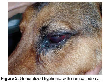
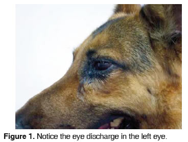
Clinical neurological examination revealed no alterations. However, clinical ophthalmic examination found miotic pupil, a marked blepharospasm and eye pain, mucous-purulent secretion, corneal edema, chemosis and episcleritis (<a href="#f1">Figures 1</a> and <a href="#f2">2</a>). At the obstacles test, it hit everything and menace reflex showed negative. </font></p>     ]]></body>
<body><![CDATA[<p align="center"><font face="Verdana, Arial, Helvetica, sans-serif" size="2"><img src="/img/revistas/rccp/v23n4/v23n04a09f01.jpg"></font><a name="f1"></a></p>     <p align="center"><font face="Verdana, Arial, Helvetica, sans-serif" size="2"><img src="/img/revistas/rccp/v23n4/v23n04a09f02.jpg"></font><a name="f2"></a></p>     <p><font face="Verdana, Arial, Helvetica, sans-serif" size="2">Buphthalmos was diagnosed in a second   examination of the eye, four days after the first   one. Intraocular pressure (IOP) was 67 mm Hg,   which is a greatly increased value, considering that reference values fluctuate between 15 to 20 mm Hg for the Schiotz tonometer, confirming the glaucoma diagnosis. Furthermore, widespread hyphema and surface ulcer on the 50% of cornea was found.</font></p>     <p><font face="Verdana, Arial, Helvetica, sans-serif" size="2"><I>Diagnostic aids </I></font></p>    <p><font face="Verdana, Arial, Helvetica, sans-serif" size="2">
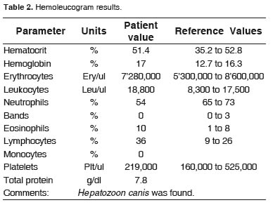
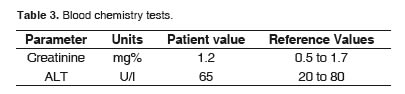
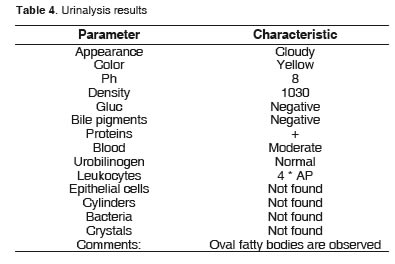
Routine laboratory examinations were performed, such as hemogram (<a href="#t2">Table 2</a>), creatinine, and ALT (Table 3), in addition to urinalysis (<a href="#t5">Table 4</a>) to rule out a systemic disease which could be the cause of uveitis, including hypertension secondary to an acute renal failure, or vasculitis for blood parasites, such as <I>Ehrlichia canis, </I>bacteria such as Leptospira, or other parasites such as Onchocerca sp. The blood chemistry tests and urine analysis found no signs that might suggest some kind of alteration (<a href="#t1">Tables 1</a>, <a href="#t2">2</a> and <a href="#t3">3</a>). The hemogram showed a mild leukocytosislymphocytosis and moderate eosinophilia.
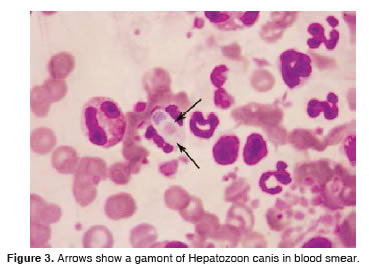
 </font></p>    <p align="center"><font face="Verdana, Arial, Helvetica, sans-serif" size="2"><img src="/img/revistas/rccp/v23n4/v23n04a09t02.jpg"></font><a name="t2"></a></p>     <p align="center"><font face="Verdana, Arial, Helvetica, sans-serif" size="2"><img src="/img/revistas/rccp/v23n4/v23n04a09t03.jpg"></font><a name="t3"></a></p>     <p align="center"><font face="Verdana, Arial, Helvetica, sans-serif" size="2"><img src="/img/revistas/rccp/v23n4/v23n04a09t04.jpg"></font><a name="t4"></a></p>     <p align="center"><font face="Verdana, Arial, Helvetica, sans-serif" size="2"><img src="/img/revistas/rccp/v23n4/v23n04a09f03.jpg"></font><a name="f3"></a></p>     <p><font face="Verdana, Arial, Helvetica, sans-serif" size="2">Three or more gamonts of <I>Hepatozoon canis </I>were found in every blood smear stained with Hemacolor (<a href="#f3">Figure 3</a>), allowing us to relate the lesions observed with the parasite.
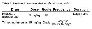
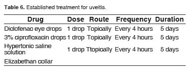
 </font></p>    ]]></body>
<body><![CDATA[<p><font face="Verdana, Arial, Helvetica, sans-serif" size="2"><I>Treatment </I></font></p>    <p><font face="Verdana, Arial, Helvetica, sans-serif" size="2">At the time of consultation, while awaiting laboratory results, and given the severity of the lesions which could cause total blindness due to functional compromise of the other eye, an uveitis and corneal edema treatment was started based on topical NSAIDs (diclofenac sodium 1.0 mg drops), ophthalmic antibiotic (Ciprofloxacin 3.0 mg) and hypertonic saline 5% (<a href="#t6">Table 6</a>) (Forlano <I>et al.</I>, 2007; Rubini <I>et al.</I>, 2006). Although there is no specific medical treatment for <I>Hepatozoon canis </I>(Aguiar <I>et al.</I>, 2004, Mateus <I>et al.</I>, 2007; Camacaro <I>et al.</I>, 1997), once the lab results were received and the parasite identified, the patient was medicated with Imidocarb dipropionate (12%) and Trimethoprimsulfa 480 mg/5ml (<a href="#t5">Table 5</a>), drugs commonly used in small animal medicine. </font></p>    <p align="center"><font face="Verdana, Arial, Helvetica, sans-serif" size="2"><img src="/img/revistas/rccp/v23n4/v23n04a09t05.jpg"></font><a name="t5"></a></p>     <p align="center"><font face="Verdana, Arial, Helvetica, sans-serif" size="2"><img src="/img/revistas/rccp/v23n4/v23n04a09t06.jpg"></font><a name="t6"></a></p>     <p><font face="Verdana, Arial, Helvetica, sans-serif" size="2">
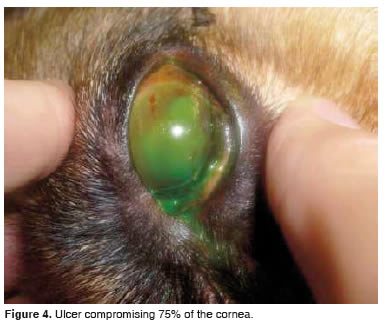
Five days after the initial treatment started, the patient showed an increase in intraocular pressure to 67 mmHg, with pain and evidence of a superficial corneal ulcer compromising 75% of the corneal surface, making it necessary to try a new therapy (<a href="#f4">Figure 4</a>). An anti-glaucomatous therapy with mannitol (20%) was started (Herrera, 2007). Given the seriousness of the problem and the risk of suffering <I>ptisis bulvi </I>because of increased intraocular pressure, 0.4 ml of aqueous humor were extracted through the iridiocorneal angle, under general anesthesia with propofol (5 mg/kg) and Diazepam (0.4 mg/kg), using a 26 gauge needle. The extracted fluid presented a bloody aspect. </font></p>    <p><font face="Verdana, Arial, Helvetica, sans-serif" size="2">The cytological analysis of the sample also resulted in the presence of <I>Hepatozoon canis </I>gamonts. After the extraction, intraocular pressure was closer to normal (52 mm/hg), preventing the loss of eye function and reducing the pain. Treatment also included ranitidine (2 mg/kg), Ampicillin-sulbactam (30 mg/kg, iv) and Tramadol (1mg/kg). </font></p>     <p align="center"><font face="Verdana, Arial, Helvetica, sans-serif" size="2"><img src="/img/revistas/rccp/v23n4/v23n04a09f04.jpg"></font><a name="f4"></a></p>     <p><font face="Verdana, Arial, Helvetica, sans-serif" size="2">
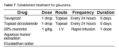
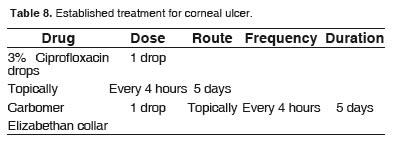
It was recommended to continue the treatment described in <a href="#t4">Table 4</a>, and medical treatment for glaucoma (<a href="#t7">Table 7</a>) and corneal ulcer (<a href="#t8">Table 8</a>) was also started. It was also recommended the use of an Elizabethan collar, and to stop administering both diclofenac drops and saline. </font></p>    <p align="center"><font face="Verdana, Arial, Helvetica, sans-serif" size="2"><img src="/img/revistas/rccp/v23n4/v23n04a09t07.jpg"></font><a name="t7"></a></p>     <p align="center"><font face="Verdana, Arial, Helvetica, sans-serif" size="2"><img src="/img/revistas/rccp/v23n4/v23n04a09t08.jpg"></font><a name="t8"></a></p>     ]]></body>
<body><![CDATA[<p><font face="Verdana, Arial, Helvetica, sans-serif" size="2">The patient was examined again 10 days later, finding that glaucoma had improved. He also showed a significant reduction of pain and intraocular pressure, but vision loss was evident, which redirected diagnosis towards a possible retinal and optic nerve degeneration that could affect up to 100% vision. </font></p>    <p><font face="Verdana, Arial, Helvetica, sans-serif" size="2">Owners were advised to hospitalize the patient and not to move him back to the farm in order to keep him under observation. Unfortunately, despite all the possible treatments offered to them, they decided to take him to another clinic for euthanasia, arguing they were not willing to continue with medical therapy for economic reasons and because the animal could not fulfill the purpose for which they were keeping him. </font></p>     <p><font face="Verdana, Arial, Helvetica, sans-serif" size="2">&nbsp;</font></p>     <p><font face="Verdana, Arial, Helvetica, sans-serif" size="3"><b>Discussion</b></font></p>    <p><font face="Verdana, Arial, Helvetica, sans-serif" size="2">Hepatozoonosis is a disease of difficult diagnosis (Johnson <I>et al.</I>, 2008, Li <I>et al.</I>, 2008; Swing <I>et al.</I>, 2003) because the clinical signs are diverse and nonspecific, resulting in nausea, malaise, uveitis, headaches and even mild to severe lameness for no apparent reason (Ettinger <I>et al.</I>, 2005). Routine laboratory examinations do not yield results that can guide to the diagnosis of the disease (Li <I>et al.</I>, 2008). </font></p>    <p><font face="Verdana, Arial, Helvetica, sans-serif" size="2">The routine laboratory method for the diagnosis of this condition is through blood smears using peripheral blood to try to visualize the gamonts (Mateus <I>et al.</I>, 2007). Nevertheless, it does not always work, because affected cells may be one or two per 1000 leukocytes (Aguiar <I>et al.</I>, 2004; Matjila <I>et al.</I>, 2008), which makes it extremely difficult to detect, generating many false negatives. </font></p>    <p><font face="Verdana, Arial, Helvetica, sans-serif" size="2">Because of this, the ideal diagnostic methods to confirm or exclude the disease include PCR, ELISA, indirect immunofluorescence or Western Blood (Aguiar <I>et al.</I>, 2004, Mateus <I>et al.</I>, 2007). But the high costs of such tests, combined with the unspecific signs, and the few reports of this condition, do not encourage their use in clinical practice (Matjila <I>et al., </I>2008). In our case, there was no possibility of performing these tests, because our laboratories do not have the required equipment. </font></p>    <p><font face="Verdana, Arial, Helvetica, sans-serif" size="2">What facilitated the accurate diagnosis in our case, was the direct visualization of <I>Hepatozoon canis </I>gamonts. It would be virtually impossible to diagnose the condition without visualizing the parasites, unless the patient had a very high parasite load. </font></p>    <p><font face="Verdana, Arial, Helvetica, sans-serif" size="2">In the case described, the parasite load was quite high, since there were three or more gamonts in each blood smear, which is very unusual and facilitated the correct diagnosis. It is important to highlight here the need of qualified laboratory personnel to properly identify the agent, avoiding false negatives. </font></p>    <p><font face="Verdana, Arial, Helvetica, sans-serif" size="2">The aqueous humor extract containing the protozoan is a very uncommon finding. No reports on <I>Hepatozoon canis </I>located in canine eyes have been found. This is difficult to explain case. For any biotic or abiotic material to go through the ocular blood-aqueous barrier it is necessary to pass through active or passive diffusion. The barrier has 40 &Aring; pores and the gamonts of <I>Hepatozoon canis </I>measure 11 &mu;m x 4 &mu;m in average (Baneth <I>et al.</I>, 2003; Karagenc <I>et al.</I>, 2006), which makes it impossible for the transit of protozoan under normal conditions. </font></p>    ]]></body>
<body><![CDATA[<p><font face="Verdana, Arial, Helvetica, sans-serif" size="2">According to Maggs <I>et al. </I>(2008), the uvea is an immunologically competent tissue. When ocular antigens are processed at distant sites, the sensitized lymphocytes migrate toward the antigen, enter the uvea and deal with the formation of antibodies or cell-mediated immune reactions. Repeated exposure to the same antigen results in a more rapid and potent response (anamnestic). This inflammation is evident because of the pain, vascular congestion and hyper-permeability. In summary the uvea acts as an accessory lymph node. </font></p>    <p><font face="Verdana, Arial, Helvetica, sans-serif" size="2">Autoimmune phenomena take place in the uvea. Any previous damage to the tissue (eg. a previous inflammation) releases specific uveal antigens normally located outside the patrol route of lymphocytes (intracellular location). These antigens promote an immune response in the uvea. It is possible that this is the mechanism participating in Canine recurrent uveitis, in which inflammatory stimulus may vary (eg, L<I>eptospira sp., Onchocerca sp</I>) (Maggs <I>et al.</I>, 2008). </font></p>    <p><font face="Verdana, Arial, Helvetica, sans-serif" size="2">A parasitic infectious process, like the one brought in this case, causes an acute inflammation which necessarily alters the structure of vascular endothelium and thus the permeability of uveal blood vessels, promoting hemoparasite transit to the anterior chamber with the local anatomical and physiological consequences previously described (Herrera, 2007; Peiffer <I>et al.</I>, 2002). </font></p>    <p><font face="Verdana, Arial, Helvetica, sans-serif" size="2">The anterior uveitis, initially manifested, produced glaucoma, since inflammation of the iris and ciliary bodies affected trabecular fibers, which together with the micro or particulate inflammatory exudates in the anterior chamber, sealed in the trabecular and ciliary cleft, favoring the accumulation of aqueous humor (Herrera, 2007). </font></p>    <p><font face="Verdana, Arial, Helvetica, sans-serif" size="2">Intraocular pressure is usually reduced during uveitis. The passage of aqueous humor can be mitigated by structural alterations that occur during uveitis, such as angle lock with inflammatory cells and debris, peripheral anterior synechia formation, cellular infiltration of the drainage angle, and pupillary blockage caused by occlusion of the pupil (Maggs <I>et al. </I>, 2008), resulting in glaucoma, secondary to anterior uveitis. </font></p>    <p><font face="Verdana, Arial, Helvetica, sans-serif" size="2">Several drugs have been used to treat hepatozoonosis. The most commonly used are trimethoprim sulfa and imidocarb dipropionate. Besides these, diminazene diaceturate, tetracyclines, primaquine phosphate, toltrazuril, clindamycin, or pyrimethamine in a combined therapy have been used with mixed results (Aguiar <I>et al.</I>, 2004; Baneth <I>et al.</I>, 2003; Mateus <I>et al.</I>, 2007). In general, these drugs do not give the expected result because the complete removal of protozoa has not been achieved (Baneth <I>et al.</I>, 2003). </font></p>    <p><font face="Verdana, Arial, Helvetica, sans-serif" size="2">In the case under discussion it was not possible to assess the patient's evolution, his response to the medical therapy, any changes in the number of gamonts, or the patient's recovery, given the owners premature decision to euthanize the patient. The autopsy would have been very helpful for understanding the pathology of the disease. </font></p>     <p><font face="Verdana, Arial, Helvetica, sans-serif" size="2">&nbsp;</font></p>     <p><font face="Verdana, Arial, Helvetica, sans-serif" size="3"><b>Conclusions</b></font></p>    <p><font face="Verdana, Arial, Helvetica, sans-serif" size="2">Hepatozoonosis is a difficult to diagnose disease. Any animal that is taken to consultation with uveitis and glaucoma should be suspected of being infected with <I>Hepatozoon canis</I>, especially if the patient has no eye compromise and other diseases compatible with the clinical signs presented have been ruled out. </font></p>    ]]></body>
<body><![CDATA[<p><font face="Verdana, Arial, Helvetica, sans-serif" size="2">Considering the complexity of diagnosing the disease, when the diagnostic tests outlined above cannot be conducted, the clinician should perform blood smears asking the clinical laboratory to look for possible parasites in the sample submitted. </font></p>    <p><font face="Verdana, Arial, Helvetica, sans-serif" size="2">Given the diversity of clinical signs and the diagnostic limitations that have been discussed, it is imperative that the medical and clinical laboratory have the sufficient knowledge about this disease, so that the clinician can have a proper approach to its diagnosis. Therefore, it is important to establish protocols to determine the prevalence and incidence of hepatozoonosis, which may be underdiagnosed. </font></p>     <p><font face="Verdana, Arial, Helvetica, sans-serif" size="2">&nbsp;</font></p>     <p><font face="Verdana, Arial, Helvetica, sans-serif" size="3"><b>Acknowledgements </b></font></p>    <p><font face="Verdana, Arial, Helvetica, sans-serif" size="2">The authors thank bacteriologist Clemencia Correa Restrepo from the Veterinary Clinical Laboratory, Faculty of Agricultural Sciences, University of Antioquia, for her assistance in the identification of the parasite, and to veterinarian H&eacute;ctor A. Jim&eacute;nez Arboleda, for reviewing and proofreading this paper. </font></p>     <p><font face="Verdana, Arial, Helvetica, sans-serif" size="2">&nbsp;</font></p>     <p><font face="Verdana, Arial, Helvetica, sans-serif" size="3"><b>Referencias </b></font></p>     <!-- ref --><p><font face="Verdana, Arial, Helvetica, sans-serif" size="2">1. Aguiar DM, Ribeiro MG, Silva WB, Dias JG, Megid J, Megid AC<I>. </I>Hepatozoonose canina: achados cl&iacute;nico-epidemiol&oacute;gicos emtr&ecirc;s casos. Arq Bras Med Vet Zootec 2004; 56, suppl 3:411-413. </font>&nbsp;&nbsp;&nbsp;&nbsp;&nbsp;&nbsp;&nbsp;&nbsp;[&#160;<a href="javascript:void(0);" onclick="javascript: window.open('/scielo.php?script=sci_nlinks&ref=000090&pid=S0120-0690201000040000900001&lng=','','width=640,height=500,resizable=yes,scrollbars=1,menubar=yes,');">Links</a>&#160;]<!-- end-ref --><!-- ref --><p><font face="Verdana, Arial, Helvetica, sans-serif" size="2">2. Allen KE, Li Y, Kaltenboeck B, Jonson EM, Reichard MV, Roger JP, Susan EL<I>. </I>Diversity of Hepatozoon species in naturally infected dogs in the southern United Status. Vet Parasitol 2008; 154:220-225. </font>&nbsp;&nbsp;&nbsp;&nbsp;&nbsp;&nbsp;&nbsp;&nbsp;[&#160;<a href="javascript:void(0);" onclick="javascript: window.open('/scielo.php?script=sci_nlinks&ref=000091&pid=S0120-0690201000040000900002&lng=','','width=640,height=500,resizable=yes,scrollbars=1,menubar=yes,');">Links</a>&#160;]<!-- end-ref --><!-- ref --><p><font face="Verdana, Arial, Helvetica, sans-serif" size="2">3. Baneth G, Mathew JS, Shkap V, Macintire D K, Barta JR, Sidney AE. Swing. Canine hepatozoonosis: two disease syndromes caused by separate Hepatozoonspp. Trends Parasitol 2003; 19 suppl1:27-31. </font>&nbsp;&nbsp;&nbsp;&nbsp;&nbsp;&nbsp;&nbsp;&nbsp;[&#160;<a href="javascript:void(0);" onclick="javascript: window.open('/scielo.php?script=sci_nlinks&ref=000092&pid=S0120-0690201000040000900003&lng=','','width=640,height=500,resizable=yes,scrollbars=1,menubar=yes,');">Links</a>&#160;]<!-- end-ref --><!-- ref --><p><font face="Verdana, Arial, Helvetica, sans-serif" size="2">4. Camacaro I, P&eacute;rez JR, Hern&aacute;ndez R. Hepatozoonosis y erlichiosis en un perro. Informe de un caso. Vet Tropic 1997; 22 suppl 1:77-81. </font>&nbsp;&nbsp;&nbsp;&nbsp;&nbsp;&nbsp;&nbsp;&nbsp;[&#160;<a href="javascript:void(0);" onclick="javascript: window.open('/scielo.php?script=sci_nlinks&ref=000093&pid=S0120-0690201000040000900004&lng=','','width=640,height=500,resizable=yes,scrollbars=1,menubar=yes,');">Links</a>&#160;]<!-- end-ref --><!-- ref --><p><font face="Verdana, Arial, Helvetica, sans-serif" size="2">5. Eiras DF, J Basabeb J, Scodellaro CF, Banach DB, Matos ML, Krimer A, Baneth G. First molecular characterization of canine hepatozoonosis in Argentina: evaluation of asymptomatic Hepatozooncanis infection in dogs from Buenos Aires. Vet Parasitol 2007; 149:275-279. </font>&nbsp;&nbsp;&nbsp;&nbsp;&nbsp;&nbsp;&nbsp;&nbsp;[&#160;<a href="javascript:void(0);" onclick="javascript: window.open('/scielo.php?script=sci_nlinks&ref=000094&pid=S0120-0690201000040000900005&lng=','','width=640,height=500,resizable=yes,scrollbars=1,menubar=yes,');">Links</a>&#160;]<!-- end-ref --><!-- ref --><p><font face="Verdana, Arial, Helvetica, sans-serif" size="2">6. Ettinger SJ, Feldman CE (eds). Textbook of veterinary internal medicine. 6 ed. St. Louis: Saunders; 2005. </font>&nbsp;&nbsp;&nbsp;&nbsp;&nbsp;&nbsp;&nbsp;&nbsp;[&#160;<a href="javascript:void(0);" onclick="javascript: window.open('/scielo.php?script=sci_nlinks&ref=000095&pid=S0120-0690201000040000900006&lng=','','width=640,height=500,resizable=yes,scrollbars=1,menubar=yes,');">Links</a>&#160;]<!-- end-ref --><!-- ref --><p><font face="Verdana, Arial, Helvetica, sans-serif" size="2">7. Forlano MD, Teixeira KRS, Scofield A, Elisei C, Yotoko KSC, Fernandes KR, Linhares GF, Ewing SA, Massard CL. Molecular characterization of Hepatozoonsp. from Brazilian dogs and its phylogenetic relationship with other Hepatozoonspp. Vet Parasitol 2007; 145:21-30. </font>&nbsp;&nbsp;&nbsp;&nbsp;&nbsp;&nbsp;&nbsp;&nbsp;[&#160;<a href="javascript:void(0);" onclick="javascript: window.open('/scielo.php?script=sci_nlinks&ref=000096&pid=S0120-0690201000040000900007&lng=','','width=640,height=500,resizable=yes,scrollbars=1,menubar=yes,');">Links</a>&#160;]<!-- end-ref --><!-- ref --><p><font face="Verdana, Arial, Helvetica, sans-serif" size="2">8. Herrera DH. Oftalmolog&iacute;a cl&iacute;nica en animales de compa&ntilde;&iacute;a. 2nd ed. Buenos Aires. Editorial Inter-m&eacute;dica; 2007. </font>&nbsp;&nbsp;&nbsp;&nbsp;&nbsp;&nbsp;&nbsp;&nbsp;[&#160;<a href="javascript:void(0);" onclick="javascript: window.open('/scielo.php?script=sci_nlinks&ref=000097&pid=S0120-0690201000040000900008&lng=','','width=640,height=500,resizable=yes,scrollbars=1,menubar=yes,');">Links</a>&#160;]<!-- end-ref --><!-- ref --><p><font face="Verdana, Arial, Helvetica, sans-serif" size="2">9. Jonson EM, Allen KE, Panciera RJ, Little SE, Swing SA. Infectivity of Hepatozoonamericanum cystozoites for a dog. Vet Parasitol 2008; 154:148-150. </font>&nbsp;&nbsp;&nbsp;&nbsp;&nbsp;&nbsp;&nbsp;&nbsp;[&#160;<a href="javascript:void(0);" onclick="javascript: window.open('/scielo.php?script=sci_nlinks&ref=000098&pid=S0120-0690201000040000900009&lng=','','width=640,height=500,resizable=yes,scrollbars=1,menubar=yes,');">Links</a>&#160;]<!-- end-ref --><!-- ref --><p><font face="Verdana, Arial, Helvetica, sans-serif" size="2">10. Karagenc TI, Pasa S, Kirli G, Hosgor M, Bilgic HB, Ozon YH, Atasoy A, Eren H<I>. </I>A parasitological, molecular and serological survey of <I>Hepatozoon canis </I>infection in dogs around the Aegean coast of Turkey. Vet Parasitol 2006; 135:113-119. </font>&nbsp;&nbsp;&nbsp;&nbsp;&nbsp;&nbsp;&nbsp;&nbsp;[&#160;<a href="javascript:void(0);" onclick="javascript: window.open('/scielo.php?script=sci_nlinks&ref=000099&pid=S0120-0690201000040000900010&lng=','','width=640,height=500,resizable=yes,scrollbars=1,menubar=yes,');">Links</a>&#160;]<!-- end-ref --><!-- ref --><p><font face="Verdana, Arial, Helvetica, sans-serif" size="2">11. Li Y, Wang C , Allen KE, Little SE, Ahluwalia SK, Gao D, Macintire DK, Blagburn DL, Kaltenboeck B. Diagnosis of canine <I>Hepatozoon spp</I>. infection by quantitative PCR. Vet Parasitol 2008; 157:50-58. </font>&nbsp;&nbsp;&nbsp;&nbsp;&nbsp;&nbsp;&nbsp;&nbsp;[&#160;<a href="javascript:void(0);" onclick="javascript: window.open('/scielo.php?script=sci_nlinks&ref=000100&pid=S0120-0690201000040000900011&lng=','','width=640,height=500,resizable=yes,scrollbars=1,menubar=yes,');">Links</a>&#160;]<!-- end-ref --><!-- ref --><p><font face="Verdana, Arial, Helvetica, sans-serif" size="2">12. Maggs DJ, Miller PE, Ofri R. Slatter`s Fundamentals of Veterinary Opthalmology. 4 edici&oacute;n. St. Louis, Missouri. Ed. Saunders Elsevier; 2008. </font>&nbsp;&nbsp;&nbsp;&nbsp;&nbsp;&nbsp;&nbsp;&nbsp;[&#160;<a href="javascript:void(0);" onclick="javascript: window.open('/scielo.php?script=sci_nlinks&ref=000101&pid=S0120-0690201000040000900012&lng=','','width=640,height=500,resizable=yes,scrollbars=1,menubar=yes,');">Links</a>&#160;]<!-- end-ref --><!-- ref --><p><font face="Verdana, Arial, Helvetica, sans-serif" size="2">13. Mateus A, Cala F, Vargas G, Arcila V, Castellanos V. Reporte de casos cl&iacute;nicos con <I>Hepatozoon canis </I>en el Centro M&eacute;dico Quir&uacute;rgico Veterinario de la Universidad Cooperativa de Colombia. Rev electr&oacute;nica Vet 2007; 8 suppl 5:1695-7504. </font>&nbsp;&nbsp;&nbsp;&nbsp;&nbsp;&nbsp;&nbsp;&nbsp;[&#160;<a href="javascript:void(0);" onclick="javascript: window.open('/scielo.php?script=sci_nlinks&ref=000102&pid=S0120-0690201000040000900013&lng=','','width=640,height=500,resizable=yes,scrollbars=1,menubar=yes,');">Links</a>&#160;]<!-- end-ref --><!-- ref --><p><font face="Verdana, Arial, Helvetica, sans-serif" size="2">14. Matjila PT, Andrew L, Leisewitz, Jongejan F, Henk J, Penzhorn BL<I>. </I>Molecular detection of Babesia rossi and <I>Hepatozoon </I>sp. In African wild dogs (Lycaon pictus) in South Africa. Vet Parasitol 2008; 157:123-127. </font>&nbsp;&nbsp;&nbsp;&nbsp;&nbsp;&nbsp;&nbsp;&nbsp;[&#160;<a href="javascript:void(0);" onclick="javascript: window.open('/scielo.php?script=sci_nlinks&ref=000103&pid=S0120-0690201000040000900014&lng=','','width=640,height=500,resizable=yes,scrollbars=1,menubar=yes,');">Links</a>&#160;]<!-- end-ref --><!-- ref --><p><font face="Verdana, Arial, Helvetica, sans-serif" size="2">15. Mundim AV, Morais IA, Tavares M, Cury MC, Mundim MJS. Clinical and hematological signs associated with dogs naturally infected by <I>Hepatozoon sp</I>. and with other hematozoa: A retrospective study in Uberla ndia, Minas Gerais, Brazil. Vet Parasitol 2008; 153:3-8 </font>&nbsp;&nbsp;&nbsp;&nbsp;&nbsp;&nbsp;&nbsp;&nbsp;[&#160;<a href="javascript:void(0);" onclick="javascript: window.open('/scielo.php?script=sci_nlinks&ref=000104&pid=S0120-0690201000040000900015&lng=','','width=640,height=500,resizable=yes,scrollbars=1,menubar=yes,');">Links</a>&#160;]<!-- end-ref --><!-- ref --><p><font face="Verdana, Arial, Helvetica, sans-serif" size="2">16. O'Dwyer LH, Saito ME, Hasegawa MY, Kohayagawa A. Prevalence, hematology and serum biochemistry in stray dogs naturally infected by <I>Hepatozoon canis </I>in S&atilde;o Paulo. Arq Bras Med Vet Zootec 2006; 58 suppl 4:688- 690. </font>&nbsp;&nbsp;&nbsp;&nbsp;&nbsp;&nbsp;&nbsp;&nbsp;[&#160;<a href="javascript:void(0);" onclick="javascript: window.open('/scielo.php?script=sci_nlinks&ref=000105&pid=S0120-0690201000040000900016&lng=','','width=640,height=500,resizable=yes,scrollbars=1,menubar=yes,');">Links</a>&#160;]<!-- end-ref --><!-- ref --><p><font face="Verdana, Arial, Helvetica, sans-serif" size="2">17. Peiffer RL, Petersen SM. Oftalmolog&iacute;a de peque&ntilde;os animales. 2nd ed. Madrid: Ediciones Harcourt; 2002. </font>&nbsp;&nbsp;&nbsp;&nbsp;&nbsp;&nbsp;&nbsp;&nbsp;[&#160;<a href="javascript:void(0);" onclick="javascript: window.open('/scielo.php?script=sci_nlinks&ref=000106&pid=S0120-0690201000040000900017&lng=','','width=640,height=500,resizable=yes,scrollbars=1,menubar=yes,');">Links</a>&#160;]<!-- end-ref --><!-- ref --><p><font face="Verdana, Arial, Helvetica, sans-serif" size="2">18. Rubini AS, Paduan KS, Perez RR, Ribolla PEM, O'Dwyer LH. Molecular characterization of feline Hepatozoon species from Brazil. Vet Parasitol 2006; 137:168-171. </font>&nbsp;&nbsp;&nbsp;&nbsp;&nbsp;&nbsp;&nbsp;&nbsp;[&#160;<a href="javascript:void(0);" onclick="javascript: window.open('/scielo.php?script=sci_nlinks&ref=000107&pid=S0120-0690201000040000900018&lng=','','width=640,height=500,resizable=yes,scrollbars=1,menubar=yes,');">Links</a>&#160;]<!-- end-ref --><!-- ref --><p><font face="Verdana, Arial, Helvetica, sans-serif" size="2">19. Stades FC, Boev&eacute; MH, Neumann W, Wyman M. Oftalmolog&iacute;a para el veterinario pr&aacute;ctico. 1ed. Buenos Aires. Editorial Intermedica; 1999. </font>&nbsp;&nbsp;&nbsp;&nbsp;&nbsp;&nbsp;&nbsp;&nbsp;[&#160;<a href="javascript:void(0);" onclick="javascript: window.open('/scielo.php?script=sci_nlinks&ref=000108&pid=S0120-0690201000040000900019&lng=','','width=640,height=500,resizable=yes,scrollbars=1,menubar=yes,');">Links</a>&#160;]<!-- end-ref --><!-- ref --><p><font face="Verdana, Arial, Helvetica, sans-serif" size="2">20. Swing SA, Panciera RJ. American Canine Hepatozoonosis. Clin microbiol reviews 2003, 16 suppl 4:688-697.EscucharLeer fon&eacute;ticamenteDiccionario - Ver diccionario detallado </font>&nbsp;&nbsp;&nbsp;&nbsp;&nbsp;&nbsp;&nbsp;&nbsp;[&#160;<a href="javascript:void(0);" onclick="javascript: window.open('/scielo.php?script=sci_nlinks&ref=000109&pid=S0120-0690201000040000900020&lng=','','width=640,height=500,resizable=yes,scrollbars=1,menubar=yes,');">Links</a>&#160;]<!-- end-ref --> ]]></body><back>
<ref-list>
<ref id="B1">
<label>1</label><nlm-citation citation-type="journal">
<person-group person-group-type="author">
<name>
<surname><![CDATA[Aguiar]]></surname>
<given-names><![CDATA[DM]]></given-names>
</name>
<name>
<surname><![CDATA[Ribeiro]]></surname>
<given-names><![CDATA[MG]]></given-names>
</name>
<name>
<surname><![CDATA[Silva]]></surname>
<given-names><![CDATA[WB]]></given-names>
</name>
<name>
<surname><![CDATA[Dias]]></surname>
<given-names><![CDATA[JG]]></given-names>
</name>
<name>
<surname><![CDATA[Megid]]></surname>
<given-names><![CDATA[J]]></given-names>
</name>
<name>
<surname><![CDATA[Megid]]></surname>
<given-names><![CDATA[AC]]></given-names>
</name>
</person-group>
<article-title xml:lang="es"><![CDATA[Hepatozoonose canina: achados clínico-epidemiológicos emtrês casos]]></article-title>
<source><![CDATA[Arq Bras Med Vet Zootec]]></source>
<year>2004</year>
<volume>56</volume>
<numero>^s3</numero>
<issue>^s3</issue>
<supplement>3</supplement>
<page-range>411-413</page-range></nlm-citation>
</ref>
<ref id="B2">
<label>2</label><nlm-citation citation-type="journal">
<person-group person-group-type="author">
<name>
<surname><![CDATA[Allen]]></surname>
<given-names><![CDATA[KE]]></given-names>
</name>
<name>
<surname><![CDATA[Li]]></surname>
<given-names><![CDATA[Y]]></given-names>
</name>
<name>
<surname><![CDATA[Kaltenboeck]]></surname>
<given-names><![CDATA[B]]></given-names>
</name>
<name>
<surname><![CDATA[Jonson]]></surname>
<given-names><![CDATA[EM]]></given-names>
</name>
<name>
<surname><![CDATA[Reichard]]></surname>
<given-names><![CDATA[MV]]></given-names>
</name>
<name>
<surname><![CDATA[Roger]]></surname>
<given-names><![CDATA[JP]]></given-names>
</name>
<name>
<surname><![CDATA[Susan]]></surname>
<given-names><![CDATA[EL]]></given-names>
</name>
</person-group>
<article-title xml:lang="en"><![CDATA[Diversity of Hepatozoon species in naturally infected dogs in the southern United Status]]></article-title>
<source><![CDATA[Vet Parasitol]]></source>
<year>2008</year>
<volume>154</volume>
<page-range>220-225</page-range></nlm-citation>
</ref>
<ref id="B3">
<label>3</label><nlm-citation citation-type="journal">
<person-group person-group-type="author">
<name>
<surname><![CDATA[Baneth]]></surname>
<given-names><![CDATA[G]]></given-names>
</name>
<name>
<surname><![CDATA[Mathew]]></surname>
<given-names><![CDATA[JS]]></given-names>
</name>
<name>
<surname><![CDATA[Shkap]]></surname>
<given-names><![CDATA[V]]></given-names>
</name>
<name>
<surname><![CDATA[Macintire]]></surname>
<given-names><![CDATA[D K]]></given-names>
</name>
<name>
<surname><![CDATA[Barta]]></surname>
<given-names><![CDATA[JR]]></given-names>
</name>
<name>
<surname><![CDATA[Sidney]]></surname>
<given-names><![CDATA[AE]]></given-names>
</name>
</person-group>
<article-title xml:lang="en"><![CDATA[Canine hepatozoonosis: two disease syndromes caused by separate Hepatozoonspp]]></article-title>
<source><![CDATA[Trends Parasitol]]></source>
<year>2003</year>
<volume>19 suppl1</volume>
<page-range>27-31</page-range></nlm-citation>
</ref>
<ref id="B4">
<label>4</label><nlm-citation citation-type="journal">
<person-group person-group-type="author">
<name>
<surname><![CDATA[Camacaro]]></surname>
<given-names><![CDATA[I]]></given-names>
</name>
<name>
<surname><![CDATA[Pérez]]></surname>
<given-names><![CDATA[JR]]></given-names>
</name>
<name>
<surname><![CDATA[Hernández]]></surname>
<given-names><![CDATA[R]]></given-names>
</name>
</person-group>
<article-title xml:lang="es"><![CDATA[Hepatozoonosis y erlichiosis en un perro]]></article-title>
<source><![CDATA[Informe de un caso. Vet Tropic]]></source>
<year>1997</year>
<volume>22 suppl 1</volume>
<page-range>77-81</page-range></nlm-citation>
</ref>
<ref id="B5">
<label>5</label><nlm-citation citation-type="journal">
<person-group person-group-type="author">
<name>
<surname><![CDATA[Eiras]]></surname>
<given-names><![CDATA[DF]]></given-names>
</name>
<name>
<surname><![CDATA[J]]></surname>
<given-names><![CDATA[Basabeb J]]></given-names>
</name>
<name>
<surname><![CDATA[Scodellaro]]></surname>
<given-names><![CDATA[CF]]></given-names>
</name>
<name>
<surname><![CDATA[Banach]]></surname>
<given-names><![CDATA[DB]]></given-names>
</name>
<name>
<surname><![CDATA[Matos]]></surname>
<given-names><![CDATA[ML]]></given-names>
</name>
<name>
<surname><![CDATA[Krimer]]></surname>
<given-names><![CDATA[A]]></given-names>
</name>
<name>
<surname><![CDATA[Baneth]]></surname>
<given-names><![CDATA[G]]></given-names>
</name>
</person-group>
<article-title xml:lang="en"><![CDATA[First molecular characterization of canine hepatozoonosis in Argentina: evaluation of asymptomatic Hepatozooncanis infection in dogs from Buenos Aires]]></article-title>
<source><![CDATA[Vet Parasitol]]></source>
<year>2007</year>
<volume>149</volume>
<page-range>275-279</page-range></nlm-citation>
</ref>
<ref id="B6">
<label>6</label><nlm-citation citation-type="book">
<person-group person-group-type="author">
<name>
<surname><![CDATA[Ettinger]]></surname>
<given-names><![CDATA[SJ]]></given-names>
</name>
<name>
<surname><![CDATA[Feldman]]></surname>
<given-names><![CDATA[CE]]></given-names>
</name>
</person-group>
<source><![CDATA[Textbook of veterinary internal medicine]]></source>
<year>2005</year>
<edition>6</edition>
<publisher-loc><![CDATA[St. Louis ]]></publisher-loc>
<publisher-name><![CDATA[Saunders]]></publisher-name>
</nlm-citation>
</ref>
<ref id="B7">
<label>7</label><nlm-citation citation-type="journal">
<person-group person-group-type="author">
<name>
<surname><![CDATA[Forlano]]></surname>
<given-names><![CDATA[MD]]></given-names>
</name>
<name>
<surname><![CDATA[Teixeira]]></surname>
<given-names><![CDATA[KRS]]></given-names>
</name>
<name>
<surname><![CDATA[Scofield]]></surname>
<given-names><![CDATA[A]]></given-names>
</name>
<name>
<surname><![CDATA[Elisei]]></surname>
<given-names><![CDATA[C]]></given-names>
</name>
<name>
<surname><![CDATA[Yotoko]]></surname>
<given-names><![CDATA[KSC]]></given-names>
</name>
<name>
<surname><![CDATA[Fernandes]]></surname>
<given-names><![CDATA[KR]]></given-names>
</name>
<name>
<surname><![CDATA[Linhares]]></surname>
<given-names><![CDATA[GF]]></given-names>
</name>
<name>
<surname><![CDATA[Ewing]]></surname>
<given-names><![CDATA[SA]]></given-names>
</name>
<name>
<surname><![CDATA[Massard]]></surname>
<given-names><![CDATA[CL]]></given-names>
</name>
</person-group>
<article-title xml:lang="en"><![CDATA[Molecular characterization of Hepatozoonsp]]></article-title>
<source><![CDATA[from Brazilian dogs and its phylogenetic relationship with other Hepatozoonspp. Vet Parasitol]]></source>
<year>2007</year>
<volume>145</volume>
<page-range>21-30</page-range></nlm-citation>
</ref>
<ref id="B8">
<label>8</label><nlm-citation citation-type="book">
<person-group person-group-type="author">
<name>
<surname><![CDATA[Herrera]]></surname>
<given-names><![CDATA[DH]]></given-names>
</name>
</person-group>
<source><![CDATA[Oftalmología clínica en animales de compañía]]></source>
<year>2007</year>
<publisher-loc><![CDATA[2Buenos Aires ]]></publisher-loc>
<publisher-name><![CDATA[Editorial Inter-médica]]></publisher-name>
</nlm-citation>
</ref>
<ref id="B9">
<label>9</label><nlm-citation citation-type="journal">
<person-group person-group-type="author">
<name>
<surname><![CDATA[Jonson]]></surname>
<given-names><![CDATA[EM]]></given-names>
</name>
<name>
<surname><![CDATA[Allen]]></surname>
<given-names><![CDATA[KE]]></given-names>
</name>
<name>
<surname><![CDATA[Panciera]]></surname>
<given-names><![CDATA[RJ]]></given-names>
</name>
<name>
<surname><![CDATA[Little]]></surname>
<given-names><![CDATA[SE]]></given-names>
</name>
<name>
<surname><![CDATA[Swing]]></surname>
<given-names><![CDATA[SA]]></given-names>
</name>
</person-group>
<article-title xml:lang="en"><![CDATA[Infectivity of Hepatozoonamericanum cystozoites for a dog]]></article-title>
<source><![CDATA[Vet Parasitol]]></source>
<year>2008</year>
<volume>154</volume>
<page-range>148-150</page-range></nlm-citation>
</ref>
<ref id="B10">
<label>10</label><nlm-citation citation-type="journal">
<person-group person-group-type="author">
<name>
<surname><![CDATA[Karagenc]]></surname>
<given-names><![CDATA[TI]]></given-names>
</name>
<name>
<surname><![CDATA[Pasa]]></surname>
<given-names><![CDATA[S]]></given-names>
</name>
<name>
<surname><![CDATA[Kirli]]></surname>
<given-names><![CDATA[G]]></given-names>
</name>
<name>
<surname><![CDATA[Hosgor]]></surname>
<given-names><![CDATA[M]]></given-names>
</name>
<name>
<surname><![CDATA[Bilgic]]></surname>
<given-names><![CDATA[HB]]></given-names>
</name>
<name>
<surname><![CDATA[Ozon]]></surname>
<given-names><![CDATA[YH]]></given-names>
</name>
<name>
<surname><![CDATA[Atasoy]]></surname>
<given-names><![CDATA[A]]></given-names>
</name>
<name>
<surname><![CDATA[Eren]]></surname>
<given-names><![CDATA[H]]></given-names>
</name>
</person-group>
<article-title xml:lang="en"><![CDATA[A parasitological, molecular and serological survey of Hepatozoon canis infection in dogs around the Aegean coast of Turkey]]></article-title>
<source><![CDATA[Vet Parasitol]]></source>
<year>2006</year>
<volume>135</volume>
<page-range>113-119</page-range></nlm-citation>
</ref>
<ref id="B11">
<label>11</label><nlm-citation citation-type="journal">
<person-group person-group-type="author">
<name>
<surname><![CDATA[Li]]></surname>
<given-names><![CDATA[Y]]></given-names>
</name>
<name>
<surname><![CDATA[Wang]]></surname>
<given-names><![CDATA[C]]></given-names>
</name>
<name>
<surname><![CDATA[Allen]]></surname>
<given-names><![CDATA[KE]]></given-names>
</name>
<name>
<surname><![CDATA[Little]]></surname>
<given-names><![CDATA[SE]]></given-names>
</name>
<name>
<surname><![CDATA[Ahluwalia]]></surname>
<given-names><![CDATA[SK]]></given-names>
</name>
<name>
<surname><![CDATA[Gao]]></surname>
<given-names><![CDATA[D]]></given-names>
</name>
<name>
<surname><![CDATA[Macintire]]></surname>
<given-names><![CDATA[DK]]></given-names>
</name>
<name>
<surname><![CDATA[Blagburn]]></surname>
<given-names><![CDATA[DL]]></given-names>
</name>
<name>
<surname><![CDATA[Kaltenboeck]]></surname>
<given-names><![CDATA[B]]></given-names>
</name>
</person-group>
<article-title xml:lang="en"><![CDATA[Diagnosis of canine Hepatozoon spp. infection by quantitative PCR]]></article-title>
<source><![CDATA[Vet Parasitol]]></source>
<year>2008</year>
<volume>157</volume>
<page-range>50-58</page-range></nlm-citation>
</ref>
<ref id="B12">
<label>12</label><nlm-citation citation-type="book">
<person-group person-group-type="author">
<name>
<surname><![CDATA[Maggs]]></surname>
<given-names><![CDATA[DJ]]></given-names>
</name>
<name>
<surname><![CDATA[Miller]]></surname>
<given-names><![CDATA[PE]]></given-names>
</name>
<name>
<surname><![CDATA[Ofri]]></surname>
<given-names><![CDATA[R]]></given-names>
</name>
</person-group>
<source><![CDATA[Slatter`s Fundamentals of Veterinary Opthalmology]]></source>
<year>2008</year>
<edition>4</edition>
<publisher-loc><![CDATA[St. Louis^eMissouri Missouri]]></publisher-loc>
<publisher-name><![CDATA[Ed. Saunders Elsevier]]></publisher-name>
</nlm-citation>
</ref>
<ref id="B13">
<label>13</label><nlm-citation citation-type="journal">
<person-group person-group-type="author">
<name>
<surname><![CDATA[Mateus]]></surname>
<given-names><![CDATA[A]]></given-names>
</name>
<name>
<surname><![CDATA[Cala]]></surname>
<given-names><![CDATA[F]]></given-names>
</name>
<name>
<surname><![CDATA[Vargas]]></surname>
<given-names><![CDATA[G]]></given-names>
</name>
<name>
<surname><![CDATA[Arcila]]></surname>
<given-names><![CDATA[V]]></given-names>
</name>
<name>
<surname><![CDATA[Castellanos]]></surname>
<given-names><![CDATA[V]]></given-names>
</name>
</person-group>
<article-title xml:lang="es"><![CDATA[Reporte de casos clínicos con Hepatozoon canis en el Centro Médico Quirúrgico Veterinario de la Universidad Cooperativa de Colombia]]></article-title>
<source><![CDATA[Rev electrónica Vet]]></source>
<year>2007</year>
<volume>8 suppl 5</volume>
<page-range>1695-7504</page-range></nlm-citation>
</ref>
<ref id="B14">
<label>14</label><nlm-citation citation-type="journal">
<person-group person-group-type="author">
<name>
<surname><![CDATA[Matjila]]></surname>
<given-names><![CDATA[PT]]></given-names>
</name>
<name>
<surname><![CDATA[Andrew]]></surname>
<given-names><![CDATA[L]]></given-names>
</name>
<name>
<surname><![CDATA[Leisewitz,]]></surname>
<given-names><![CDATA[Jongejan F]]></given-names>
</name>
<name>
<surname><![CDATA[Henk]]></surname>
<given-names><![CDATA[J]]></given-names>
</name>
<name>
<surname><![CDATA[Penzhorn]]></surname>
<given-names><![CDATA[BL]]></given-names>
</name>
</person-group>
<article-title xml:lang="en"><![CDATA[Molecular detection of Babesia rossi and Hepatozoon sp]]></article-title>
<source><![CDATA[In African wild dogs (Lycaon pictus) in South Africa. Vet Parasitol]]></source>
<year>2008</year>
<volume>157</volume>
<page-range>123-127</page-range></nlm-citation>
</ref>
<ref id="B15">
<label>15</label><nlm-citation citation-type="journal">
<person-group person-group-type="author">
<name>
<surname><![CDATA[Mundim]]></surname>
<given-names><![CDATA[AV]]></given-names>
</name>
<name>
<surname><![CDATA[Morais]]></surname>
<given-names><![CDATA[IA]]></given-names>
</name>
<name>
<surname><![CDATA[Tavares]]></surname>
<given-names><![CDATA[M]]></given-names>
</name>
<name>
<surname><![CDATA[Cury]]></surname>
<given-names><![CDATA[MC]]></given-names>
</name>
<name>
<surname><![CDATA[Mundim]]></surname>
<given-names><![CDATA[MJS]]></given-names>
</name>
</person-group>
<article-title xml:lang="en"><![CDATA[Clinical and hematological signs associated with dogs naturally infected by Hepatozoon sp. and with other hematozoa: A retrospective study in Uberla ndia, Minas Gerais, Brazil]]></article-title>
<source><![CDATA[Vet Parasitol]]></source>
<year>2008</year>
<volume>153</volume>
<page-range>3-8</page-range></nlm-citation>
</ref>
<ref id="B16">
<label>16</label><nlm-citation citation-type="journal">
<person-group person-group-type="author">
<name>
<surname><![CDATA[O'Dwyer]]></surname>
<given-names><![CDATA[LH]]></given-names>
</name>
<name>
<surname><![CDATA[Saito]]></surname>
<given-names><![CDATA[ME]]></given-names>
</name>
<name>
<surname><![CDATA[Hasegawa]]></surname>
<given-names><![CDATA[MY]]></given-names>
</name>
<name>
<surname><![CDATA[Kohayagawa]]></surname>
<given-names><![CDATA[A]]></given-names>
</name>
</person-group>
<article-title xml:lang="en"><![CDATA[Prevalence, hematology and serum biochemistry in stray dogs naturally infected by Hepatozoon canis in São Paulo]]></article-title>
<source><![CDATA[Arq Bras Med Vet Zootec]]></source>
<year>2006</year>
<volume>58</volume>
<numero>^s4</numero>
<issue>^s4</issue>
<supplement>4</supplement>
<page-range>688- 690</page-range></nlm-citation>
</ref>
<ref id="B17">
<label>17</label><nlm-citation citation-type="book">
<person-group person-group-type="author">
<name>
<surname><![CDATA[Peiffer]]></surname>
<given-names><![CDATA[RL]]></given-names>
</name>
<name>
<surname><![CDATA[Petersen]]></surname>
<given-names><![CDATA[SM]]></given-names>
</name>
</person-group>
<source><![CDATA[Oftalmología de pequeños animales]]></source>
<year>2002</year>
<edition>2</edition>
<publisher-loc><![CDATA[Madrid ]]></publisher-loc>
<publisher-name><![CDATA[Ediciones Harcourt]]></publisher-name>
</nlm-citation>
</ref>
<ref id="B18">
<label>18</label><nlm-citation citation-type="journal">
<person-group person-group-type="author">
<name>
<surname><![CDATA[Rubini]]></surname>
<given-names><![CDATA[AS]]></given-names>
</name>
<name>
<surname><![CDATA[Paduan]]></surname>
<given-names><![CDATA[KS]]></given-names>
</name>
<name>
<surname><![CDATA[Perez]]></surname>
<given-names><![CDATA[RR]]></given-names>
</name>
<name>
<surname><![CDATA[Ribolla]]></surname>
<given-names><![CDATA[PEM]]></given-names>
</name>
<name>
<surname><![CDATA[O'Dwyer]]></surname>
<given-names><![CDATA[LH]]></given-names>
</name>
</person-group>
<article-title xml:lang="en"><![CDATA[Molecular characterization of feline Hepatozoon species from Brazil]]></article-title>
<source><![CDATA[Vet Parasitol]]></source>
<year>2006</year>
<volume>137</volume>
<page-range>168-171</page-range></nlm-citation>
</ref>
<ref id="B19">
<label>19</label><nlm-citation citation-type="book">
<person-group person-group-type="author">
<name>
<surname><![CDATA[Stades]]></surname>
<given-names><![CDATA[FC]]></given-names>
</name>
<name>
<surname><![CDATA[Boevé]]></surname>
<given-names><![CDATA[MH]]></given-names>
</name>
<name>
<surname><![CDATA[Neumann]]></surname>
<given-names><![CDATA[W]]></given-names>
</name>
<name>
<surname><![CDATA[Wyman]]></surname>
<given-names><![CDATA[M]]></given-names>
</name>
</person-group>
<source><![CDATA[Oftalmología para el veterinario práctico]]></source>
<year>1999</year>
<edition>1</edition>
<publisher-name><![CDATA[Editorial Intermedica]]></publisher-name>
</nlm-citation>
</ref>
<ref id="B20">
<label>20</label><nlm-citation citation-type="journal">
<person-group person-group-type="author">
<name>
<surname><![CDATA[Swing]]></surname>
<given-names><![CDATA[SA]]></given-names>
</name>
<name>
<surname><![CDATA[Panciera]]></surname>
<given-names><![CDATA[RJ]]></given-names>
</name>
</person-group>
<article-title xml:lang="en"><![CDATA[American Canine Hepatozoonosis]]></article-title>
<source><![CDATA[Clin microbiol reviews]]></source>
<year>2003</year>
<volume>16</volume>
<numero>^s4</numero>
<issue>^s4</issue>
<supplement>4</supplement>
<page-range>688-697</page-range></nlm-citation>
</ref>
</ref-list>
</back>
</article>
