Paraplejia en gatos domésticos compatible con el nemátodo Gurltia paralysans. Primer reporte de casos en Colombia
Paraplegia em gatos domésticos compatíveis com o Gurltia paralysans. Primeiro reporte de casos na Colômbia.
Gildardo Alzate Gómez1*, MV, Esp C, MSc; Diego Aranzazu Taborda1, MV, Esp C, MSc; Andrés Alzate G2, MV; Jenny J Chaparro Gutiérrez1, MV, MSc, DrSc
1Centauro (Grupo de Investigación en Ciencias Veterinarias), Escuela de Medicina Veterinaria. Universidad de Antioquia A.A. 1226, Medellín, Colombia
2Clínica Veterinaria. Caninos y Felinos, Velódromo. Medellín Colombia
(Recibido: 19 mayo, 2011; aceptado: 11 octubre, 2011)
Summary
]]> Six cases of cat paraplegia diagnosed in years 2001 and 2002 in Antioquia province (Colombia) are reported in this paper. Diagnosis was supported on clinical exams, radiography, myelogram, necropsy, and histopathology. Clinical signs where ataxia, decreased superficial sensitivity, loss of deep sensitivity, progressive paralysis, hind limb atrophy, and urinary and fecal retention. Necropsy and histopathology analysis revealed the presence of a nematode in the spinal cord meningeal blood vessels at the T10-L4 segment level, causing medullar compression and myelomalacia. Differential diagnose was conducted through the analysis of the parasite's histopathological cuts. Its morphological characteristics differed from those of other possible nematodes such as filarial erratic larve, Ancylostome sp, and Ascaris sp. It was concluded that the nematode is compatible with the one described as Gurltia paralysans.Key words: ataxia, metastrongyloidea, myelomalacia, nematode.
Resumen
En este trabajo se reportan seis casos de paraplejia en gatos, presentados durante los años 2001 y 2002 en el departamento de Antioquia (Colombia). El diagnóstico se realizó mediante examen clínico, radiografía, mielografía, necropsia e histopatología. Los signos clínicos fueron ataxia, disminución de la sensibilidad superficial, pérdida de la sensibilidad profunda, parálisis progresiva, atrofia de músculos del tren posterior, así como retención urinaria y fecal. La necropsia y el análisis histopatológico mostraron la presencia de un nematodo ubicado en los vasos sanguíneos de las meninges de la médula espinal, a nivel del segmento T10-L4, y el cual produjo compresión medular y mielomalacia. Al hacer diagnóstico diferencial mediante análisis de los cortes histopatológicos del parásito, se encontró que sus características morfológicas diferían de las de otros nematodos posibles, como es el caso de las larvas erráticas de filaria, Ancylostoma sp. y Ascaris sp, por lo cual se concluyó que el nematodo presente es compatible con el descrito como Gurltia paralysans.
Palabras clave: ataxia, metastrongyloidea, mielomalacia, nematodo.
Resumo
Neste trabalho reportam-se seis casos de paraplegia em gatos, apresentados durante os anos 2001 e 2002 no departamento de Antioquia (Colômbia). O diagnóstico foi feito pelo exame clínico, radiografia, mielografia, necropsia e histopatologia. As sinais clínicas foram: ataxia, diminuição da sensibilidade ao toque leve, perda sensorial profunda, paralisia progressiva, atrofia dos músculos na parte traseira, bem como retenção urinária e fecal. A necropsia e a análise histopatológica mostrou a presença de um nematóide localizados nos vasos sanguíneos das meninges da medula espinhal no segmento T10-L4, e que provocou a compressão da medula espinhal e mielomalácia. Ao fazer um diagnóstico diferencial pela análise das amostras do parasita, verificou-se que as suas características morfológicas diferentes às de outros nematóides possíveis, como é o caso das larvas erráticas de filaria, Ancylostoma sp e Ascaris sp, por isso, concluiu-se que este nematóide é compatível com o descrito como Gurltia paralysans.
Palavras chave: ataxia, metastrongyloidea, mielomalácia, nematóide.
]]> ¤ To cite this article: Gómez G, Aranzazu D, Alzate A, Chaparro JJ. Domestic cat paraplegia compatible with Gurltia paralysans nematode. First cases reported in Colombia. Rev Colomb Cienc Pecu 2011; 24:663-669.* Corresponding author: Gildardo Alzate Gómez. Clínica Veterinaria Caninos y Felinos, Medellín, Colombia. E-mail: alzategomez@gmail.com
Introduction
The distribution and importance of injuries produced by the Metastrongyloidea family of nematodes in domestic carnivores is well documented (Oliver et al., 1997; Bowman, 2002). These parasites are frequently associated with pulmonary tissue, although it is usually found some parasites stages in blood vessels. There are four of these parasites reported in domestic cats. Aelurostrongylus abstrusus is the most common among them. The other three species are: Troglostrongylus subcrenatus, Oslerus rostratus, and Gurltia paralysans, are sporadically reported (Bowman et al., 2002). In years 2001 and 2002 six cats with signs of hindlimb weakness; paraparesis; mobility difficultly, and progressive hindlimb muscle atrophy, they were taken for medical attention to the veterinary medical center, "Caninos y Felinos". All of them underwent support therapy. Only one evidenced medical improvement and survived, but it did not show complete recovery. After making the clinical and neurological examination, complementary laboratory tests, simple and contrast radiographies, euthanasia was performed in the remaining cats due to the severity of the disease. Histopathological studies in four of the five dead cats showed a nematode parasite compatible with Gurtlia paralysans, located in the spinal cord's venous plexus. Myelomalacia and a local hemorrhage in the T10 – L4 segment were also reported.
Case description
Clinical examination
Five family related Siamese cats [a six month old male (A); an eight month old female (B); an adult male (C); an adult female (D), and an adult female (E)], coming from the municipality of Tarso (state of Antioquia), and a common adult male of no specific breed (F) coming from the municipality of Amagá (state of Antioquia), were taken to the Veterinary clinic "Caninos y Felinos", located in the city of Medellín, state of Antioquia (Colombia), in order to receive medical attention. They had signs of hindlimb weakness (A,B,C,D,E,F); paresis (B,F); paraparesis (D,E); ataxia (A,B,D,E,F); signs of pain at the touch of the spinal cord (A, B, C, D, E, F); evidence of constant anxiety and restlessness (A,B). The clinical examination showed that only the locomotive system was affected (A, B, D, E). Neurological examinations (Boyd, 1992) showed signs associated with upper motor neuron degeneration (A, B, C, D, E, F); presence of normal thoracic spinal and pelvic reflexes. (A, B, D, E, F); hindlimb proprioception partial loss (C, D, E, F); decreased superficial sensitivity and deep sensitivity loss (A, B, D, E, F); paraplegia (B, F). Light abdominal distention (C and F) and leg muscle atrophy (C, D, E, F) were found in some of the animals.
Paraclinical tests
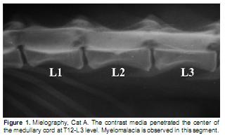
]]> Laboratory test, hemograms, blood chemistry, and stool examinations were made. Cats A, B, C and E did not show significant alterations. Cat D evidenced anemia and the presence of Ancylostomes in the stool examination. Cat F showed a mild decrease in the hemoglobin and hematocrit levels, and leukocytosis with neutrophilia. A modified Knott test was performed on this animal in order to search for microfilaria in circulation, nonetheless the result was negative.The symptoms suggested a medullar injury in the thoracolumbar region, therefore lateral and ventral- dorsal simple column radiographies were taken. The differential diagnoses were medullar trauma vs. degenerative bone injury. Vertebrae bone structure alterations were not found in any of the cases. In order to locate the injury with higher accuracy, a mielography was made (Hernández, 1992) in animals A, B, D, E, and F. 0.3 ml/kg Lopamidol were administered between spaces L5-L6, and radiography showed signs compatible with a myelomamacia at T12-L3 (A, B, D, F) levels and at T11-L4 segment (E); this explains deep sensitivity loss (Figure 1, cat A).
Medical treatment
Cat A's medical treatment consisted of cortocosteroids administration (Dexametasone, descending dose, starting with 1 mg/kg per day) for fifteen days. After this period of time no favorable results were observed, on the contrary, paraplegia manifestation increased by the loss of sphincter control. For cat C a B vitamin complex and ivermectin (400 mg/kg, subcutaneous administration) based therapy was established and administered twice within a two week period. The animal was hospitalized for a month. During this period of time it recovered its hindlimb mobility and the level of ataxia was reduced. This cat returned to its natural habitat. A pyrantel pamoate treatment was started with cat D, and an ivermectine dose was also administered (400 mg/kg, subcutaneous administration). A new evaluation was done fifteen days later. An obvious improvement in the hematocrit and hemoglobin levels was seen, while the stool examination showed negative results for parasite eggs. Cats D and E underwent B complex therapy, 75 mg/day/animal, for twenty days, and ketoprofen (2 mg/kg) for 6 days. No positive change was seen in the locomotive system's symptomatology (cats A, D, E and F). Cats B and F did not undergo any treatment due the severity of the cats' symptoms.
Necropsy findings
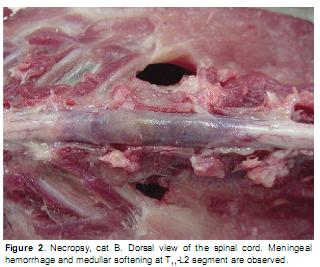
Due to the seriousness of the cats'symptoms, the cat owners (A, B, D, E and F) decided to practice euthanasia in the animals. The recommended product for the case was used (Sodium Pentobarbital and Fenilhidantoine, 1 mL/5 kg animal weight dose). The significant findings in necropsy were: meningeal hemorrhage at T10-L2 medullar segment level and softening of this region's spinal cord (Figure 2). The hemorrhage showed morphological suffusion characteristics, and it affected the spinal cord in different ways at T10 and L2 segment levels, with projection towards the medullar segments L1-L4 in cats A and B and towards segments L5-L6, in the remaining cats (cats E and F). Cat D showed less significant macroscopic injuries, only a low myelomalacia was observed in segment T10 -L2.
Histopathology study
Samples were taken from different organs and from the affected spinal cord. All tissue was placed in formaline 10% and processed for histopathology tests with hematoxiline and eosine staining. Histopathology tests showed a parasite with nematode characteristics (cats A, B, E and F) located in the meningeal blood vessels, which were dilated and compressing the medullar cord. No parasite was found in cat D's spinal cord.
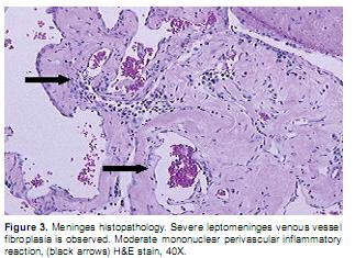
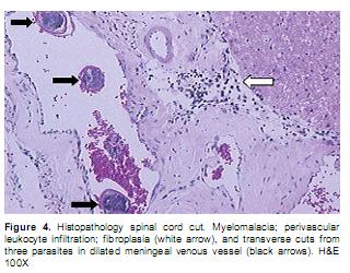
]]> Nematode parasite structures, similar to the descriptions made by Wolffhugel (1933) for Gurltia paralysans were observed in the four positive cases. When transversal serial cuts were made to the cord, hemorrhages mainly involved peripheral areas related to the meninges; they frequently affected the spinal cord's white and gray substances. Other organs did not show significant injuries, or any other lesion related to the parasitic disease. Cat F showed evident bladder strain with severe bladder wall hypertrophy.In general terms, the microscopic lesions found in the spinal cord, in the cases that are being studied, were characterized by the presence of hemorrhagic deposits, related with extended necrosis of both white and gray substances (panmyelomalacia). There was also evidence of distention; axonal fragmentation and massive neuronal necrosis; severe congestion and moderate mononuclear perivascular infiltration, mainly linfoplasmocitary type (Figure 3). Leptomeninges (piamater and arachnoidea) showed a chronic inflammatory process, still active with moderated fibroplasia, vascular congestion with mixed inflammatory agents and perivascular location. Large venous clots surrounded by macrophage charged with abundant hematic pigment, having similar characteristics to those of hemosiderin (Figure 4).
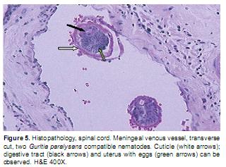
In all of the studied cases, besides the changes previously described, inside the piamater's venous blood vessels, nematode transverse cuts were observed. In them it was possible to differentiate the cuticle, the digestive tract, and the uterus, having a large number of eggs in its interior. Although these characteristics correspond to general nematodes, this parasite had an exclusive intravascular location in the spinal cord. No parasites with cardiac, pulmonary or enteral location, such as erratic grub, were observed. These parasites were generally accompanied by abundant fibrin and eosinophil (Figure 5).
Discussion
Wolffhugel (1933), in Chile, described the parasite Gurltia paralysans as a nematode with filiform body, belonging to the Metastrongylidae superfamily, having a lipless mouth, and papillae with no oral capsule. The male shows bursa copulatrix. The edge of the bursa is held by sickle shaped chitinous reinforcements. It has two bristle shaped spicules with gubernaculum. At the exit of the spicules there are two chitinous spines. The female has cranial vulva to the anus in the terminal extremity. It is also oviparous. The female is up to 23 mm long, having a 0.15 mm diameter; the male is 12 mm long, and up to 0.102 mm wide.
This species is called paralysans because of its pathogen action and the clinical signs it produces (Wolffhugel, 1933). In the cases described in this report, it was not possible to isolate whole parasites from the examined spinal cords in order to define, using morphological criteria, the gender and species of the parasite present in the meningeal blood vessels. Nonetheless, the tissue's transversal cuts that were observed are similar to the ones published for Gurltia paralysans like cases (Gómez et al., 2010; Wolffhugel, 1933). They are also significantly different from the cuts described for erratic nematodes located in blood vessels and especially in this animals' and other carnivores' nervous systems (Sprent, 1955; Dvir et al., 2007; Wessmann et al., 2006). Apart from the alteration found in the medullar cord, in the studied cases, no parasites were observed in other tissues.
On the other hand, in the different cases described in this report, there are clinical results similar to the ones described in literature (Gomez et al., 2010). The first symptoms reported are weakness of the hind legs, followed by uncoordination and ataxia. The symptoms remain for several months before the appearance of muscle atrophy of the hind legs, distension of the abdomen, urinary and fecal retention. Clinical examination showed abdominal muscle hypotonia, pelvic muscle hyporeflexia, and evidence of superior motor neuron (SMN) and inferior motor neuron (IMN) damage, probably because of the progressive myelomalacia in the spinal cord. Signs such as ascending dysfunction and analgesia, hind legs nociception and propioception loss, dilated anus, decreased perineal reflex, distended bladder, and urethra tone decrease also appear. Finally, intercostal muscles fail causing death (Bojrab, 1996; Bonagura, 2000; Negrin et al., 2009).
]]> A large number of the pathological processes in the spinal cord are located in the T3-L3 thoracolumbar segments, both in dogs and cats. Because they are defined syndromes, these processes have different etiologies, such as: trauma, intervertebral disc alteration, neoplasia, degenerative myelopathies, and infections (Ettinger and Feldman, 2005; Marioni-Henry, 2010). Most of these symptoms are chronic and degenerative in nature. The most significant signs are related to a normal pace in the front legs combined with parapesia, paralysis, or ataxia in the hind legs.The spinal reflexes in the thoracic extremities are normal or exaggerated. Muscle atrophy is not present in the front legs either, but is evident in the hind legs to a different degree, depending on the problem and especially on disuse, as seen in cat F. Bladder activity varies; bladder distension because of sphincter control loss is usually present, but evacuation occurs when it is filled with urine. In a similar manner, defecation occurs when the rectum is filled, independent of time and place. All of these signs conform to SMN symptoms. In the parasite reported cases, they have appeared in different degrees, but follow a similar behavior pattern (Bojrab, 1996; Bonagura, 2000; Ettinger y Feldman, 2005). Besides hind legs nociception and propioception loss, these signs where described by Wolffhuggel (1933) as consequences of the pathogen action of the G. paralysans nematode. This author reports that there is no treatment and infected animals die in one or two years. This data is related to the information given by the inhabitants of the region where the infected cats came from. The disease is known as the "renguera" plague, and the infected cats that are not checked by a veterinarian die approximately a year after the beginning of the symptoms.
Ettinger and Feldman (2005) bring up the existence of spinal nematodiasis caused by parasite erratic migration inside the rachidian canal. Its entrance route is not known, there have been reported cases both in dogs and cats , and it has caused different types of damage in the medullar cord due to infarcts and myelocompression due to the formation of granuloma. The parasites that were isolated in those cases were Dirofilaria inmitis, Toxacara canis, and Ancylostoma caninum. It is important to highlight that these parasites do not have regular behavior regarding localization and type of injury. They have been identified based on their particular morphologic characteristics. In the cases reported in this article, parasites were identified by histopathology studies, and then a differential study of the parasites' morphologic characteristics was made. The comparison with the erratic parasites' morphology mentioned by Ettinger and Feldman (2005), taking into account their localization in the same medullar segment, makes it possible to conclude that the parasite that was found is compatible with the descriptions made for Gurltia paralysans by Wolffhugel (1933).
There is a viral entity called the Borna disease; disease that affects cats, especially the ones that are young and have a country life style. Even though this disease shows some clinical signs similar to the ones described in the cases that were previously described (ataxia and loss of coordination and jump capacity) due to the spinal cord disorder, this disease also affects the encephalon, developing symptoms such as depression and changes of conduct (Ettinger and Feldman, 2005). Clinical expressions such as these and non suppurative encephalitis, typical of this disease, were not observed in the cases reported in this paper.
Conclusion
This is the first time that the presence of a Gurltia paralysans compatible parasite is documented in Colombia. Albeit this parasite was identified and reported in year 1933 by Wolffhugel in Chile, there is not much information about it. There have been only a few cases reported around the world (Gomez et al., 2010), all of them showing a clinical and injury pattern similar to the cases studied in this report. Unfortunately, the lack of complete samples from the nematode found in the studied spinal cords made it impossible to verify the identity of the parasite by its morphologic characteristics.
Besides the clinical and pathological aspects of the disease, it is necessary to investigate and define other relevant aspects, such as its epidemiological behavior, the parasite's life cycle, and its means of transmission. In order to do so, an investigation project is being developed in the Veterinary Medicine School of Universidad de Antioquia by an interdisciplinary team of veterinary doctors, epidemiologists, biologists, and parasitologists.
Referencias
]]>1. Bojrab MJ. Fisiopatología y clínica quirúrgica en animales pequeños. Argentina: Intermedica, 1996. 1173. [ Links ]
2. Bonagura JS. Kirk´s current veterinary therapy, small animal practice.13 ed. USA: WB.Saunders, 2000, 976-977. [ Links ]
3. Bowman DD, Hendrix CH, Lindsay DS, Stephen CB. Feline Clinical Parasitology. Blackwell Publishing. USA 2002, p 267- 273. [ Links ]
4. Boyd JS. Atlas de anatomía clínica canina y felina. España: Grass Ediciones, 1992. 46,49. [ Links ]
5. Dvir E, Perl S, Loeb E, Shklar-Hirsch S, Chai O, Mazaki- Tovi M, Aroch I, Shamir MH.. Spinal intramedullary aberrant Spirocerca lupi migration in 3 dogs. J Vet Int Med 2007; 21:860-864. [ Links ]
6. Ettinger SJ, Feldman EC. Textbook of Veterinary Internal Medicine. Diseases of the dog and cat. 6th Ed. Elsevier Saunders 2005, p 842-849. [ Links ]
7. Gomez M, Mieres M, Moronic, Mora A, Barrios N, Simeonee C, Lindsay D. Meningomyelitis due to nematode infection in four cats. Vet Parasitol 2010; 24:327-330. [ Links ]
8. Hernández MM. Radiología veterinaria, pequeños animales. España: Interamericana Mc Graw-Hill, 1992. 46-463. [ Links ]
9. Marioni-Henry K. Feline spinal cord diseases. Vet Clin North Am Small Anim Pract 2010; 40:1011-28. [ Links ]
10. Negrin A, Schatzberg S, Platt SR. The paralyzed cat. Neuroanatomic diagnosis and specific spinal. J Feline Medicine & Surgery 2009; 11:361-372 [ Links ]
11. Oliver JE, Lorenz MD, Kornegay JN. Handbook of veterinary neurology. USA: WB Saunders, 1997. 129. [ Links ]
12. Sprent JF. On the invasion of the central nervous system by nematodes I. The incidence and pathological significance of nematodes in the central nervous system. Parasitology 1955; 45:1-40. [ Links ]
13. Wessmann A, Lu D, Lamb CR, Smyth B, Mantis P, Chandler K, Boag A, Cherubini GB, Cappello R.. Brain and spinal cord haemorrhages associated with Angiostrongylus vasorum infection in four dogs. Vet Rec 2006; 158:858-863. [ Links ]
14. Wolffhugel K. Paraplejia cruralis parasitaria felis causada por Gurltia paralysans nov. gen., n. sp. (nematodes). Rev Chil Hist Nat 1933; 37:190-192. [ Links ] ]]>