<?xml version="1.0" encoding="ISO-8859-1"?><article xmlns:mml="http://www.w3.org/1998/Math/MathML" xmlns:xlink="http://www.w3.org/1999/xlink" xmlns:xsi="http://www.w3.org/2001/XMLSchema-instance">
<front>
<journal-meta>
<journal-id>0120-0690</journal-id>
<journal-title><![CDATA[Revista Colombiana de Ciencias Pecuarias]]></journal-title>
<abbrev-journal-title><![CDATA[Rev Colom Cienc Pecua]]></abbrev-journal-title>
<issn>0120-0690</issn>
<publisher>
<publisher-name><![CDATA[Facultad de Ciencias Agrarias, Universidad de Antioquia]]></publisher-name>
</publisher>
</journal-meta>
<article-meta>
<article-id>S0120-06902012000200016</article-id>
<title-group>
<article-title xml:lang="en"><![CDATA[Posterior paralysis in a Holstein cow with Enzootic Bovine Leukosis]]></article-title>
<article-title xml:lang="es"><![CDATA[Parálisis posterior en una vaca Holstein con Leucosis Enzoótica Bovina]]></article-title>
<article-title xml:lang="pt"><![CDATA[Paralise posterior numa vaca holandesa com Leucose Enzoótica Bovina]]></article-title>
</title-group>
<contrib-group>
<contrib contrib-type="author">
<name>
<surname><![CDATA[Villar]]></surname>
<given-names><![CDATA[David]]></given-names>
</name>
<xref ref-type="aff" rid="A01"/>
<xref ref-type="aff" rid="A06"/>
</contrib>
<contrib contrib-type="author">
<name>
<surname><![CDATA[Duque]]></surname>
<given-names><![CDATA[Leonardo]]></given-names>
</name>
<xref ref-type="aff" rid="A02"/>
</contrib>
<contrib contrib-type="author">
<name>
<surname><![CDATA[Giraldo]]></surname>
<given-names><![CDATA[Carlos]]></given-names>
</name>
<xref ref-type="aff" rid="A03"/>
</contrib>
<contrib contrib-type="author">
<name>
<surname><![CDATA[Pérez Montes]]></surname>
<given-names><![CDATA[Juan Esteban]]></given-names>
</name>
<xref ref-type="aff" rid="A01"/>
</contrib>
<contrib contrib-type="author">
<name>
<surname><![CDATA[Pallares]]></surname>
<given-names><![CDATA[Francisco]]></given-names>
</name>
<xref ref-type="aff" rid="A04"/>
</contrib>
<contrib contrib-type="author">
<name>
<surname><![CDATA[Schwartz]]></surname>
<given-names><![CDATA[Kent]]></given-names>
</name>
<xref ref-type="aff" rid="A05"/>
</contrib>
</contrib-group>
<aff id="A06">
<institution><![CDATA[,University of Antioquia American Board of V eterinary T oxicology ]]></institution>
<addr-line><![CDATA[ ]]></addr-line>
</aff>
<aff id="A01">
<institution><![CDATA[,University of Antioquia Facultad de Ciencias Agrarias ]]></institution>
<addr-line><![CDATA[Medellín ]]></addr-line>
<country>Colombia</country>
</aff>
<aff id="A02">
<institution><![CDATA[,Corporación Universitaria Remington Facultad de Medicina Veterinaria Grupo de Investigación GINVER]]></institution>
<addr-line><![CDATA[ ]]></addr-line>
</aff>
<aff id="A03">
<institution><![CDATA[,Universidad de Antioquia Facultad de Ciencias Agrarias Grupo de investigación VERICEL]]></institution>
<addr-line><![CDATA[Medellín ]]></addr-line>
<country>Colombia</country>
</aff>
<aff id="A04">
<institution><![CDATA[,Universidad de Murcia Facultad de Veterinaria Departamento de Anatomía y Patología]]></institution>
<addr-line><![CDATA[ ]]></addr-line>
<country>España</country>
</aff>
<aff id="A05">
<institution><![CDATA[,Iowa State University College of Veterinary Medicine Veterinary Diagnostic Laboratory]]></institution>
<addr-line><![CDATA[ ]]></addr-line>
<country>USA</country>
</aff>
<pub-date pub-type="pub">
<day>00</day>
<month>06</month>
<year>2012</year>
</pub-date>
<pub-date pub-type="epub">
<day>00</day>
<month>06</month>
<year>2012</year>
</pub-date>
<volume>25</volume>
<numero>2</numero>
<fpage>325</fpage>
<lpage>329</lpage>
<copyright-statement/>
<copyright-year/>
<self-uri xlink:href="http://www.scielo.org.co/scielo.php?script=sci_arttext&amp;pid=S0120-06902012000200016&amp;lng=en&amp;nrm=iso"></self-uri><self-uri xlink:href="http://www.scielo.org.co/scielo.php?script=sci_abstract&amp;pid=S0120-06902012000200016&amp;lng=en&amp;nrm=iso"></self-uri><self-uri xlink:href="http://www.scielo.org.co/scielo.php?script=sci_pdf&amp;pid=S0120-06902012000200016&amp;lng=en&amp;nrm=iso"></self-uri><abstract abstract-type="short" xml:lang="en"><p><![CDATA[A 6 year-old Holstein cow was euthanized after a 3 week course of progressive paraplegia. In spite of the increasing difficulties to rise and walk, the animal remained bright, alert, afebrile and with good appetite throughout most of the clinical course. Complete blood counts, biochemical profiles and analysis of the cerebrospinal fluid were reported within normal limits. Antibody was detected for bovine leukaemia virus using an enzyme-linked immunosorbent assay, supporting a tentative diagnosis of bovine leukosis. Post-mortem examination revealed a localized form of lymphosarcoma with few 2-5 cm nodular tumors confined to the walls of the gastrointestinal tract, particularly in the abomasum. In addition, soft grey tumors were found within the vertebral canal surrounding the lumbar spinal cord and associated nerve roots. Microscopic examination revealed the nodular masses were composed of neoplastic lymphocytes. Mass in the lumbar vertebral canal had extradural neoplastic lymphocytes infiltrating connective tissues around the spinal cord and spinal nerve roots. Unlike the more common chronic and wasting presentation of the disease with widespread lymphadenopathy, the rapid progression of the disease to total paraplegia in this animal could be explained by the localized presence of tumors in the spinal canal.]]></p></abstract>
<abstract abstract-type="short" xml:lang="es"><p><![CDATA[Una vaca Holstein de 6 años de edad fue sacrificada después de un curso de paraplejía progresiva de 3 semanas de duración. A pesar de las crecientes dificultades para levantarse y caminar, el animal se mantuvo alerta, sin fiebre y con buen apetito casi todo el transcurso de la enfermedad. Los análisis del hemograma, perfil bioquímico y líquido cefalorraquídeo no mostraron alteraciones fuera del rango normal. El análisis de ELISA frente al virus de la Leucosis Bovina resultó positivo, apoyando el diagnóstico diferencial de leucemia. El examen post-mortem reveló una forma localizada de linfosarcoma, con escasas y pequeñas tumoraciones nodulares de 2-5 cm confinadas a las paredes del tracto gastrointestinal, sobre todo en el abomaso. Además, los tumores estaban presentes en el canal vertebral rodeando la médula lumbar espinal y raíces nerviosas adyacentes. El examen microscópico reveló que las masas nodulares estaban compuestas de linfocitos neoplásicos; igualmente, el tejido conectivo rodeando la medula lumbar presentaba gran infiltración de dichos linfocitos. A diferencia de la presentación más corriente y crónica de la enfermedad en que existe un desgaste progresivo del animal asociado a una linfadenopatía generalizada, en este caso la rápida progresión de la enfermedad hacia una paraplejia total se podría explicar por la presencia de tumores localizados en el canal espinal.]]></p></abstract>
<abstract abstract-type="short" xml:lang="pt"><p><![CDATA[Uma vaca holandesa de seis anos de idade foi abatida após um curso de paraplegia progressiva de três semanas de duração. Ainda que a vaca teve muitas e crescentes dificuldades para levantarse e caminhar, o animal se manteve alerta, sem febre e com bom apetite quase todo o transcurso da doença. A análise do resultado de um hemograma, do perfil bioquímico e do líquido cefalorraquiano não mostraram alterações fora do padrão normal. A análise feita pelo teste de ELISA para o vírus da leucose bovina foi positivo, apoiando o diagnostico diferencial de leucemia. O exame post-mortem revelou uma forma localizada de linfosarcoma, com escassas e pequenas tumorações nodulares de 2-5 cm confinadas às paredes do trato gastrointestinal, principalmente no abomaso. Alem, os tumores estavam presentes no canal vertebral rodeando a medula espinhal na região lombar e as raízes nervosas adjacentes. A avaliação microscópica revelou que as massas nodulares estavam compostas de linfócitos neoplásicos; igualmente, o tecido conjuntivo ao redor da medula espinhal na região lombar apresentava uma grande infiltração desses linfócitos. A diferença deste caso com a forma mais comum e crônica da doença na qual existe um desgaste progressivo do animal associado a uma linfadenopatia generalizada, foi que neste caso houve uma rápida progressão da doença que evoluiu para uma paraplegia total que poderia explicar-se pela presença de tumores localizados na medula espinhal.]]></p></abstract>
<kwd-group>
<kwd lng="en"><![CDATA[bovine leucosis]]></kwd>
<kwd lng="en"><![CDATA[linfadenopathy]]></kwd>
<kwd lng="en"><![CDATA[paraplegia]]></kwd>
<kwd lng="en"><![CDATA[tumour]]></kwd>
<kwd lng="es"><![CDATA[leucosis bovina]]></kwd>
<kwd lng="es"><![CDATA[linfadenopatia]]></kwd>
<kwd lng="es"><![CDATA[paraplegia]]></kwd>
<kwd lng="es"><![CDATA[tumor]]></kwd>
<kwd lng="pt"><![CDATA[leucose bovina]]></kwd>
<kwd lng="pt"><![CDATA[linfadenopatia]]></kwd>
<kwd lng="pt"><![CDATA[paraplegia]]></kwd>
<kwd lng="pt"><![CDATA[tumor]]></kwd>
</kwd-group>
</article-meta>
</front><body><![CDATA[ <p align="right"><b><font size="2" face="Verdana, Arial, Helvetica, sans-serif">CASOS CL&Iacute;NICOS </font></b></p>     <p align="right">&nbsp;</p>     <p align="center"><font face="Verdana, Arial, Helvetica, sans-serif" size="4"><b>Posterior paralysis in a Holstein cow with   Enzootic Bovine Leukosis<sup><a href="#*">&curren;</a><a name="*b"></a></sup></b></font></p>     <p align="center">&nbsp;</p>     <p align="center"><font face="Verdana, Arial, Helvetica, sans-serif" size="3"><b>Par&aacute;lisis posterior en una vaca Holstein con Leucosis Enzo&oacute;tica Bovina</b></font></p>     <p align="center">&nbsp;</p>     <p align="center"><font face="Verdana, Arial, Helvetica, sans-serif" size="3"><b>Paralise posterior numa vaca holandesa com Leucose Enzo&oacute;tica Bovina</b></font></p>     <p align="center">&nbsp;</p>     <p align="center">&nbsp;</p>     <p><font face="Verdana, Arial, Helvetica, sans-serif" size="2"><b>David Villar<sup>1*</sup>, MV, PhD; Leonardo Duque<sup>2</sup>, MV, MSc; Carlos Giraldo<sup>3</sup>, MV, MSc; Juan Esteban P&eacute;rez Montes<sup>1</sup>, MV, MSc; Francisco Pallares<sup>4</sup>, MV, PhD; Kent Schwartz<sup>5</sup>, MV, MSc</b></font></p>     ]]></body>
<body><![CDATA[<p>&nbsp;</p>     <p><font face="Verdana, Arial, Helvetica, sans-serif" size="2">*	 	 Corresponding 	 author: 	 David 	 V illar, 	 DVM, 	 PhD, 	 University 	 of 	 Antioquia. 	 American 	 Board 	 of 	 V eterinary	 T oxicology, 	 Diplomate 	 (<A href="www.abvt.org" target="_blank">www.abvt.org</a>).	 Phone:(574) 	 2199133, 	 (57) 	 317 	 804 	 7381. 	 Fax:	 (574) 	 2199149. 	 <a href="mailto:davidvillar2003@yahoo.com">davidvillar2003@yahoo.com</a>	 or 	 <a href="mailto:dvillar@agronica.udea.edu.co">dvillar@agronica.udea.edu.co</a></font></p>     <p><font face="Verdana, Arial, Helvetica, sans-serif" size="2"><sup>1</sup> Facultad de Ciencias Agrarias, Carrera 75, No. 65&#8211;87, Ciudadela Robledo, Universidad de Antioquia, Medell&iacute;n, Colombia.</font></p>     <p><font face="Verdana, Arial, Helvetica, sans-serif" size="2">   <sup>2</sup> Grupo de Investigaci&oacute;n GINVER, Facultad de Medicina Veterinaria, Corporaci&oacute;n Universitaria Remington.</font></p>     <p><font face="Verdana, Arial, Helvetica, sans-serif" size="2">   <sup>3</sup> Grupo de investigaci&oacute;n VERICEL, Facultad de Ciencias Agrarias, Carrera 75, No. 65&#8211;87, Ciudadela Robledo, Universidad de Antioquia,   Medell&iacute;n, Colombia.</font></p>     <p><font face="Verdana, Arial, Helvetica, sans-serif" size="2">   <sup>4</sup> Departamento de Anatom&iacute;a y Patolog&iacute;a, Facultad de Veterinaria, Universidad de Murcia, Espa&ntilde;a.</font></p>     <p><font face="Verdana, Arial, Helvetica, sans-serif" size="2">   <sup>5</sup> Veterinary Diagnostic Laboratory, College of Veterinary Medicine, Iowa State University, Ames IA 50011, USA. </font></p>     <p>&nbsp;</p>     <p><font face="Verdana, Arial, Helvetica, sans-serif" size="2">(Recibido: febrero 12 de 2012; aceptado: mayo 8 de 2012)</font></p>     <p>&nbsp;</p> <hr size="1" />     ]]></body>
<body><![CDATA[<p><font face="Verdana, Arial, Helvetica, sans-serif" size="2"><b>Summary</b></font></p>     <p><font face="Verdana, Arial, Helvetica, sans-serif" size="2">A 6 year&#8211;old Holstein cow was euthanized after a 3 week course of progressive paraplegia. In spite   of the increasing difficulties to rise and walk, the animal remained bright, alert, afebrile and with good   appetite throughout most of the clinical course. Complete blood counts, biochemical profiles and analysis   of the cerebrospinal fluid were reported within normal limits. Antibody was detected for bovine leukaemia   virus using an enzyme&#8211;linked immunosorbent assay, supporting a tentative diagnosis of bovine leukosis.   Post&#8211;mortem examination revealed a localized form of lymphosarcoma with few 2&#8211;5 cm nodular tumors   confined to the walls of the gastrointestinal tract, particularly in the abomasum. In addition, soft grey   tumors were found within the vertebral canal surrounding the lumbar spinal cord and associated nerve   roots. Microscopic examination revealed the nodular masses were composed of neoplastic lymphocytes.   Mass in the lumbar vertebral canal had extradural neoplastic lymphocytes infiltrating connective tissues   around the spinal cord and spinal nerve roots. Unlike the more common chronic and wasting presentation   of the disease with widespread lymphadenopathy, the rapid progression of the disease to total paraplegia in this animal could be explained by the localized presence of tumors in the spinal canal.</font></p>     <p><font face="Verdana, Arial, Helvetica, sans-serif" size="2"><b>Key words:</b> bovine leucosis, linfadenopathy, paraplegia, tumour.</font></p> <hr size="1" />     <p><font face="Verdana, Arial, Helvetica, sans-serif" size="2"><b>Resumen </b></font></p>     <p><font face="Verdana, Arial, Helvetica, sans-serif" size="2">Una vaca Holstein de 6 a&ntilde;os de edad fue sacrificada despu&eacute;s de un curso de paraplej&iacute;a progresiva de 3   semanas de duraci&oacute;n. A pesar de las crecientes dificultades para levantarse y caminar, el animal se mantuvo   alerta, sin fiebre y con buen apetito casi todo el transcurso de la enfermedad. Los an&aacute;lisis del hemograma,   perfil bioqu&iacute;mico y l&iacute;quido cefalorraqu&iacute;deo no mostraron alteraciones fuera del rango normal. El an&aacute;lisis   de ELISA frente al virus de la Leucosis Bovina result&oacute; positivo, apoyando el diagn&oacute;stico diferencial de   leucemia. El examen post&#8211;mortem revel&oacute; una forma localizada de linfosarcoma, con escasas y peque&ntilde;as   tumoraciones nodulares de 2&#8211;5 cm confinadas a las paredes del tracto gastrointestinal, sobre todo en el   abomaso. Adem&aacute;s, los tumores estaban presentes en el canal vertebral rodeando la m&eacute;dula lumbar espinal   y ra&iacute;ces nerviosas adyacentes. El examen microsc&oacute;pico revel&oacute; que las masas nodulares estaban compuestas   de linfocitos neopl&aacute;sicos; igualmente, el tejido conectivo rodeando la medula lumbar presentaba gran   infiltraci&oacute;n de dichos linfocitos. A diferencia de la presentaci&oacute;n m&aacute;s corriente y cr&oacute;nica de la enfermedad   en que existe un desgaste progresivo del animal asociado a una linfadenopat&iacute;a generalizada, en este caso   la r&aacute;pida progresi&oacute;n de la enfermedad hacia una paraplejia total se podr&iacute;a explicar por la presencia de tumores localizados en el canal espinal.</font></p>     <p><font face="Verdana, Arial, Helvetica, sans-serif" size="2"><b>Palabras clave:</b> leucosis bovina, linfadenopatia, paraplegia, tumor.</font></p> <hr size="1" />     <p><font face="Verdana, Arial, Helvetica, sans-serif" size="2"><b>Resumo</b></font></p>     <p><font face="Verdana, Arial, Helvetica, sans-serif" size="2">Uma vaca holandesa de seis anos de idade foi abatida ap&oacute;s um curso de paraplegia progressiva   de tr&ecirc;s semanas de dura&ccedil;&atilde;o. Ainda que a vaca teve muitas e crescentes dificuldades para levantarse   e caminhar, o animal se manteve alerta, sem febre e com bom apetite quase todo o transcurso da   doen&ccedil;a. A an&aacute;lise do resultado de um hemograma, do perfil bioqu&iacute;mico e do l&iacute;quido cefalorraquiano   n&atilde;o mostraram altera&ccedil;&otilde;es fora do padr&atilde;o normal. A an&aacute;lise feita pelo teste de ELISA para o v&iacute;rus   da leucose bovina foi positivo, apoiando o diagnostico diferencial de leucemia. O exame post&#8211;mortem   revelou uma forma localizada de linfosarcoma, com escassas e pequenas tumora&ccedil;&otilde;es nodulares de 2&#8211;5   cm confinadas &agrave;s paredes do trato gastrointestinal, principalmente no abomaso. Alem, os tumores   estavam presentes no canal vertebral rodeando a medula espinhal na regi&atilde;o lombar e as ra&iacute;zes nervosas   adjacentes. A avalia&ccedil;&atilde;o microsc&oacute;pica revelou que as massas nodulares estavam compostas de linf&oacute;citos   neopl&aacute;sicos; igualmente, o tecido conjuntivo ao redor da medula espinhal na regi&atilde;o lombar apresentava   uma grande infiltra&ccedil;&atilde;o desses linf&oacute;citos. A diferen&ccedil;a deste caso com a forma mais comum e cr&ocirc;nica da   doen&ccedil;a na qual existe um desgaste progressivo do animal associado a uma linfadenopatia generalizada,   foi que neste caso houve uma r&aacute;pida progress&atilde;o da doen&ccedil;a que evoluiu para uma paraplegia total que poderia explicar&#8211;se pela presen&ccedil;a de tumores localizados na medula espinhal.</font></p>     <p><font face="Verdana, Arial, Helvetica, sans-serif" size="2">   <b>Palavras chave:</b> leucose bovina, linfadenopatia, paraplegia, tumor.</font></p> <hr size="1" />     <p>&nbsp;</p>     ]]></body>
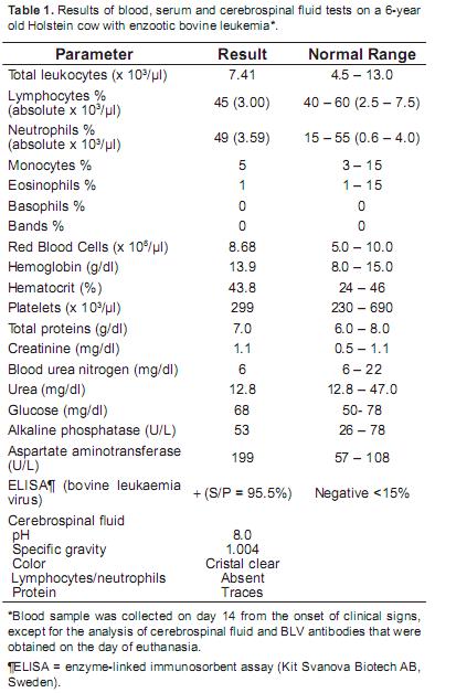
<body><![CDATA[<p>&nbsp;</p>     <p><font face="Verdana, Arial, Helvetica, sans-serif" size="3"><b>Introduction</b></font></p>     <p><font face="Verdana, Arial, Helvetica, sans-serif" size="2">   The classification of bovine lymphoproliferative   neoplasms has been widely accepted to fall into   one of four distinct forms based on age and site   of tumor development (Angelos and Thurmond,   2009). The adult multicentric form is also   known as Enzootic Bovine Leukosis (EBL). The   other three are grouped under the term sporadic   bovine leukosis (SBL) and include the juvenile   multicentric, thymic, and cutaneous forms. The   cause of EBL is bovine leukemia virus (BLV), a   retrovirus that integrates itself into the genome   of bovine B cells with the potential to induce a   lymphosarcoma and/or a persistent lymphocytosis (Yin <i>et al.</i>, 2003). EBL affects cattle over 3 years   of age and the clinical signs may vary depending   on the location and development of neoplasia.   Most reports describe a progressive loss of weight,   decreased milk production, anorexia, diarrhea or   constipation, which is associated with a generalized   lymphadenopathy (Ohshima <i>et al.</i>, 1980; Reed,   1981; Thompson <i>et al.</i>, 1993). Cases with a history   of posterior paralysis have been mostly reported   in younger cattle with SBL (Grimshaw <i>et al.</i>,   1979; Doige, 1987; Oliver&#8211;Espinosa <i>et al.</i>, 1994)   but it does not appear to be the main complain in   adult cows with EBL. The following clinical case   describes the presentation of progressive posterior   paresis and ataxia caused by EBL.</font></p>     <p>&nbsp;</p>     <p><font face="Verdana, Arial, Helvetica, sans-serif" size="3"><b> Patient's examination</b></font></p>     <p><font face="Verdana, Arial, Helvetica, sans-serif" size="2"><i>   Anamnesis and clinical findings</i></font></p>     <p><font face="Verdana, Arial, Helvetica, sans-serif" size="2">A 6&#8211;year&#8211;old and 730 kg nulliparous Holstein   female with an implanted rumen fistula for the   collection of ruminal fluid was initially observed   by the caretakers with slight ataxia and urinary   incontinence. Upon physical examination by   the veterinarian, a differential diagnosis list was   made which in order of decreasing probabilities   included: spinal cord lesion by a vertebral abscess   or infection, parasitic reaction following deworming   protocols, limphosarcoma, delayed neuropathy   by neurotoxicants, and rare but possible forms   of rabies, botulism, and bovine spongiform   encephalopathy. Summarizing the clinical course,   the slight ataxia progressed to paresis, paraplegia,   and complete ataxia over a 21 day period, culminating in humane euthanasia.</font></p>     <p>&nbsp;</p>     <p><font face="Verdana, Arial, Helvetica, sans-serif" size="3"><b>Treatment schedule</b></font></p>     <p><font face="Verdana, Arial, Helvetica, sans-serif" size="2">During the first week, medical treatment   with thiamine (3 mg/kg/day IM for 5 days, and   dexamethasone (20 mg/day IM for 5 days, resulted   in transient improvement followed by relapse and   continued progression in severity of clinical signs.   According to the daily observations by caretakers   during the first 2 weeks, the animal was not febrile,   remained bright, alert, and showed normal appetite   and defecation. However, on the third week she   became anorectic and had severe difficulty for   rising and walking, making painful grunts whenever   attempts were made to stand up or lay down. By the   end of the third week, the cow was unable to stand   on her hind legs and had complete loss of sensation   of the posterior extremities. Haematological   parameters and analysis of cerebrospinal fluid   (collected by cistern puncture) did not reveal any   abnormalities (<a href="#t1">Table 1</a>). The animal was euthanized   on day 21 after onset of clinical signs using an   intracisternal overdose of xylazine, followed within   minutes by lidocaine. Serum was then collected   and submitted for the detection of antibodies   against BLV envelope glycoprotein gp51 and was   found positive (<a href="#t1">Table 1</a>) using an enzyme&#8211;linked   immunosorbent assay (Kit Svanova, Biotech AB, Sweden).</font></p>     ]]></body>
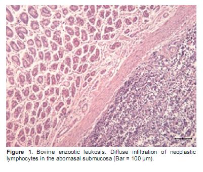
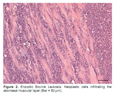
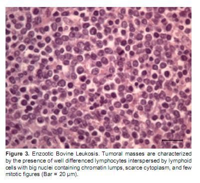
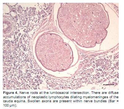
<body><![CDATA[<p align="center"><a name="t1"><img src="/img/revistas/rccp/v25n2/v25n2a16t1.jpg"></a></p>     <p>&nbsp;</p>     <p><font face="Verdana, Arial, Helvetica, sans-serif" size="3"><b>Findings at necropsy</b></font></p>     <p><font face="Verdana, Arial, Helvetica, sans-serif" size="2">   Gross post&#8211;mortem findings revealed the   presence of 4 to 6 small nodular masses, ranging in   size from 1 to 5 cm, which were embedded in the   walls of the small intestines but did not seem to   adhere to adjacent tissues and did not appear to have   perforated the mucosa or caused visible ulcers, or   areas of haemorrhage. On cut surface, these masses   were cream&#8211;colored with scarce areas of superficial   haemorrhage and had a semi&#8211;firm consistency. The   abomasal wall had several similar nodules as well   as areas of diffuse wall thickening associated with   an irregularly shaped eroded reddened mucosa. The   adipose tissue surrounding segments of the lumbar   spinal cord was interspersed with darker grey firm   areas and small haemorrhages.</font></p>     <p><font face="Verdana, Arial, Helvetica, sans-serif" size="2">The mediastinal and mesenteric lymph   nodes examined were not enlarged nor were   other abnormal lymph nodes detected by gross   examination. No masses were observed in other   organs including the spleen, liver, brain, lungs, heart and kidneys.</font></p>     <p><font face="Verdana, Arial, Helvetica, sans-serif" size="2">Nodular masses and sections of the spinal cord   were fixed in 10% formalin, embedded in paraffin,   sectioned at 4 &mu;m, and stained with hematoxylin &amp;   eosin. There was a diffuse infiltration of neoplastic   lymphocytes in the abomasal submucosa and   muscular layers (Figures <a href="#f1">1</a>, <a href="#f2">2</a> and <a href="#f3">3</a>). Histological   examination of the tissue surrounding the dura   mater of the lumbar spine revealed abundant infiltration of pleomorphic lymphocytes (<a href="#f4">Figure 4</a>).</font></p>     <p><font face="Verdana, Arial, Helvetica, sans-serif" size="2">The neoplastic cells aggregated as space   occupying infiltrations of the connective and   adipose tissue in extradural portions of the affected   segments of the spinal cord and spinal nerve roots.   Focal haemorrhages were present within neuropyle   of the spinal cord, with mild axonal swelling   observed in ventral cord horns as well as in spinal   nerves. The changes were considered to be the result   of pressure from the space&#8211;occupying neoplastic masses within the vertebral canal.</font></p>     <p align="center"><a name="f1"><img src="/img/revistas/rccp/v25n2/v25n2a16f1.jpg"></a></p>     <p align="center"><a name="f2"><img src="/img/revistas/rccp/v25n2/v25n2a16f2.jpg"></a></p>     <p align="center"><a name="f3"><img src="/img/revistas/rccp/v25n2/v25n2a16f3.jpg"></a></p>     ]]></body>
<body><![CDATA[<p align="center"><a name="f4"><img src="/img/revistas/rccp/v25n2/v25n2a16f4.jpg"></a></p>     <p>&nbsp;</p>     <p><font face="Verdana, Arial, Helvetica, sans-serif" size="3"><b>Discussion </b></font></p>     <p><font face="Verdana, Arial, Helvetica, sans-serif" size="2">Neither clinical nor paraclinical examinations   confirmed the diagnosis during the clinical course   of the disease in this animal. Approximately 30% of   cows that seroconvert to BLV develop a persistent   lymphocytosis (Angelos and Thurmond, 2009),   but this animal was not leukemic and peripheral   lymphocyte counts where within the normal range   (2.5 &#8211; 7.5 x 10<sup>3</sup> &mu;L). In these cases, it has been shown   that a careful morphological examination of the blood   smear may distinguish between atypical mononuclear   cells from other normal lymphocytes, thus providing   an early diagnosis (Ohshima <i>et al.</i>, 1980). The   present cow had very few and small lesions   detectable at necropsy for what would be expected   with typical EBL, and they could have been easily missed by a quick and superficial rectal palpation.</font></p>     <p><font face="Verdana, Arial, Helvetica, sans-serif" size="2">In a pathology study (Ohshima <i>et al.</i>, 1982) of   13 bovines euthanized in the early stages of EBL   based on persistent lymphocytosis and/or atypical   mononuclear cells, but no definitive enlargement   of the lymph nodes, it was found that the histologic   lesions preceding the tumor detection were marked   follicular hyperplasia accompanied by atypical   lymphoblastic cells in the sinuses and paracortical   areas of the lymph nodes. In the current case, it is   likely that insufficient time elapsed for tumors to   grow&#8211;up to a size of antemortem clinical detection   because of the rapid progression of the spinal   cord compromise and posterior paralysis. Careful   microscopic observations of lymph node biopsies   may have revealed early generalized neoplastic   infiltrations without gross enlargement of the lymph nodes, but was not performed in this case.</font></p>     <p><font face="Verdana, Arial, Helvetica, sans-serif" size="2">This cow came from a dairy herd of 250 cows   in the high tropics of Antioquia, Colombia. Routine   serological screening of aborted cows in this herd   revealed that 6 out of 10 cows were seropositive   for BLV during 2009. This is important because   several reports have evaluated the economical   implications of different rates of BLV infection   in dairy herds (Pelzer, 1997; Rhodes <i>et al.</i>, 2003).   For a prevalence of 50%, herds with 100 cows are   predicted to have 0.66 cases of lymphosarcoma   per year, with an expectation of 1&#8211;2 cases every 2 years (Rhodes <i>et al.</i>, 2003). However, the mean annual cost of a subclinical infection due to premature culling and loss in milk production at 50% prevalence was estimated at $6,400 (Rhodes <i>et al.</i>, 2003; Thurmond <i>et al.</i>, 1985). Considering that various epidemiological studies in major dairy areas of Colombia are quoting prevalence ranging between 21 and 62% (Alfonso <i>et al.</i>, 1998; Betancur <i>et al.</i>, 2008) basic control programs would likely be of significant economical benefit to producers.</font></p>     <p>&nbsp;</p>     <p><font face="Verdana, Arial, Helvetica, sans-serif" size="3"><b>References </b></font></p>     <!-- ref --><p><font face="Verdana, Arial, Helvetica, sans-serif" size="2">1. Alfonso R, Almansa JE, Barrera, J.C. Prevalencia serol&oacute;gica y   evaluaci&oacute;n de los factores de riesgo de Leucosis Bovina Enzo&oacute;tica   en la Sabana de Bogot&aacute; y los Valles de Ubat&eacute; y de Chiquinquir&aacute;,   Colombia. Rev Sci Tech Off Int Epiz 1998; 17:723&#8211;732. </font>&nbsp;&nbsp;&nbsp;&nbsp;&nbsp;&nbsp;&nbsp;&nbsp;[&#160;<a href="javascript:void(0);" onclick="javascript: window.open('/scielo.php?script=sci_nlinks&ref=000061&pid=S0120-0690201200020001600001&lng=','','width=640,height=500,resizable=yes,scrollbars=1,menubar=yes,');">Links</a>&#160;]<!-- end-ref --><!-- ref --><p><font face="Verdana, Arial, Helvetica, sans-serif" size="2">2. Angelos JA, Thurmond MC. Diseases of the Hematopoietic and   hemolymphatic systems &#8211; bovine lymphoma. In Large Animal   Internal Medicine. Ed. Bradford P Smith. 4th Edition, Mosby,   Elsevier. 2009; 1173&#8211;1176. </font>&nbsp;&nbsp;&nbsp;&nbsp;&nbsp;&nbsp;&nbsp;&nbsp;[&#160;<a href="javascript:void(0);" onclick="javascript: window.open('/scielo.php?script=sci_nlinks&ref=000062&pid=S0120-0690201200020001600002&lng=','','width=640,height=500,resizable=yes,scrollbars=1,menubar=yes,');">Links</a>&#160;]<!-- end-ref --><!-- ref --><p><font face="Verdana, Arial, Helvetica, sans-serif" size="2">3. Betancur CH, Rodas JG. Seroprevalencia del virus de la Leucosis   Viral Bovina en animales con trastornos reproductivos de   Monter&iacute;a. Rev MVZ C&oacute;rdoba. 2008; 13:1197&#8211;1204. </font>&nbsp;&nbsp;&nbsp;&nbsp;&nbsp;&nbsp;&nbsp;&nbsp;[&#160;<a href="javascript:void(0);" onclick="javascript: window.open('/scielo.php?script=sci_nlinks&ref=000063&pid=S0120-0690201200020001600003&lng=','','width=640,height=500,resizable=yes,scrollbars=1,menubar=yes,');">Links</a>&#160;]<!-- end-ref --><!-- ref --><p><font face="Verdana, Arial, Helvetica, sans-serif" size="2">4. Doige CE. Bone and bone marrow necrosis associated with the calf   form of sporadic bovine leukosis. Vet Pathol 1987; 24:186&#8211;188. </font>&nbsp;&nbsp;&nbsp;&nbsp;&nbsp;&nbsp;&nbsp;&nbsp;[&#160;<a href="javascript:void(0);" onclick="javascript: window.open('/scielo.php?script=sci_nlinks&ref=000064&pid=S0120-0690201200020001600004&lng=','','width=640,height=500,resizable=yes,scrollbars=1,menubar=yes,');">Links</a>&#160;]<!-- end-ref --><!-- ref --><p><font face="Verdana, Arial, Helvetica, sans-serif" size="2">5. Grimshaw WTR, Wiseman A, Petrie L, Selman IE. Bovine   Leucosis (lymphosarcoma): a clinical study of 60 pathological   confirmed cases. Vet Rec 1979; 105:267&#8211;272. </font>&nbsp;&nbsp;&nbsp;&nbsp;&nbsp;&nbsp;&nbsp;&nbsp;[&#160;<a href="javascript:void(0);" onclick="javascript: window.open('/scielo.php?script=sci_nlinks&ref=000065&pid=S0120-0690201200020001600005&lng=','','width=640,height=500,resizable=yes,scrollbars=1,menubar=yes,');">Links</a>&#160;]<!-- end-ref --><!-- ref --><p><font face="Verdana, Arial, Helvetica, sans-serif" size="2">6. Ohshima K, Ozai Y, Okada K, Numakunai S. Pathological studies   on aleukemic case of bovine leucosis. Jpn J Vet Sci 1980; 42:297&#8211;   309. </font>&nbsp;&nbsp;&nbsp;&nbsp;&nbsp;&nbsp;&nbsp;&nbsp;[&#160;<a href="javascript:void(0);" onclick="javascript: window.open('/scielo.php?script=sci_nlinks&ref=000066&pid=S0120-0690201200020001600006&lng=','','width=640,height=500,resizable=yes,scrollbars=1,menubar=yes,');">Links</a>&#160;]<!-- end-ref --><!-- ref --><p><font face="Verdana, Arial, Helvetica, sans-serif" size="2">7. Ohshima K, Sato S, Okada K. A pathologic study on initial lesions   of enzootic bovine leukosis. Jpn J Vet Sci. 1982; 44:249&#8211;257. </font>&nbsp;&nbsp;&nbsp;&nbsp;&nbsp;&nbsp;&nbsp;&nbsp;[&#160;<a href="javascript:void(0);" onclick="javascript: window.open('/scielo.php?script=sci_nlinks&ref=000067&pid=S0120-0690201200020001600007&lng=','','width=640,height=500,resizable=yes,scrollbars=1,menubar=yes,');">Links</a>&#160;]<!-- end-ref --><!-- ref --><p><font face="Verdana, Arial, Helvetica, sans-serif" size="2">8. Oliver&#8211;Espinosa O, Physic&#8211;Sheard PW, Wollenberg GK, Taylor   J. Sporadic bovine leukosis associated with ataxia and tibiotarsal   joint swelling: a case report. Can Vet J 1994; 35:777&#8211;779. </font>&nbsp;&nbsp;&nbsp;&nbsp;&nbsp;&nbsp;&nbsp;&nbsp;[&#160;<a href="javascript:void(0);" onclick="javascript: window.open('/scielo.php?script=sci_nlinks&ref=000068&pid=S0120-0690201200020001600008&lng=','','width=640,height=500,resizable=yes,scrollbars=1,menubar=yes,');">Links</a>&#160;]<!-- end-ref --><!-- ref --><p><font face="Verdana, Arial, Helvetica, sans-serif" size="2">9. Pelzer KD. Economics of Bovine Leukaemia Virus Infection. Vet   Clin North Am Food Anim Pract 1997; 13:129&#8211;141. </font>&nbsp;&nbsp;&nbsp;&nbsp;&nbsp;&nbsp;&nbsp;&nbsp;[&#160;<a href="javascript:void(0);" onclick="javascript: window.open('/scielo.php?script=sci_nlinks&ref=000069&pid=S0120-0690201200020001600009&lng=','','width=640,height=500,resizable=yes,scrollbars=1,menubar=yes,');">Links</a>&#160;]<!-- end-ref --><!-- ref --><p><font face="Verdana, Arial, Helvetica, sans-serif" size="2">10. Reed VI. 1981. Enzootic bovine leukosis. Can Vet J 1981; 22:95&#8211;   102. </font>&nbsp;&nbsp;&nbsp;&nbsp;&nbsp;&nbsp;&nbsp;&nbsp;[&#160;<a href="javascript:void(0);" onclick="javascript: window.open('/scielo.php?script=sci_nlinks&ref=000070&pid=S0120-0690201200020001600010&lng=','','width=640,height=500,resizable=yes,scrollbars=1,menubar=yes,');">Links</a>&#160;]<!-- end-ref --><!-- ref --><p><font face="Verdana, Arial, Helvetica, sans-serif" size="2">11. Rhodes JK, Pelzer KD, Johnson YJ. Economic implications of   bovine leukaemia virus infection in mid&#8211;Atlantic dairy herds.   JAVMA 2003; 223:346&#8211;352. </font>&nbsp;&nbsp;&nbsp;&nbsp;&nbsp;&nbsp;&nbsp;&nbsp;[&#160;<a href="javascript:void(0);" onclick="javascript: window.open('/scielo.php?script=sci_nlinks&ref=000071&pid=S0120-0690201200020001600011&lng=','','width=640,height=500,resizable=yes,scrollbars=1,menubar=yes,');">Links</a>&#160;]<!-- end-ref --><!-- ref --><p><font face="Verdana, Arial, Helvetica, sans-serif" size="2">12. Thompson KG, Johnstone AC, Hilbink F. Enzootic bovine leucosis   in New Zealand &#8211; a case report and update. N Z Vet J 1993;   41:190&#8211;194. </font>&nbsp;&nbsp;&nbsp;&nbsp;&nbsp;&nbsp;&nbsp;&nbsp;[&#160;<a href="javascript:void(0);" onclick="javascript: window.open('/scielo.php?script=sci_nlinks&ref=000072&pid=S0120-0690201200020001600012&lng=','','width=640,height=500,resizable=yes,scrollbars=1,menubar=yes,');">Links</a>&#160;]<!-- end-ref --><!-- ref --><p><font face="Verdana, Arial, Helvetica, sans-serif" size="2">13. Thurmond MC, Maden CB, Carter RL. Cull rates of dairy cattle   with antibodies to bovine leukaemia virus. Cancer Res 1985: 45:1967&#8211;1989.</font>&nbsp;&nbsp;&nbsp;&nbsp;&nbsp;&nbsp;&nbsp;&nbsp;[&#160;<a href="javascript:void(0);" onclick="javascript: window.open('/scielo.php?script=sci_nlinks&ref=000073&pid=S0120-0690201200020001600013&lng=','','width=640,height=500,resizable=yes,scrollbars=1,menubar=yes,');">Links</a>&#160;]<!-- end-ref --><!-- ref --><p><font face="Verdana, Arial, Helvetica, sans-serif" size="2">   14. Yin S, Makara M, Pan Y, Ishiguro H, Ikeda M, Numakunai S,   Goryo M, Okada K. Relation between phenotype of tumor cells   and clinicopathology in bovine leukosis. J Vet Med Sci 2003;   65:599&#8211;606.</font>&nbsp;&nbsp;&nbsp;&nbsp;&nbsp;&nbsp;&nbsp;&nbsp;[&#160;<a href="javascript:void(0);" onclick="javascript: window.open('/scielo.php?script=sci_nlinks&ref=000074&pid=S0120-0690201200020001600014&lng=','','width=640,height=500,resizable=yes,scrollbars=1,menubar=yes,');">Links</a>&#160;]<!-- end-ref --><p>&nbsp;</p>     <p>&nbsp;</p>     <p><font face="Verdana, Arial, Helvetica, sans-serif" size="2">&nbsp;</font><font face="Verdana, Arial, Helvetica, sans&ndash;serif" size="3"><b>Notas</b></font></p>     <p><font face="Verdana, Arial, Helvetica, sans&ndash;serif" size="2"><a href="#*"></a><a href="#*b" name="*"><sup>&curren; </sup></a></font><font face="Verdana, Arial, Helvetica, sans-serif" size="2">To cite this article: Villar D, Duque L, Giraldo CA, P&eacute;rez JE, Pallares F, Schwartz K. Posterior paralysis in a Holstein cow suffering Enzootic Bovine   Leukosis. Rev Colomb Cienc Pecu 2012; 25:325&#8211;329.</font></p>      ]]></body><back>
<ref-list>
<ref id="B1">
<label>1</label><nlm-citation citation-type="journal">
<person-group person-group-type="author">
<name>
<surname><![CDATA[Alfonso]]></surname>
<given-names><![CDATA[R]]></given-names>
</name>
<name>
<surname><![CDATA[Almansa]]></surname>
<given-names><![CDATA[JE]]></given-names>
</name>
<name>
<surname><![CDATA[Barrera,]]></surname>
<given-names><![CDATA[J.C]]></given-names>
</name>
</person-group>
<article-title xml:lang="es"><![CDATA[Prevalencia serológica y evaluación de los factores de riesgo de Leucosis Bovina Enzoótica en la Sabana de Bogotá y los Valles de Ubaté y de Chiquinquirá, Colombia]]></article-title>
<source><![CDATA[Rev Sci Tech Off Int Epiz]]></source>
<year>1998</year>
<volume>17</volume>
<page-range>723-732</page-range></nlm-citation>
</ref>
<ref id="B2">
<label>2</label><nlm-citation citation-type="book">
<person-group person-group-type="author">
<name>
<surname><![CDATA[Angelos]]></surname>
<given-names><![CDATA[JA]]></given-names>
</name>
<name>
<surname><![CDATA[Thurmond]]></surname>
<given-names><![CDATA[MC]]></given-names>
</name>
</person-group>
<article-title xml:lang="en"><![CDATA[Diseases of the Hematopoietic and hemolymphatic systems &#8211; bovine lymphoma]]></article-title>
<source><![CDATA[Large Animal Internal Medicine]]></source>
<year>2009</year>
<edition>4</edition>
<page-range>1173-1176</page-range><publisher-name><![CDATA[Elsevier]]></publisher-name>
</nlm-citation>
</ref>
<ref id="B3">
<label>3</label><nlm-citation citation-type="journal">
<person-group person-group-type="author">
<name>
<surname><![CDATA[Betancur]]></surname>
<given-names><![CDATA[CH]]></given-names>
</name>
<name>
<surname><![CDATA[Rodas]]></surname>
<given-names><![CDATA[JG]]></given-names>
</name>
</person-group>
<article-title xml:lang="es"><![CDATA[Seroprevalencia del virus de la Leucosis Viral Bovina en animales con trastornos reproductivos de Montería]]></article-title>
<source><![CDATA[Rev MVZ Córdoba]]></source>
<year>2008</year>
<volume>13</volume>
<page-range>1197-1204</page-range></nlm-citation>
</ref>
<ref id="B4">
<label>4</label><nlm-citation citation-type="journal">
<person-group person-group-type="author">
<name>
<surname><![CDATA[Doige]]></surname>
<given-names><![CDATA[CE]]></given-names>
</name>
</person-group>
<article-title xml:lang="en"><![CDATA[Bone and bone marrow necrosis associated with the calf form of sporadic bovine leukosis]]></article-title>
<source><![CDATA[Vet Pathol]]></source>
<year>1987</year>
<volume>24</volume>
<page-range>186-188</page-range></nlm-citation>
</ref>
<ref id="B5">
<label>5</label><nlm-citation citation-type="journal">
<person-group person-group-type="author">
<name>
<surname><![CDATA[Grimshaw]]></surname>
<given-names><![CDATA[WTR]]></given-names>
</name>
<name>
<surname><![CDATA[Wiseman]]></surname>
<given-names><![CDATA[A]]></given-names>
</name>
<name>
<surname><![CDATA[Petrie]]></surname>
<given-names><![CDATA[L]]></given-names>
</name>
<name>
<surname><![CDATA[Selman]]></surname>
<given-names><![CDATA[IE]]></given-names>
</name>
</person-group>
<article-title xml:lang="en"><![CDATA[Bovine Leucosis (lymphosarcoma): a clinical study of 60 pathological confirmed cases]]></article-title>
<source><![CDATA[Vet Rec]]></source>
<year>1979</year>
<volume>105</volume>
<page-range>267-272</page-range></nlm-citation>
</ref>
<ref id="B6">
<label>6</label><nlm-citation citation-type="journal">
<person-group person-group-type="author">
<name>
<surname><![CDATA[Ohshima]]></surname>
<given-names><![CDATA[K]]></given-names>
</name>
<name>
<surname><![CDATA[Ozai]]></surname>
<given-names><![CDATA[Y]]></given-names>
</name>
<name>
<surname><![CDATA[Okada]]></surname>
<given-names><![CDATA[K]]></given-names>
</name>
<name>
<surname><![CDATA[Numakunai]]></surname>
<given-names><![CDATA[S]]></given-names>
</name>
</person-group>
<article-title xml:lang="en"><![CDATA[Pathological studies on aleukemic case of bovine leucosis]]></article-title>
<source><![CDATA[Jpn J Vet Sci]]></source>
<year>1980</year>
<volume>42</volume>
<page-range>297- 309</page-range></nlm-citation>
</ref>
<ref id="B7">
<label>7</label><nlm-citation citation-type="journal">
<person-group person-group-type="author">
<name>
<surname><![CDATA[Ohshima]]></surname>
<given-names><![CDATA[K]]></given-names>
</name>
<name>
<surname><![CDATA[Sato]]></surname>
<given-names><![CDATA[S]]></given-names>
</name>
<name>
<surname><![CDATA[Okada]]></surname>
<given-names><![CDATA[K]]></given-names>
</name>
</person-group>
<article-title xml:lang="en"><![CDATA[A pathologic study on initial lesions of enzootic bovine leukosis]]></article-title>
<source><![CDATA[Jpn J Vet Sci]]></source>
<year>1982</year>
<volume>44</volume>
<page-range>249-257</page-range></nlm-citation>
</ref>
<ref id="B8">
<label>8</label><nlm-citation citation-type="journal">
<person-group person-group-type="author">
<name>
<surname><![CDATA[Oliver-Espinosa]]></surname>
<given-names><![CDATA[O]]></given-names>
</name>
<name>
<surname><![CDATA[Physic-Sheard]]></surname>
<given-names><![CDATA[PW]]></given-names>
</name>
<name>
<surname><![CDATA[Wollenberg]]></surname>
<given-names><![CDATA[GK]]></given-names>
</name>
<name>
<surname><![CDATA[Taylor]]></surname>
<given-names><![CDATA[J]]></given-names>
</name>
</person-group>
<article-title xml:lang="en"><![CDATA[Sporadic bovine leukosis associated with ataxia and tibiotarsal joint swelling: a case report]]></article-title>
<source><![CDATA[Can Vet J]]></source>
<year>1994</year>
<volume>35</volume>
<page-range>777-779</page-range></nlm-citation>
</ref>
<ref id="B9">
<label>9</label><nlm-citation citation-type="journal">
<person-group person-group-type="author">
<name>
<surname><![CDATA[Pelzer]]></surname>
<given-names><![CDATA[KD]]></given-names>
</name>
</person-group>
<article-title xml:lang="en"><![CDATA[Economics of Bovine Leukaemia Virus Infection]]></article-title>
<source><![CDATA[Vet Clin North Am Food Anim Pract]]></source>
<year>1997</year>
<volume>13</volume>
<page-range>129-141</page-range></nlm-citation>
</ref>
<ref id="B10">
<label>10</label><nlm-citation citation-type="journal">
<person-group person-group-type="author">
<name>
<surname><![CDATA[Reed]]></surname>
<given-names><![CDATA[VI]]></given-names>
</name>
</person-group>
<article-title xml:lang="en"><![CDATA[Enzootic bovine leukosis]]></article-title>
<source><![CDATA[Can Vet J]]></source>
<year>1981</year>
<volume>22</volume>
<page-range>95- 102</page-range></nlm-citation>
</ref>
<ref id="B11">
<label>11</label><nlm-citation citation-type="journal">
<person-group person-group-type="author">
<name>
<surname><![CDATA[Rhodes]]></surname>
<given-names><![CDATA[JK]]></given-names>
</name>
<name>
<surname><![CDATA[Pelzer]]></surname>
<given-names><![CDATA[KD]]></given-names>
</name>
<name>
<surname><![CDATA[Johnson]]></surname>
<given-names><![CDATA[YJ]]></given-names>
</name>
</person-group>
<article-title xml:lang="en"><![CDATA[Economic implications of bovine leukaemia virus infection in mid-Atlantic dairy herds]]></article-title>
<source><![CDATA[JAVMA]]></source>
<year>2003</year>
<volume>223</volume>
<page-range>346-352</page-range></nlm-citation>
</ref>
<ref id="B12">
<label>12</label><nlm-citation citation-type="journal">
<person-group person-group-type="author">
<name>
<surname><![CDATA[Thompson]]></surname>
<given-names><![CDATA[KG]]></given-names>
</name>
<name>
<surname><![CDATA[Johnstone]]></surname>
<given-names><![CDATA[AC]]></given-names>
</name>
<name>
<surname><![CDATA[Hilbink]]></surname>
<given-names><![CDATA[F]]></given-names>
</name>
</person-group>
<article-title xml:lang="en"><![CDATA[Enzootic bovine leucosis in New Zealand &#8211; a case report and update]]></article-title>
<source><![CDATA[N Z Vet J]]></source>
<year>1993</year>
<volume>41</volume>
<page-range>190-194</page-range></nlm-citation>
</ref>
<ref id="B13">
<label>13</label><nlm-citation citation-type="journal">
<person-group person-group-type="author">
<name>
<surname><![CDATA[Thurmond]]></surname>
<given-names><![CDATA[MC]]></given-names>
</name>
<name>
<surname><![CDATA[Maden]]></surname>
<given-names><![CDATA[CB]]></given-names>
</name>
<name>
<surname><![CDATA[Carter]]></surname>
<given-names><![CDATA[RL]]></given-names>
</name>
</person-group>
<article-title xml:lang="en"><![CDATA[Cull rates of dairy cattle with antibodies to bovine leukaemia virus]]></article-title>
<source><![CDATA[Cancer Res]]></source>
<year>1985</year>
<volume>45</volume>
<page-range>1967-1989</page-range></nlm-citation>
</ref>
<ref id="B14">
<label>14</label><nlm-citation citation-type="journal">
<person-group person-group-type="author">
<name>
<surname><![CDATA[Yin]]></surname>
<given-names><![CDATA[S]]></given-names>
</name>
<name>
<surname><![CDATA[Makara]]></surname>
<given-names><![CDATA[M]]></given-names>
</name>
<name>
<surname><![CDATA[Pan]]></surname>
<given-names><![CDATA[Y]]></given-names>
</name>
<name>
<surname><![CDATA[Ishiguro]]></surname>
<given-names><![CDATA[H]]></given-names>
</name>
<name>
<surname><![CDATA[Ikeda]]></surname>
<given-names><![CDATA[M]]></given-names>
</name>
<name>
<surname><![CDATA[Numakunai]]></surname>
<given-names><![CDATA[S]]></given-names>
</name>
<name>
<surname><![CDATA[Goryo]]></surname>
<given-names><![CDATA[M]]></given-names>
</name>
<name>
<surname><![CDATA[Okada]]></surname>
<given-names><![CDATA[K]]></given-names>
</name>
</person-group>
<article-title xml:lang="en"><![CDATA[Relation between phenotype of tumor cells and clinicopathology in bovine leukosis]]></article-title>
<source><![CDATA[J Vet Med Sci]]></source>
<year>2003</year>
<volume>65</volume>
<page-range>599-606</page-range></nlm-citation>
</ref>
</ref-list>
</back>
</article>