<?xml version="1.0" encoding="ISO-8859-1"?><article xmlns:mml="http://www.w3.org/1998/Math/MathML" xmlns:xlink="http://www.w3.org/1999/xlink" xmlns:xsi="http://www.w3.org/2001/XMLSchema-instance">
<front>
<journal-meta>
<journal-id>0120-0690</journal-id>
<journal-title><![CDATA[Revista Colombiana de Ciencias Pecuarias]]></journal-title>
<abbrev-journal-title><![CDATA[Rev Colom Cienc Pecua]]></abbrev-journal-title>
<issn>0120-0690</issn>
<publisher>
<publisher-name><![CDATA[Facultad de Ciencias Agrarias, Universidad de Antioquia]]></publisher-name>
</publisher>
</journal-meta>
<article-meta>
<article-id>S0120-06902013000300009</article-id>
<title-group>
<article-title xml:lang="en"><![CDATA[Detection of macrophages infected with Mycobacterium avium subspecies paratuberculosis in a cow with clinical stage IV of Johne's disease. A case report]]></article-title>
<article-title xml:lang="es"><![CDATA[Detección de macrófagos infectados con Mycobacterium avium subspecies paratuberculosis en una vaca en fase clínica IV de la enfermedad de Johne. Reporte de un caso]]></article-title>
<article-title xml:lang="pt"><![CDATA[Detecção de macrófagos infectados com Mycobacterium avium paratuberculosis subespécie em uma vaca com uma fase clínica IV da doença de Johne. Um relato de caso]]></article-title>
</title-group>
<contrib-group>
<contrib contrib-type="author">
<name>
<surname><![CDATA[Ramírez-García]]></surname>
<given-names><![CDATA[René]]></given-names>
</name>
</contrib>
<contrib contrib-type="author">
<name>
<surname><![CDATA[Maldonado-Estrada]]></surname>
<given-names><![CDATA[Juan G]]></given-names>
</name>
<xref ref-type="aff" rid="A01"/>
</contrib>
</contrib-group>
<aff id="A01">
<institution><![CDATA[,Universidad de Antioquia  ]]></institution>
<addr-line><![CDATA[Medellín ]]></addr-line>
<country>Colombia</country>
</aff>
<pub-date pub-type="pub">
<day>00</day>
<month>09</month>
<year>2013</year>
</pub-date>
<pub-date pub-type="epub">
<day>00</day>
<month>09</month>
<year>2013</year>
</pub-date>
<volume>26</volume>
<numero>3</numero>
<fpage>219</fpage>
<lpage>225</lpage>
<copyright-statement/>
<copyright-year/>
<self-uri xlink:href="http://www.scielo.org.co/scielo.php?script=sci_arttext&amp;pid=S0120-06902013000300009&amp;lng=en&amp;nrm=iso"></self-uri><self-uri xlink:href="http://www.scielo.org.co/scielo.php?script=sci_abstract&amp;pid=S0120-06902013000300009&amp;lng=en&amp;nrm=iso"></self-uri><self-uri xlink:href="http://www.scielo.org.co/scielo.php?script=sci_pdf&amp;pid=S0120-06902013000300009&amp;lng=en&amp;nrm=iso"></self-uri><abstract abstract-type="short" xml:lang="en"><p><![CDATA[Anamnesis: a six-year-old milking Holstein cow (Bos taurus taurus) was diagnosed with stage IV Johne's Disease (JD). Clinical and Laboratory findings: the cow suffered intermittent diarrhea during 6 months with no response to antibiotic treatment. Consequently, the cow was subjected to euthanasia. Treatment approach: antemortem milk and peripheral blood samples and postmortem colon, mediastinic, mesenteric lymph nodes, and spleen samples were processed for macrophages isolation. Total DNA was extracted from macrophages and used to diagnose IS900 of Mycobacterium avium paratuberculosis (MAP) through real time PCR. The MAP IS900 segment was successfully amplified from cells of all samples, indicating that these cells were MAP-infected macrophages. Conclusion: macrophages of cows suffering from JD can be used for amplification of the MAP IS900 segment.]]></p></abstract>
<abstract abstract-type="short" xml:lang="es"><p><![CDATA[Anamnesis: una vaca Holstein de 6 años de edad (Bos taurus taurus) presentó sintomatología de la fase IV de la enfermedad de Johne. Hallazgos clínicos y de laboratorio: la vaca presentó diarrea intermitente durante 6 meses sin respuesta al tratamiento con antibióticos. En consecuencia, la vaca fue sometida a eutanasia. Esquema de tratamiento: muestras de leche y de sangre periférica se tomaron ante-mortem; muestras de la mucosa del intestino, bazo y linfonodos mediastínico y mesentérico, se tomaron post-mortem, todas para aislamiento de macrófagos. El ADN total de los macrófagos fue usado para la amplificación del segmento IS900 de Mycobacterium avium paratuberculosis (MAP) por PCR en tiempo real. Conclusión: los macrófagos de vacas con la enfermedad de Johne pueden ser usados para la amplificación del segmento IS900 de MAP.]]></p></abstract>
<abstract abstract-type="short" xml:lang="pt"><p><![CDATA[Anamnese: uma vaca Holstein com 6 anos de idade (Bos taurus taurus) apresentou os sintomas da fase IV da doença de Johne. Achados clínicos e laboratoriais: a vaca teve diarreia intermitente por seis meses sem resposta ao tratamento com antibióticos. Por conseguinte, a vaca foi submetida à eutanásia. Abordagem de tratamento: amostras de leite e de sangue periférico foram retiradas ante-mortem, enquanto as amostras da mucosa intestinal, baço e linfonodos mesentéricos e mediastinais foram tomadas todas post-mortem para o isolamento de macrófagos. O ADN total de macrófagos foi utilizado para amplificação do segmento IS900 do Mycobacterium avium paratuberculosis (MAP) por PCR em tempo real. Conclusão: macrófagos isolados a partir de vacas com doença de Johne podem ser utilizados para a amplificação da segmento IS900 do MAP.]]></p></abstract>
<kwd-group>
<kwd lng="en"><![CDATA[intracellular bacteria]]></kwd>
<kwd lng="en"><![CDATA[real time PCR]]></kwd>
<kwd lng="en"><![CDATA[resident macrophages]]></kwd>
<kwd lng="en"><![CDATA[susceptible host]]></kwd>
<kwd lng="es"><![CDATA[bacteria intracelular]]></kwd>
<kwd lng="es"><![CDATA[hospedero susceptible]]></kwd>
<kwd lng="es"><![CDATA[macrófagos residentes]]></kwd>
<kwd lng="es"><![CDATA[PCR en tiempo real]]></kwd>
<kwd lng="pt"><![CDATA[bactérias intracelulares]]></kwd>
<kwd lng="pt"><![CDATA[hospedeiro suscetível]]></kwd>
<kwd lng="pt"><![CDATA[macrófagos residentes]]></kwd>
<kwd lng="pt"><![CDATA[PCR em tempo real]]></kwd>
</kwd-group>
</article-meta>
</front><body><![CDATA[  <font size="2" face="Verdana, Arial, Helvetica, sans-serif">     <p align="right"><b>CLINICAL CASES</b></p>     <p align="right">&nbsp;</p>     <p align="center"><b><font size="4">Detection of macrophages infected with Mycobacterium avium   subspecies paratuberculosis in a cow with clinical stage IV of Johne's disease. A case report </font></b> <b><font size="4"><sup><a name="b1" id="b1"></a><a href="#1">&curren;</a></sup></font></b></p>     <p>&nbsp;  </p>     <p align="center"><b><font size="3">Detecci&oacute;n de macr&oacute;fagos infectados con Mycobacterium avium subspecies paratuberculosis en una vaca   en fase cl&iacute;nica IV de la enfermedad de Johne. Reporte de un caso</font></b></p>     <p>&nbsp;  </p>     <p align="center"><b><font size="3">Detec&ccedil;&atilde;o de macr&oacute;fagos infectados com Mycobacterium avium paratuberculosis subesp&eacute;cie em uma   vaca com uma fase cl&iacute;nica IV da doen&ccedil;a de Johne. Um relato de caso</font></b></p>     <p>&nbsp;  </p>     <p>&nbsp;</p>     ]]></body>
<body><![CDATA[<p><b>Ren&eacute; Ram&iacute;rez-Garc&iacute;a, MV, MSc; Juan G Maldonado-Estrada*, MVZ, MS, PhD.</b> </p>     <p>* Corresponding author: Juan G Maldonado-Estrada. Escuela de Medicina Veterinaria, and Sede de Investigaci&oacute;n Universitaria (SIU) Calle 62 # 52-59, Lab-233. Universidad de Antioquia. 050011 Medell&iacute;n, Colombia. E-mail: <a href="mailto:juanguimal@gmail.com">juanguimal@gmail.com</a></p>     <p>Grupo de investigaci&oacute;n CENTAURO, Escuela de Medicina Veterinaria, Facultad de Ciencias Agrarias. Universidad de   Antioquia, Medell&iacute;n, Colombia. </p>     <p>&nbsp;</p>     <p>(Received: October 22, 2012; accepted: January, 29, 2013) </p>     <p>&nbsp;</p> <hr size="1" />     <p><b>Summary</b></p>     <p>   <b>Anamnesis:</b> a six-year-old milking Holstein cow (<i>Bos taurus taurus</i>) was diagnosed with stage IV Johne's   Disease (JD). <b>Clinical and Laboratory findings:</b> the cow suffered intermittent diarrhea during 6 months   with no response to antibiotic treatment. Consequently, the cow was subjected to euthanasia. <b>Treatment   approach:</b> antemortem milk and peripheral blood samples and postmortem colon, mediastinic, mesenteric   lymph nodes, and spleen samples were processed for macrophages isolation. Total DNA was extracted from   macrophages and used to diagnose IS<i>900</i> of <i>Mycobacterium avium paratuberculosis</i> (MAP) through real time   PCR. The MAP IS<i>900</i> segment was successfully amplified from cells of all samples, indicating that these   cells were MAP-infected macrophages. Conclusion: macrophages of cows suffering from JD can be used for   amplification of the MAP IS<i>900</i> segment.  </p>     <p><b>Key words:</b> intracellular bacteria, real time PCR, resident macrophages, susceptible host.</p> <hr size="1" />     <p>   <b>Resumen</b></p>     ]]></body>
<body><![CDATA[<p>   <b>Anamnesis:</b> una vaca Holstein de 6 a&ntilde;os de edad (<i>Bos taurus taurus</i>) present&oacute; sintomatolog&iacute;a de la fase   IV de la enfermedad de Johne. <b>Hallazgos cl&iacute;nicos y de laboratorio:</b> la vaca present&oacute; diarrea intermitente   durante 6 meses sin respuesta al tratamiento con antibi&oacute;ticos. En consecuencia, la vaca fue sometida a eutanasia. <b>Esquema de tratamiento:</b> muestras de leche y de sangre perif&eacute;rica se tomaron ante-mortem;   muestras de la mucosa del intestino, bazo y linfonodos mediast&iacute;nico y mesent&eacute;rico, se tomaron post-mortem,   todas para aislamiento de macr&oacute;fagos. El ADN total de los macr&oacute;fagos fue usado para la amplificaci&oacute;n del   segmento IS<i>900</i> de <i>Mycobacterium avium paratuberculosis</i> (MAP) por PCR en tiempo real. <b>Conclusi&oacute;n:</b>  los macr&oacute;fagos de vacas con la enfermedad de Johne pueden ser usados para la amplificaci&oacute;n del segmento   IS900 de MAP.</p>     <p>   <b>Palabras clave:</b> bacteria intracelular, hospedero susceptible, macr&oacute;fagos residentes, PCR en tiempo real.</p> <hr size="1" />     <p>   <b>Resumo</b></p>     <p>   <b>Anamnese:</b> uma vaca Holstein com 6 anos de idade (<i>Bos taurus taurus</i>) apresentou os sintomas da fase   IV da doen&ccedil;a de Johne. <b>Achados cl&iacute;nicos e laboratoriais:</b> a vaca teve diarreia intermitente por seis meses   sem resposta ao tratamento com antibi&oacute;ticos. Por conseguinte, a vaca foi submetida &agrave; eutan&aacute;sia. <b>Abordagem   de tratamento:</b> amostras de leite e de sangue perif&eacute;rico foram retiradas ante-mortem, enquanto as amostras   da mucosa intestinal, ba&ccedil;o e linfonodos mesent&eacute;ricos e mediastinais foram tomadas todas post-mortem para   o isolamento de macr&oacute;fagos. O ADN total de macr&oacute;fagos foi utilizado para amplifica&ccedil;&atilde;o do segmento IS<i>900</i>  do <i>Mycobacterium avium paratuberculosis</i> (MAP) por PCR em tempo real. <b>Conclus&atilde;o:</b> macr&oacute;fagos isolados   a partir de vacas com doen&ccedil;a de Johne podem ser utilizados para a amplifica&ccedil;&atilde;o da segmento IS<i>900</i> do MAP.</p>     <p>   <b>Palavras chave:</b> bact&eacute;rias intracelulares, hospedeiro suscet&iacute;vel, macr&oacute;fagos residentes, PCR em tempo   real.</p> <hr size="1" />     <p>&nbsp;</p>     <p>&nbsp;</p>     <p><b><font size="3">Introduction</font></b></p>     <p>   Paratuberculosis or Johne's disease (JD) is caused   by the intracellular bacterium <i>Mycobacterium avium</i>  subspecies <i>paratuberculosis</i> (MAP). MAP causes a   devastating chronic disease in the affected animal.   The prevalence and incidence of JD ranges from   5% to 100% (McNab <i>et al.</i>, 1991). Our research   group documented the presence of JD in the northern   dairy region of Antioquia province (Colombia)   based on clinical, pathological (Ramirez <i>et al.</i>,   2011), and molecular results (Zapata-Restrepo <i>et al.</i>,   2010; Fernandez-Silva <i>et al.</i>, 2011b). Macrophages   infected with MAP can circulate and re-circulate   from Peyer's patches of infected intestinal mucosa   to other tissues via blood and lymph compartments   (Mutharia <i>et al.</i>, 2010; Bimczok and Rothk&ouml;tter,   2006). Consequently, we wondered if it would be   possible to take advantage of this recirculation   process for early detection and diagnosis in cows.   The main objective of this work was to evaluate the   usefulness of circulating and resident macrophages   for amplification of the IS<i>900</i> sequence of MAP.</p>     <p>&nbsp;</p>     ]]></body>
<body><![CDATA[<p><b><font size="3">Patient examination</font></b></p>     <p>   <i>Anamnesis</i></p>     <p>  A six-year-old lactating Holstein cow (<i>Bos   taurus taurus</i>) from the University of Antioquia's   dairy herd was diagnosed with stage IV JD. The   farm is located at 2200 m above sea level in a   high-humidity region at San Pedro de los Milagros   (Antioquia, Colombia). The farm is enzootic for   JD as reported by Ram&iacute;rez <i>et al.</i> (2011). The cow   suffered from recurrent episodes of profuse diarrhea   with progressive weight loss for 6 months.</p>     <p>   <i>Clinical findings</i></p>     <p>   
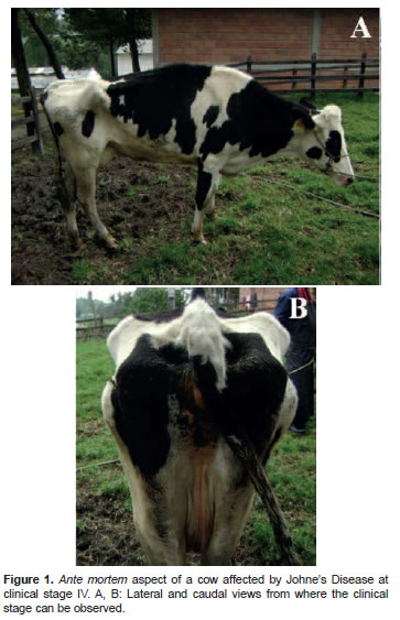
The cow's body condition score (BCS) was   1.5 (scale 1 to 5) and showed severe depression.   Cardiac frequency was 80 beats/min. The animal   was tachypneic and evidenced diarrhea. It had   not eaten for several days (<a href="#f1">Figure 1</a>). Rectal   temperature was 39.5 &deg;C. Mucosae were pale and   dry. Lack of intestinal motility was also evidenced   at auscultation of abdominal quadrants. The last milking record of the cow&#8212;taken 1 month before   the clinical exam&#8212;indicated a yield of 20 kg   milk per day. Based on the enzootic status of the   farm, anamnesis, and clinical exam, the cow was   diagnosed as clinically compatible with stage   IV JD. In addition, the cow had tested positive   when evaluated for antibodies against MAP by   ELISA test. This test was performed as reported   by Fern&aacute;ndez-Silva <i>et al.</i> (2011a) and the specific   result of this cow was taken from the official record   reported to the staff of the farm. The farm and   veterinarian staff decided to euthanize the cow.   Blood samples were taken ante-mortem in sterile   vacutainer tubes containing citrate as anticoagulant.   A milk sample was obtained by manual milking in   sterile 50 ml Coning centrifuge tubes (Corning, NY,   USA). Samples were transported under refrigeration   to the laboratory and processed to isolate   mononuclear cells as previously reported (Zapata-   Restrepo <i>et al.</i>, 2010).</p>     <p align="center"><a name="f1"></a><img src="/img/revistas/rccp/v26n3/v26n3a9f1.jpg"/></p>     <p>
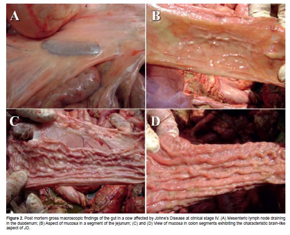
The cow was then euthanized by intrathecal   injection of 25 ml lydocaine through the <i>foramen   magnum</i>. Significant carcass alterations were not   observed at the macroscopic examination during   necropsy but a generalized pale condition suggested   anemia. Abundant brown fat was observed around   the pericardium near the insertion of the aorta   adventitia. Adherences were present at the left   costal side of the pericardium. Lung atelectasis   was present. The heart was cardiomegalic and   marked atrophy of the spleen was observed. The   liver borders were rounded, bile ducts were thick,   and several adult forms of <i>Fasciola hepatica</i>  were observed in the bile ducts. Mediastinal and   mesenteric lymph nodes were hypertrophic (<a href="#f2">Figure   2A</a>). Jejunum and ileum were normal (<a href="#f2">Figure 2</a>B),   but the terminal ileum and the colon were thickened   and the macroscopic aspect of intestinal mucosa was typical of JD (<a href="#f2">Figures 2</a>C and <a href="#f2">2</a>D).</p>     <p align="center"><a name="f2"></a><img src="/img/revistas/rccp/v26n3/v26n3a9f2.jpg"/></p>     <p>Several mediastinic and mesenteric lymph   nodes, a spleen sample (5 x 5 cm), and transversal   colon segments were taken during post-mortem   examination. Samples were imbibed in Phosphate   Buffer Saline (PBS, 1mM, pH = 7.2; Sigma-   Aldrich-Aldrich, St. Louis, MO, USA) containing   1% penicillin/streptomycin and amphotericin B.   Samples were kept on ice and transported to the   laboratory in 75 cm<sup>2</sup> Falcon culture flasks (Falcon,   San Jose, CA, USA). After necropsy the carcass was   deposited into a septic fossa according to the farm's   sanitary protocol, which includes deep underground   disposition (1.5 m) followed by abundant lime covering and complete carcass burial.</p>     <p>Tissue samples and secondary lymphoid tissues   were cut and processed by mincing with dissecting   scissors sterilized in PBS with added antibiotics as   previously indicated. Samples were processed for   macrophage isolation according to the protocol by   Ramirez-Garcia <i>et al.</i> (personal communication,   2012). Tissues were minced with surgical scissors   in petri dishes with added PBS and RPMI-1640   complete medium. The supernatant was recovered   and centrifuged in a 1.077 density Ficoll gradient   (Sigma, St. Louis, MO, USA) at 800 x g/40 min.   Blood and milk samples were processed directly for   isolation of mononuclear cells by centrifugation in 1.077 Ficoll gradient at 800 x g/40 min. After gradient centrifugation, the buffy-coats were recovered with a sterile plastic Pasteur pipette and washed three times in PBS at 400 x g/10 min. Pellets were recovered from mesenteric lymph nodes, spleen, mediastinal lymph nodes, blood, and milk. Cell pellets were resuspended in RPMI-1640 complete media (Mediatech Inc., Herndon, VA, USA) and cultured at 37 &deg;C/2 hours in sterile polypropylene petri dishes. The nonadherent mononuclear cells were removed after this incubation period while the adherent cells were washed twice with RPMI-1640 complete media and cultured under standard conditions at 37 &deg;C/5% CO2 in a humidified atmosphere.</p>     ]]></body>
<body><![CDATA[<p>The cells obtained by this protocol were bovine   macrophages according to CD14 expression (PEconjugated   mouse anti-human CD14 moAb &#91;clone   61D3&#93;) (Biosurce, San Diego, CA, USA) and   phagocytosis of polypropylene spheres coupled   to Phycoerythrin (CaliBRITE PE-beads; Becton-   Dickinson, CA, USA). Plastic-adherent cells were   cultured for at least 48 hours, after which time cells   showed the characteristic pattern of macrophages.   After 48 hours of macrophages culture cells were   processed for total DNA isolation using the DNA   Qiagen-mini kit (Qiagen, TX, USA) according to   the manufacturer's instructions. The DNA purity   and concentration was evaluated in a Nanodrop&reg;   ND1000 spectrophotometer (Thermo Fisher   Scientific, Wilmington, DE, USA); samples were stored at -80 &deg;C until processing.</p>     <p>Amplification of IS900 MAP fragment by real   time PCR was performed using DNA samples   obtained from all sources of macrophages as   previously indicated, according to the protocol by   O'Mahony and Hill (2002), with modifications in the   thermal profile as follows: hot start at 15 min/95&deg;C   (instead of 10 min); 40 cycles (instead of 50) of   amplification phase at 30 sec/95&deg;C (instead of 15   sec), 10 sec/55&deg;C (instead of 30 sec/60&deg;C) and 30   sec/72&deg; (instead of 12 sec). Primers used for IS900   amplification were: (FW) 5'CGA CGT GTC CTT   ACA CAG C 3'; and reverse (RW) 5G'GT ATG GTT   TCA TGT GGT T 3'. Real time PCR were processed   in a Rotor-GeneTM 6000 real time rotary analyzer   (Corbett Research, Australia). The PCR curves and   florescence normalization were established by the   Threshold value (Ct). LinREGPCRTM software was   used for the efficiency calculation. All samples were   processed in triplicate and data were analyzed by the   &#8211;(&Delta;&Delta;ct) method (Livak and Schmittgen, 2001). The   GAPDH gene was used as control for calculation   of &Delta;Ct. The results were analyzed using the Rotor-   Gene 6000 series 1.7 software (Corbett Research,   Sydney, Australia). The IS900 segment of MAP was   amplified from all samples of macrophages evaluated (<a href="#f3">Figure 3</a>).</p>     <p align="center"><a name="f3"></a><img src="/img/revistas/rccp/v26n3/v26n3a9f3.jpg"/></p>     <p>The strongest real time-PCR signal of IS900   MAP fragment was observed in macrophages   isolated from mesenteric lymph nodes (<a href="#f3">Figure 3</a>B)   and colon mucosa (<a href="#f3">Figure 3</a>D), whereas the lowest   signal was obtained from mediastinal lymph nodes   (<a href="#f3">Figure 3</a>A), milk (Figure 3C) or peripheral blood   macrophages (data not shown). The Ct value for   the IS<i>900</i> sequence was 0.003 whereas it was 0.001   for the GAPDH gene. Ct was 9 and 16 for 1/10   and 1/100 positive control samples, respectively,   whereas Ct of samples from macrophages ranged between 16 and 24.</p>     <p>&nbsp;</p>     <p><b><font size="3">Treatment approach</font></b></p>     <p>   The cow was subjected to euthanasia due to   the advanced clinical stage of the disease. For this   reason the treatment schedule is not reported here.</p>     <p>&nbsp;</p>     <p><b><font size="3">Discussion</font></b></p>     <p>   Typical findings of stage IV JD were observed   at the clinical exam and gross findings at necropsy,   confirmed by amplification of the IS<i>900</i> fragment   in DNA samples isolated of macrophages from   all tissues, peripheral blood, and milk. The cow   was bred in a dairy farm that was enzootic for JD   as demonstrated by clinical, histopathological   (Ramirez <i>et al.</i>, 2011), serological (Fernandez   <i>et al.</i>, 2011b), and molecular (Zapata <i>et al.</i>,   2010; Fernandez <i>et al.</i>, 2011a) evidence. During   6 months the cow had presented intermittent   episodes of stage III and IV JD that were treated   according to standard procedures with apparent   transient recovery for several weeks, followed by   episodes of diarrhea. The cow had also a <i>Fasciola   hepatica</i> infection that could be related to an   immunosuppressive condition (considering its   chronicity, evidenced by the gross findings in the   biliary ducts and liver).</p>     ]]></body>
<body><![CDATA[<p>The IS<i>900</i> MAP fragment was successfully   amplified in DNA obtained from circulating   macrophages (peripheral blood and milk) and   resident macrophages (lymph nodes, colon, and   spleen), suggesting that MAP uses the key processes   in the physiology of leukocyte trafficking (Bimczok   and Rothk&ouml;tter, 2006) for its dissemination through   all tissues in the affected animal (Cheville <i>et al.</i>,   2001). Colon mucosa and mesenteric lymph nodes   displayed the strongest signal of MAP genome   amplification (<a href="#f3">Figure 3</a>). This finding probably   represents the normal consequence of a high   amplification rate of MAP in macrophages present   in the lamina propria of the bowel and suggests that   circulation and recirculation of macrophages is a   mechanism by which MAP disseminates throughout the organism.</p>     <p>&nbsp;</p>     <p><b><font size="3">Conclusions</font></b></p>     <p>   Bovine macrophages isolated from lymphoid   tissues, peripheral blood, and milk samples obtained   from cows suffering stage IV JD can be used for the   amplification of the MAP IS900 segment.</p>     <p>&nbsp;</p> </font> <hr size="1" /> <font size="2" face="Verdana, Arial, Helvetica, sans-serif">     <p><b><font size="3">Notas</font></b></p>     <p><a name="1"></a><a href="#b1">&curren;</a>To cite this article: Ram&iacute;rez-Garc&iacute;a R, Maldonado-Estrada JG. Detection of macrophages infected with Mycobacterium avium subspecies paratuberculosis in a cow with clinical stage IV of the disease. A case report. Rev Colomb Cienc Pecu 2013; 26:219-225.<br /> </p>  </font> <hr size="1" /> <font size="2" face="Verdana, Arial, Helvetica, sans-serif">     <p>&nbsp;</p>     <p>&nbsp;</p>     <p><b><font size="3">Acknowledgements</font></b></p>     ]]></body>
<body><![CDATA[<p>   The authors wish to thank Dr. Carlos Muskus   (PECET Group, University of Antioquia), The   Colombian Institute for Tropical Medicine (CES   University), Giovanny Torres, and Ronald Pelaez   (Medell&iacute;n, Colombia). Gratitude is also extended to   the University of Antioquia (CODI, Convocatoria   Mediana Cuant&iacute;a 2007) for the financial support of   this work.</p>     <p>&nbsp;</p>     <p><b><font size="3">References</font></b></p>     <!-- ref --><p>   Bimczok D, Rothk&ouml;tter HJ. Lymphocyte migration studies. Vet   Res 2006; 37:325-338.    &nbsp;&nbsp;&nbsp;&nbsp;&nbsp;&nbsp;&nbsp;&nbsp;[&#160;<a href="javascript:void(0);" onclick="javascript: window.open('/scielo.php?script=sci_nlinks&ref=000066&pid=S0120-0690201300030000900001&lng=','','width=640,height=500,resizable=yes,scrollbars=1,menubar=yes,');">Links</a>&#160;]<!-- end-ref --></p>     <!-- ref --><p>   Cheville NF, Hostetter J, Thomsen BV, Simutis F, Vanloubbeeck   Y, Steadham E. Intracellular trafficking of <i>Mycobacterium   avium</i> ss. <i>paratuberculosis</i> in macrophages. Dtsch Tierarztl   Wochenschr 2001; 108:236-243.    &nbsp;&nbsp;&nbsp;&nbsp;&nbsp;&nbsp;&nbsp;&nbsp;[&#160;<a href="javascript:void(0);" onclick="javascript: window.open('/scielo.php?script=sci_nlinks&ref=000068&pid=S0120-0690201300030000900002&lng=','','width=640,height=500,resizable=yes,scrollbars=1,menubar=yes,');">Links</a>&#160;]<!-- end-ref --></p>     <!-- ref --><p>   Fern&aacute;ndez-Silva JA, Abdulmawjood A, Akineden O, B&uuml;lte M.   Serological and molecular detection of <i>Mycobacterium avium</i>  subsp. <i>paratuberculosis</i> in cattle of dairy herds in Colombia.   Trop Anim Health Prod 2011a; 43:1501-1507.    &nbsp;&nbsp;&nbsp;&nbsp;&nbsp;&nbsp;&nbsp;&nbsp;[&#160;<a href="javascript:void(0);" onclick="javascript: window.open('/scielo.php?script=sci_nlinks&ref=000070&pid=S0120-0690201300030000900003&lng=','','width=640,height=500,resizable=yes,scrollbars=1,menubar=yes,');">Links</a>&#160;]<!-- end-ref --></p>     <!-- ref --><p>   Fern&aacute;ndez-Silva JA, Abdulmawjood A, B&uuml;lte M. Diagnosis   and molecular characterization of <i>Mycobacterium avium</i> subsp.   <i>paratuberculosis</i> from dairy cows in Colombia. Vet Med Int   2011b; 2011:352-561.    &nbsp;&nbsp;&nbsp;&nbsp;&nbsp;&nbsp;&nbsp;&nbsp;[&#160;<a href="javascript:void(0);" onclick="javascript: window.open('/scielo.php?script=sci_nlinks&ref=000072&pid=S0120-0690201300030000900004&lng=','','width=640,height=500,resizable=yes,scrollbars=1,menubar=yes,');">Links</a>&#160;]<!-- end-ref --></p>     <!-- ref --><p>   Livak KJ, Schmittgen TD. Analysis of relative gene expression   data using real-time quantitative PCR and the 2(-Delta Delta   C(T)) Method. Methods 2001; 25:402-408.    &nbsp;&nbsp;&nbsp;&nbsp;&nbsp;&nbsp;&nbsp;&nbsp;[&#160;<a href="javascript:void(0);" onclick="javascript: window.open('/scielo.php?script=sci_nlinks&ref=000074&pid=S0120-0690201300030000900005&lng=','','width=640,height=500,resizable=yes,scrollbars=1,menubar=yes,');">Links</a>&#160;]<!-- end-ref --></p>     <!-- ref --><p>   McNab WB, Meek AH, Martin SW, Duncan JR. Associations   between dairy production indices and lipoarabinomannan   enzyme-immunoassay results for paratuberculosis. Can J Vet   Res 1991; 55:356-361.    &nbsp;&nbsp;&nbsp;&nbsp;&nbsp;&nbsp;&nbsp;&nbsp;[&#160;<a href="javascript:void(0);" onclick="javascript: window.open('/scielo.php?script=sci_nlinks&ref=000076&pid=S0120-0690201300030000900006&lng=','','width=640,height=500,resizable=yes,scrollbars=1,menubar=yes,');">Links</a>&#160;]<!-- end-ref --></p>     <!-- ref --><p>   Mutharia LM, Klassen MD, Fairles J, Barbut S, Gill CO.     <i>Mycobacterium avium</i> subsp. <i>paratuberculosis</i> in muscle,   lymphatic and organ tissues from cows with advanced Johne's   disease. Int J Food Microbiol 2010; 136:340-344.    &nbsp;&nbsp;&nbsp;&nbsp;&nbsp;&nbsp;&nbsp;&nbsp;[&#160;<a href="javascript:void(0);" onclick="javascript: window.open('/scielo.php?script=sci_nlinks&ref=000078&pid=S0120-0690201300030000900007&lng=','','width=640,height=500,resizable=yes,scrollbars=1,menubar=yes,');">Links</a>&#160;]<!-- end-ref --></p>     <!-- ref --><p>   O'Mahony J, Hill C. A real time PCR assay for the   detection and quantitation of <i>Mycobacterium avium</i> subsp <i>paratuberculosis</i> using SYBR Green and the Light Cycler. J   Microbiol Methods 2002; 51:283-293.    &nbsp;&nbsp;&nbsp;&nbsp;&nbsp;&nbsp;&nbsp;&nbsp;[&#160;<a href="javascript:void(0);" onclick="javascript: window.open('/scielo.php?script=sci_nlinks&ref=000080&pid=S0120-0690201300030000900008&lng=','','width=640,height=500,resizable=yes,scrollbars=1,menubar=yes,');">Links</a>&#160;]<!-- end-ref --></p>     <!-- ref --><p>   Ram&iacute;rez NF, Rodr&iacute;guez B, Fernandez JA. Diagn&oacute;stico cl&iacute;nico   e histopatol&oacute;gico de paratuberculosis bovina en un hato lechero   en Colombia. Rev MVZ C&oacute;rdoba 2011; 16:2742-2753.    &nbsp;&nbsp;&nbsp;&nbsp;&nbsp;&nbsp;&nbsp;&nbsp;[&#160;<a href="javascript:void(0);" onclick="javascript: window.open('/scielo.php?script=sci_nlinks&ref=000082&pid=S0120-0690201300030000900009&lng=','','width=640,height=500,resizable=yes,scrollbars=1,menubar=yes,');">Links</a>&#160;]<!-- end-ref --></p>     <!-- ref --><p>   Zapata-Restrepo M, Arroyave-Henao O, Ram&iacute;rez-Garc&iacute;a   R, Piedrahita-Ochoa C, Rodas-Gonz&aacute;lez JD, Maldonado-   Estrada JG. Identification of <i>Mycobacterium avium</i> subspecies   <i>paratuberculosis</i> by PCR techniques and establishment of   control programs for bovine paratuberculosis in a dairy herds.   Rev Colomb Cienc Pecu 2010; 23:17-27.    &nbsp;&nbsp;&nbsp;&nbsp;&nbsp;&nbsp;&nbsp;&nbsp;[&#160;<a href="javascript:void(0);" onclick="javascript: window.open('/scielo.php?script=sci_nlinks&ref=000084&pid=S0120-0690201300030000900010&lng=','','width=640,height=500,resizable=yes,scrollbars=1,menubar=yes,');">Links</a>&#160;]<!-- end-ref --></p> </font>      ]]></body><back>
<ref-list>
<ref id="B1">
<nlm-citation citation-type="journal">
<person-group person-group-type="author">
<name>
<surname><![CDATA[Bimczok]]></surname>
<given-names><![CDATA[D]]></given-names>
</name>
<name>
<surname><![CDATA[Rothkötter]]></surname>
<given-names><![CDATA[HJ]]></given-names>
</name>
</person-group>
<article-title xml:lang="en"><![CDATA[Lymphocyte migration studies]]></article-title>
<source><![CDATA[Vet Res]]></source>
<year>2006</year>
<volume>37</volume>
<page-range>325-338</page-range></nlm-citation>
</ref>
<ref id="B2">
<nlm-citation citation-type="journal">
<person-group person-group-type="author">
<name>
<surname><![CDATA[Cheville]]></surname>
<given-names><![CDATA[NF]]></given-names>
</name>
<name>
<surname><![CDATA[Hostetter]]></surname>
<given-names><![CDATA[J]]></given-names>
</name>
<name>
<surname><![CDATA[Thomsen]]></surname>
<given-names><![CDATA[BV]]></given-names>
</name>
<name>
<surname><![CDATA[Simutis]]></surname>
<given-names><![CDATA[F]]></given-names>
</name>
<name>
<surname><![CDATA[Vanloubbeeck]]></surname>
<given-names><![CDATA[Y]]></given-names>
</name>
<name>
<surname><![CDATA[Steadham]]></surname>
<given-names><![CDATA[E]]></given-names>
</name>
</person-group>
<article-title xml:lang="en"><![CDATA[Intracellular trafficking of Mycobacterium avium ss. paratuberculosis in macrophages]]></article-title>
<source><![CDATA[Dtsch Tierarztl Wochenschr]]></source>
<year>2001</year>
<volume>108</volume>
<page-range>236-243</page-range></nlm-citation>
</ref>
<ref id="B3">
<nlm-citation citation-type="journal">
<person-group person-group-type="author">
<name>
<surname><![CDATA[Fernández-Silva]]></surname>
<given-names><![CDATA[JA]]></given-names>
</name>
<name>
<surname><![CDATA[Abdulmawjood]]></surname>
<given-names><![CDATA[A]]></given-names>
</name>
<name>
<surname><![CDATA[Akineden]]></surname>
<given-names><![CDATA[O]]></given-names>
</name>
<name>
<surname><![CDATA[Bülte]]></surname>
<given-names><![CDATA[M]]></given-names>
</name>
</person-group>
<article-title xml:lang="en"><![CDATA[Serological and molecular detection of Mycobacterium avium subsp. paratuberculosis in cattle of dairy herds in Colombia]]></article-title>
<source><![CDATA[Trop Anim Health Prod]]></source>
<year>2011</year>
<volume>43</volume>
<page-range>1501-1507</page-range></nlm-citation>
</ref>
<ref id="B4">
<nlm-citation citation-type="journal">
<person-group person-group-type="author">
<name>
<surname><![CDATA[Fernández-Silva]]></surname>
<given-names><![CDATA[JA]]></given-names>
</name>
<name>
<surname><![CDATA[Abdulmawjood]]></surname>
<given-names><![CDATA[A]]></given-names>
</name>
<name>
<surname><![CDATA[Bülte]]></surname>
<given-names><![CDATA[M]]></given-names>
</name>
</person-group>
<article-title xml:lang="en"><![CDATA[Diagnosis and molecular characterization of Mycobacterium avium subsp. paratuberculosis from dairy cows in Colombia]]></article-title>
<source><![CDATA[Vet Med Int]]></source>
<year>2011</year>
<page-range>352-561</page-range></nlm-citation>
</ref>
<ref id="B5">
<nlm-citation citation-type="journal">
<person-group person-group-type="author">
<name>
<surname><![CDATA[Livak]]></surname>
<given-names><![CDATA[KJ]]></given-names>
</name>
<name>
<surname><![CDATA[Schmittgen]]></surname>
<given-names><![CDATA[TD]]></given-names>
</name>
</person-group>
<article-title xml:lang="en"><![CDATA[Analysis of relative gene expression data using real-time quantitative PCR and the 2(-Delta Delta C(T)) Method]]></article-title>
<source><![CDATA[Methods]]></source>
<year>2001</year>
<volume>25</volume>
<page-range>402-408</page-range></nlm-citation>
</ref>
<ref id="B6">
<nlm-citation citation-type="journal">
<person-group person-group-type="author">
<name>
<surname><![CDATA[McNab]]></surname>
<given-names><![CDATA[WB]]></given-names>
</name>
<name>
<surname><![CDATA[Meek]]></surname>
<given-names><![CDATA[AH]]></given-names>
</name>
<name>
<surname><![CDATA[Martin]]></surname>
<given-names><![CDATA[SW]]></given-names>
</name>
<name>
<surname><![CDATA[Duncan]]></surname>
<given-names><![CDATA[JR]]></given-names>
</name>
</person-group>
<article-title xml:lang="en"><![CDATA[Associations between dairy production indices and lipoarabinomannan enzyme-immunoassay results for paratuberculosis]]></article-title>
<source><![CDATA[Can J Vet Res]]></source>
<year>1991</year>
<volume>55</volume>
<page-range>356-361</page-range></nlm-citation>
</ref>
<ref id="B7">
<nlm-citation citation-type="journal">
<person-group person-group-type="author">
<name>
<surname><![CDATA[Mutharia]]></surname>
<given-names><![CDATA[LM]]></given-names>
</name>
<name>
<surname><![CDATA[Klassen]]></surname>
<given-names><![CDATA[MD]]></given-names>
</name>
<name>
<surname><![CDATA[Fairles]]></surname>
<given-names><![CDATA[J]]></given-names>
</name>
<name>
<surname><![CDATA[Barbut]]></surname>
<given-names><![CDATA[S]]></given-names>
</name>
<name>
<surname><![CDATA[Gill]]></surname>
<given-names><![CDATA[CO]]></given-names>
</name>
</person-group>
<article-title xml:lang="en"><![CDATA[Mycobacterium avium subsp. paratuberculosis in muscle, lymphatic and organ tissues from cows with advanced Johne's disease]]></article-title>
<source><![CDATA[Int J Food Microbiol]]></source>
<year>2010</year>
<volume>136</volume>
<page-range>340-344</page-range></nlm-citation>
</ref>
<ref id="B8">
<nlm-citation citation-type="journal">
<person-group person-group-type="author">
<name>
<surname><![CDATA[O'Mahony]]></surname>
<given-names><![CDATA[J]]></given-names>
</name>
<name>
<surname><![CDATA[Hill]]></surname>
<given-names><![CDATA[C]]></given-names>
</name>
</person-group>
<article-title xml:lang="en"><![CDATA[A real time PCR assay for the detection and quantitation of Mycobacterium avium subsp paratuberculosis using SYBR Green and the Light Cycler]]></article-title>
<source><![CDATA[J Microbiol Methods]]></source>
<year>2002</year>
<volume>51</volume>
<page-range>283-293</page-range></nlm-citation>
</ref>
<ref id="B9">
<nlm-citation citation-type="journal">
<person-group person-group-type="author">
<name>
<surname><![CDATA[Ramírez]]></surname>
<given-names><![CDATA[NF]]></given-names>
</name>
<name>
<surname><![CDATA[Rodríguez]]></surname>
<given-names><![CDATA[B]]></given-names>
</name>
<name>
<surname><![CDATA[Fernandez]]></surname>
<given-names><![CDATA[JA]]></given-names>
</name>
</person-group>
<article-title xml:lang="es"><![CDATA[Diagnóstico clínico e histopatológico de paratuberculosis bovina en un hato lechero en Colombia]]></article-title>
<source><![CDATA[Rev MVZ Córdoba]]></source>
<year>2011</year>
<volume>16</volume>
<page-range>2742-2753</page-range></nlm-citation>
</ref>
<ref id="B10">
<nlm-citation citation-type="journal">
<person-group person-group-type="author">
<name>
<surname><![CDATA[Zapata-Restrepo]]></surname>
<given-names><![CDATA[M]]></given-names>
</name>
<name>
<surname><![CDATA[Arroyave-Henao]]></surname>
<given-names><![CDATA[O]]></given-names>
</name>
<name>
<surname><![CDATA[Ramírez-García]]></surname>
<given-names><![CDATA[R]]></given-names>
</name>
<name>
<surname><![CDATA[Piedrahita-Ochoa]]></surname>
<given-names><![CDATA[C]]></given-names>
</name>
<name>
<surname><![CDATA[Rodas-González]]></surname>
<given-names><![CDATA[JD]]></given-names>
</name>
<name>
<surname><![CDATA[Maldonado- Estrada]]></surname>
<given-names><![CDATA[JG]]></given-names>
</name>
</person-group>
<article-title xml:lang="en"><![CDATA[Identification of Mycobacterium avium subspecies paratuberculosis by PCR techniques and establishment of control programs for bovine paratuberculosis in a dairy herds]]></article-title>
<source><![CDATA[Rev Colomb Cienc Pecu]]></source>
<year>2010</year>
<volume>23</volume>
<page-range>17-27</page-range></nlm-citation>
</ref>
</ref-list>
</back>
</article>
