<?xml version="1.0" encoding="ISO-8859-1"?><article xmlns:mml="http://www.w3.org/1998/Math/MathML" xmlns:xlink="http://www.w3.org/1999/xlink" xmlns:xsi="http://www.w3.org/2001/XMLSchema-instance">
<front>
<journal-meta>
<journal-id>0120-0690</journal-id>
<journal-title><![CDATA[Revista Colombiana de Ciencias Pecuarias]]></journal-title>
<abbrev-journal-title><![CDATA[Rev Colom Cienc Pecua]]></abbrev-journal-title>
<issn>0120-0690</issn>
<publisher>
<publisher-name><![CDATA[Facultad de Ciencias Agrarias, Universidad de Antioquia]]></publisher-name>
</publisher>
</journal-meta>
<article-meta>
<article-id>S0120-06902015000100010</article-id>
<title-group>
<article-title xml:lang="en"><![CDATA[Evaluation of a biopsy technique of the epiphyseal plate radius of mules with angular deformity of surgical type: case study]]></article-title>
<article-title xml:lang="es"><![CDATA[Evaluación de una técnica de biopsia de la placa epifisaria del radio de mulares con deformidad angular de tipo quirúrgica: estudio de caso]]></article-title>
<article-title xml:lang="pt"><![CDATA[Avaliação de uma técnica de biopsia da placa epifisária do rádio de muares com deformidade angular de tipo cirúrgico: estudo de caso]]></article-title>
</title-group>
<contrib-group>
<contrib contrib-type="author">
<name>
<surname><![CDATA[Zambelli Loiacono]]></surname>
<given-names><![CDATA[Bruno]]></given-names>
</name>
<xref ref-type="aff" rid="A01"/>
</contrib>
<contrib contrib-type="author">
<name>
<surname><![CDATA[Martínez Aranzales]]></surname>
<given-names><![CDATA[José R]]></given-names>
</name>
<xref ref-type="aff" rid="A02"/>
<xref ref-type="aff" rid="A04"/>
</contrib>
<contrib contrib-type="author">
<name>
<surname><![CDATA[Silveira Alves]]></surname>
<given-names><![CDATA[Geraldo E]]></given-names>
</name>
<xref ref-type="aff" rid="A03"/>
</contrib>
</contrib-group>
<aff id="A01">
<institution><![CDATA[,Hospital veterinário Estrada Real  ]]></institution>
<addr-line><![CDATA[ ]]></addr-line>
<country>Brasil</country>
</aff>
<aff id="A02">
<institution><![CDATA[,Universidad de Antioquia Facultad de Ciencias Agrarias ]]></institution>
<addr-line><![CDATA[Medelli'n ]]></addr-line>
<country>Colombia</country>
</aff>
<aff id="A03">
<institution><![CDATA[,Universidade Federal de Minas Gerais (UFMG) Escola de veterinária ]]></institution>
<addr-line><![CDATA[ ]]></addr-line>
</aff>
<aff id="A04">
<institution><![CDATA[,Universidad de Antioquia Facultad de Ciencias Agrarias ]]></institution>
<addr-line><![CDATA[Medellín ]]></addr-line>
<country>Colombia</country>
</aff>
<pub-date pub-type="pub">
<day>00</day>
<month>03</month>
<year>2015</year>
</pub-date>
<pub-date pub-type="epub">
<day>00</day>
<month>03</month>
<year>2015</year>
</pub-date>
<volume>28</volume>
<numero>1</numero>
<fpage>93</fpage>
<lpage>97</lpage>
<copyright-statement/>
<copyright-year/>
<self-uri xlink:href="http://www.scielo.org.co/scielo.php?script=sci_arttext&amp;pid=S0120-06902015000100010&amp;lng=en&amp;nrm=iso"></self-uri><self-uri xlink:href="http://www.scielo.org.co/scielo.php?script=sci_abstract&amp;pid=S0120-06902015000100010&amp;lng=en&amp;nrm=iso"></self-uri><self-uri xlink:href="http://www.scielo.org.co/scielo.php?script=sci_pdf&amp;pid=S0120-06902015000100010&amp;lng=en&amp;nrm=iso"></self-uri><abstract abstract-type="short" xml:lang="en"><p><![CDATA[Background: eight mules with angular limb deformity (ALD) type carpus varus and carpus valgus were studied. Objetive: to evaluate a biopsy technique of the distal growth plate of the radius using a Jamshidi needle. Methods: thirteen limbs with ALD were biopsied immediately before undergoing corrective surgery. The site of biopsy depended on the severity of ALD and surgical technique. The samples were preserved and processed for histopathological study. Results: of all biopsies evaluated, only three were successfully obtained. Conclusions: the biopsy technique used was not efficient (23% success rate), however it allowed us to describe the physeal dysplasia in mules with ALD. Our findings agree with those previously described in horses.]]></p></abstract>
<abstract abstract-type="short" xml:lang="es"><p><![CDATA[Antecedentes: ocho mulares con deformidad angular (ALD) de tipo varus y valgus del carpo fueron estudiados. Objetivo: evaluar una técnica de biopsia para la placa de crecimiento distal del radio con la aguja Jamshidi. Métodos: se utilizaron 13 miembros con ALD; inmediatamente antes de la cirugía correctiva. El lugar de la biopsia varió con la severidad de la ALD y con la técnica quirúrgica. Las muestras fueron conservadas y tratadas para estudio histopatológico. Resultados: se obtuvieron solamente 3 muestras exitosas. Conclusiones: la técnica de biopsia utilizada fue poca eficiente (23%), pero permitió describir la presencia de displasia fisária en mulares con ALD. Los hallazgos coinciden con los descritos para equinos.]]></p></abstract>
<abstract abstract-type="short" xml:lang="pt"><p><![CDATA[Antecedentes: oito muares com deformidade angular (ALD) de tipo varus e valgus do carpo foram estudados. Objetivo: avaliar uma técnica de biopsia para a placa de crescimento distal do rádio com a agulha de Jamshidi. Métodos: se utilizaram 13 membros com ALD, imediatamente antes da cirurgia corretiva. O lugar da biopsia dependeu da severidade de ALD e da técnica cirúrgica. As amostras foram conservadas e tratadas para estudo histopatológico. Resultados: três amostras foram obtidas com sucesso. Conclusões: a técnica de biópsia utilizada foi pouca eficiente (23%), mas permitiu descrever displasia fisária em ALD. Os achados concordam com os descritos em equinos.]]></p></abstract>
<kwd-group>
<kwd lng="en"><![CDATA[equine]]></kwd>
<kwd lng="en"><![CDATA[growth plate]]></kwd>
<kwd lng="en"><![CDATA[periosteal transection and elevation]]></kwd>
<kwd lng="en"><![CDATA[transphyseal staple]]></kwd>
<kwd lng="en"><![CDATA[valgus]]></kwd>
<kwd lng="en"><![CDATA[varus]]></kwd>
<kwd lng="es"><![CDATA[equinos]]></kwd>
<kwd lng="es"><![CDATA[grapa transfisaria]]></kwd>
<kwd lng="es"><![CDATA[placa de crecimiento]]></kwd>
<kwd lng="es"><![CDATA[transección y elevación perióstica]]></kwd>
<kwd lng="es"><![CDATA[valgus]]></kwd>
<kwd lng="es"><![CDATA[varus]]></kwd>
<kwd lng="pt"><![CDATA[equino]]></kwd>
<kwd lng="pt"><![CDATA[grampo trans-fisário]]></kwd>
<kwd lng="pt"><![CDATA[placa de crescimento]]></kwd>
<kwd lng="pt"><![CDATA[transecção e elevação de periósteo]]></kwd>
<kwd lng="pt"><![CDATA[valgus]]></kwd>
<kwd lng="pt"><![CDATA[varus]]></kwd>
</kwd-group>
</article-meta>
</front><body><![CDATA[   <font face="Verdana, Arial, Helvetica, sans-serif" size="2">     <p align="right"><b>CASE STUDY</b>     <p>&nbsp;</p>     <p align="center"><font size="4"><b>Evaluation of a biopsy technique of the epiphyseal plate radius of mules with angular deformity of surgical type: case study<a href="#0b" a name="0">&curren;</a></b></font></p>     <p>&nbsp;</p>     <p align="center"><font size="3"><i><b>Evaluaci&oacute;n de una t&eacute;cnica de biopsia de la placa epifisaria del radio de mulares con deformidad angular de tipo quir&uacute;rgica: estudio de caso</b></i></font></p>      <p>&nbsp;</p>      <p align="center"><font size="3"><i><b>Avalia&ccedil;&atilde;o de uma t&eacute;cnica de biopsia da placa epifis&aacute;ria do r&aacute;dio de muares com deformidade angular de tipo cir&uacute;rgico: estudo de caso</b></i></font></p>       <p>&nbsp;</p>       <p><b>Bruno Zambelli Loiacono<sup>1</sup>, MV, MS; Jos&eacute; R Mart&iacute;nez Aranzales<sup>2<a href="#0a" a name="a">*</a></sup>, MVZ, MS, PhD; Geraldo E Silveira Alves<sup>3</sup>, MV, MS, PhD.</b>     ]]></body>
<body><![CDATA[<p>&nbsp;</p>      <p><sup>1</sup>Hospital veterin&aacute;rio Estrada Real, R. Alencar Trist&atilde;o 28, Bairro Santa Terezinha, MG 36046-010, Juiz de Fora, Brasil.</p>     <p><sup>2</sup>L&iacute;nea de Investigaci&oacute;n en Medicina y Cirug&iacute;a Equina (LIMCE), Grupo Centauro, Escuela de Medicina Veterinaria, Facultad de Ciencias Agrarias, Universidad de Antioquia, calle 70 No. 52-21, Medelli'n, Colombia.</p>     <p><sup>3</sup>Departamento de Cl&iacute;nica e Cirurgia Veterin&aacute;rias, Escola de veterin&aacute;ria, Universidade Federal de Minas Gerais (UFMG), Av. Ant&ocirc;nio Carlos 6627, Pampulha, MG 31270-901, Belo Horizonte, Brasil.</p>        <p><sup><a href="#a" a name="0a">*</a></sup>Corresponding author: Jos&eacute; R Mart&iacute;nez Aranzales. LIMCE, Grupo Centauro, Escuela de Medicina Veterinaria, Facultad de Ciencias Agrarias, Universidad de Antioquia. Calle 70 No. 52-21, Medell&iacute;n, Colombia. Email: <a href="mailto:jrramonmvz@yahoo.com">jrramonmvz@yahoo.com</a></p>     <p>&nbsp;</p>     <p>Received: March 11 2013; accepted: June 13, 2014</p>      <p>&nbsp;</p>  <hr size="1" noshade="noshade">     <p><b>Summary</b></p>     <p><b>Background:</b> eight mules with angular limb deformity (ALD) type <i>carpus varus</i> and <i>carpus valgus</i> were studied. <b>Objetive:</b> to evaluate a biopsy technique of the distal growth plate of the radius using a Jamshidi needle. <b>Methods:</b> thirteen limbs with ALD were biopsied immediately before undergoing corrective surgery. The site of biopsy depended on the severity of ALD and surgical technique. The samples were preserved and processed for histopathological study. <b>Results:</b> of all biopsies evaluated, only three were successfully obtained. <b>Conclusions:</b> the biopsy technique used was not efficient (23% success rate), however it allowed us to describe the physeal dysplasia in mules with ALD. Our findings agree with those previously described in horses.</p>      ]]></body>
<body><![CDATA[<p><b>Key words:</b> <i>equine, growth plate, periosteal transection and elevation, transphyseal staple, valgus, varus.</i></p> <hr size="1" noshade="noshade">      <p><b>Resumen</b></p>      <p><b>Antecedentes:</b> ocho mulares con deformidad angular (ALD) de tipo <i>varus</i> y <i>valgus</i> del carpo fueron estudiados. <b>Objetivo:</b> evaluar una t&eacute;cnica de biopsia para la placa de crecimiento distal del radio con la aguja Jamshidi. <b>M&eacute;todos:</b> se utilizaron 13 miembros con ALD; inmediatamente antes de la cirug&iacute;a correctiva. El lugar de la biopsia vari&oacute; con la severidad de la ALD y con la t&eacute;cnica quir&uacute;rgica. Las muestras fueron conservadas y tratadas para estudio histopatol&oacute;gico. <b>Resultados:</b> se obtuvieron solamente 3 muestras exitosas. <b>Conclusiones:</b> la t&eacute;cnica de biopsia utilizada fue poca eficiente (23%), pero permiti&oacute; describir la presencia de displasia fis&aacute;ria en mulares con ALD. Los hallazgos coinciden con los descritos para equinos.</p>      <p><b>Palabras clave:</b> <i>equinos, grapa transfisaria, placa de crecimiento, transecci&oacute;n y elevaci&oacute;n peri&oacute;stica, valgus, varus.</i></p>  <hr size="1" noshade="noshade">     <p><b>Resumo</b></p>      <p><b>Antecedentes:</b> oito muares com deformidade angular (ALD) de tipo <i>varus</i> e <i>valgus</i> do carpo foram estudados. <b>Objetivo:</b> avaliar uma t&eacute;cnica de biopsia para a placa de crescimento distal do r&aacute;dio com a agulha de Jamshidi. <b>M&eacute;todos:</b> se utilizaram 13 membros com ALD, imediatamente antes da cirurgia corretiva. O lugar da biopsia dependeu da severidade de ALD e da t&eacute;cnica cir&uacute;rgica. As amostras foram conservadas e tratadas para estudo histopatol&oacute;gico. <b>Resultados:</b> tr&ecirc;s amostras foram obtidas com sucesso. <b>Conclus&otilde;es:</b> a t&eacute;cnica de bi&oacute;psia utilizada foi pouca eficiente (23%), mas permitiu descrever displasia fis&aacute;ria em ALD. Os achados concordam com os descritos em equinos.</p>      <p><b>Palavra chave:</b> <i>equino, grampo trans-fis&aacute;rio, placa de crescimento, transec&ccedil;&atilde;o e eleva&ccedil;&atilde;o de peri&oacute;steo, valgus, varus.</i></p> <hr size="1" noshade="noshade">      <p>&nbsp;</p>     <p>&nbsp;</p>      <p><font size="3"><b>Introduction</b></font></p>      ]]></body>
<body><![CDATA[<p>Angular limb deformities (ALD) are deviations from the axial plane of the limb (<i>varus</i> and <i>valgus</i>), described extensively in horses but rarely in mules. There are several risk factors for ALD in equines, such as excess carbohydrates, mineral deficiency and the occurrence of trauma, change the physiology of growth cartilage (Bramlage and Auer, 2006). Faults in the synthesis of extracellular matrix during chondrocyte differentiation or mineralization of the cartilaginous matrix are risk factors for physeal dysplasia (Jeffcott and Henson, 1998).</p>      <p>The most common histopathological lesions in physeal dysplasia are misalignment of chondrocyte columns (Gee <i>et al.,</i> 2005) and clusters of rounded chondrocytes (Jeffcott and Henson, 1998). In animals suffering from ALD, lesions are concentrated on the medial or lateral side. Since the side where the lesion is concentrated has a lower growth rate, the difference in growth dynamics between both sides of the epiphyseal plate leads to angular deviations (Witte and Hunt, 2009; Loiacono <i>et al.,</i> 2012).</p>      <p>Evaluative studies on equine epiphyseal plate have only been performed in cadavers. The absence of a technique to collect samples from the epiphyseal plate in living animals suffering from ALD restricts the detailed study of this condition during treatment. Additionally, information referring to physeal dysplasia in mules suffering from ALD is scarce.</p>      <p>Thus, the aim of this paper was to describe a technique for cartilage growth plate biopsies in mules suffering from moderate ALD in the carpus and undergoing surgical treatment.</p>      <p>&nbsp;</p>     <p><font size="3"><b>Materials and methods</b></font></p>      <p>This study was approved by Comit&ecirc; de &Eacute;tica em Expirementa&ccedil;&atilde;o Animal da Universidade Federal de Minas Gerais under protocol number 132/2010 on August 27, 2010.</p>      <p><i>Animals studied</i></p>      <p>Eight mules (four males and four females) suffering for at least 45 days from moderate ALD in the carpus underwent corrective surgery for these deformities. Animals were from Minas Gerais, Brazil, and averaged 16.3 months of age. From a total of 13 operated limbs, four limbs with <i>carpus varus</i> underwent placement of transphyseal staples, two limbs with <i>carpus varus</i> underwent hemicircumferential periosteal transection and elevation (HCPTE), and the remaining seven limbs with <i>carpus valgus</i> were also treated with transphyseal staples associated with HCPTED.</p>      <p><i>Biopsy technique</i></p>      ]]></body>
<body><![CDATA[<p>Biopsy of the epiphyseal plate was made during the trans-surgical period, right after doing the skin incision. Biopsies for the mules undergoing HCPTED were collected from the concave surface of the limb, while biopsies for mules with transphyseal staples were taken from the convex surface of the limb. For this procedure, a Jamshidi needle (20 cm long and 0.5 cm diameter) was used. It was placed proximal to the epiphyseal plate, in an angle of approximately 30&deg; to the axial axis of the radius. Then, the needle was inserted in a distal direction, until reduction of resistance&mdash;indicating that the epiphyseal plate has been reached&mdash;was noted. After that, the needle was inserted 1 cm further and subsequently removed.</p>      <p><i>Sample processing</i></p>      <p>Samples were removed from the needle with the aid of the mandrel and preserved in 10% buffered formalin, then decalcified for seven days in 24% formic acid (changed every 48 hours), and on the last day samples were rinsed in running water for 24 hours. Then, samples were imbedded in paraffin and stained with hematoxylin and eosin.</p>      <p><i>Histological analysis</i></p>      <p>Chondrocyte morphology and distribution of epiphyseal cartilage differentiation zones were evaluated; this is, organization of the differentiation lines of chondrocytes was observed, as well as their number and size. Cell death evidence was also evaluated.</p>      <p>&nbsp;</p>     <p><font size="3"><b>Results</b></font></p>      <p>From 13 procedures conducted to collect a fragment of distal epiphyseal plate of the radius only three were successfully obtained, despite using the same biopsy technique in all the cases. For this reason, a statistical analysis between histological findings and type of angular deformity was not conducted.</p>      <p>In the first biopsy from the limb suffering from carpal valgus and treated by HCPTE absence of differentiation lines of chondrocytes was described as a histopathological finding since these lines were found grouped and had lower than usual number and size. Besides, some of them had pyknotic nuclei. In the second biopsy, from a limb suffering from <i>carpus varus</i> and treated by HCPTE, presence of lacunae of chondrocytes was observed. This was possibly due to apoptosis of the chondrocytes (as in the histopathological findings from the first biopsy). The last biopsy was conducted in a limb suffering from <i>carpus varus</i> and treated by transphyseal staples. This biopsy showed changes in chondrocyte organization in spite of having differentiated zones of chondrocytes. Additionally, chondrocytes showed voluminous nuclei and were disposed in clusters (<a href="#f1">Figure 1</a>).</p>      <p align="center"><a name="f1"><img src="/img/revistas/rccp/v28n1/v28n1a10f1.jpg"></a></p>     ]]></body>
<body><![CDATA[<p>&nbsp;</p>      <p><font size="3"><b>Discussion</b></font></p>      <p>Angular deformities of carpus in mules are poorly described in literature compared with those in horses. In addition, information on changes of endochondral ossification process in bone surface compromised by ALD has only been reported for horses and then extrapolated to mules. To our knowledge, no technique describing a biopsy of the growth plate of the radius in mules has been reported in the literature.</p>      <p>
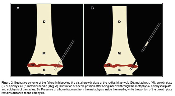
Thus, results described in this study would be the first conducted in mules and first in vivo on equids. However, the efficiency of the technique used for the biopsy of the epiphyseal plate of the radius of mules suffering from ALD was low (n = 3, 23.1%). After perforating the metaphysis, the epiphyseal plate, and part of the epiphysis, the Jamshidi needle was removed; however, in 76.9% of the cases, only a fragment of the distal metaphysis was collected (<a href="#f2">Figure 2</a>).</p>      <p align="center"><a name="f2"><img src="/img/revistas/rccp/v28n1/v28n1a10f2.jpg"></a></p>     <p>&nbsp;</p>      <p>The low effectiveness of this technique was possibly due to the following reasons: first, the needle used is not equipped to cut the cylindrical piece of tissue from the epiphysis. The fact that physeal dysplasia occurs mainly in the proximal area of the epiphyseal plate (Jeffcott and Henson, 1998) may have determined for the piece of epiphyseal plate to remain attached to the epiphysis, since the union between this and the metaphysis might be compromised. This may explain the greater proportion of samples collected from limbs treated with transphyseal staples, compared to limbs treated with HCPTE. The fact that the convex surface of the limb presents a milder physeal dysplasia than the concave surface provides better attachment between the epiphyseal plate and the metaphyseal fragment in this area, resulting in a higher success rate for biopsies taken from the convex surface. Collection of only one biopsy without any repetitions may also have influenced the low success rate of the technique used.</p>      <p>Furthermore, the difficulty in processing the biopsy material collected from the epiphyseal plate was related to the size of the fragment collected (approximately 8 mm long and 1 mm diameter). Besides, the number of sections taken from the material was limited. An average of four cuts per fragment was obtained, but all of them were broken. Finally, it was observed that the low efficiency in collecting the samples and difficulties in their processing were limiting factors, thus we do not recommend the use of the Jamshidi needle as a biopsy technique for epiphyseal plate in mules.    <p>      <p>Histopathological findings of biopsies 1 and 2 indicate a serious physeal dysplasia, since loss of microstructure of the epiphyseal plate was observed, but it was more evident in the material from biopsy 2 due to a possible occurrence of chondrocyte apoptosis. The findings in biopsy 3 are also characteristic of physeal dysplasia and agree with the observations made by Gee <i>et al.</i> (2005) on epiphyseal plates of the metatarsal III in foals. Likewise, Jeffcott and Henson (1998) also described the formation of clusters of chondrocytes in horses; however, the lesions in the epiphyseal plate of the mules were stronger when related to those described in foals.</p>      ]]></body>
<body><![CDATA[<p>The lesions found in the concave side of the limb (biopsies 1 and 2) had a stronger physeal dysplasia, even though the biopsy taken from the convex side came from a limb in which the angular deviation was greater. The difference in cellular activity of the chondrocytes in both sides of the epiphyseal plate was also observed during the induction of ALD in rabbits (Aykut <i>et al.,</i> 2005). The presence of more serious lesions in the concave side of the limbs may have caused the difference in growth dynamics between both sides of the limb, inducing the occurrence of ALD in these mules, as related by Witte and Hunt (2009). Biopsies of both sides of the same epiphyseal plate would be necessary to confirm this observation.</p>      <p>In conclusion, the biopsy technique used was not effective, but allowed to obtain useful information on physeal dysplasia in the distal epiphyseal plate of the radius of mules with carpus ALD. These histopathological findings corroborate those diagnosed elsewhere in horses.</p>      <p>&nbsp;</p>     <p><font size="3"><b>Acknowledgments</b></font></p>      <p>The authors acknowledge CNPq and CAPES/ PEC-PG, Brazil for funding this study, the Federal University of Minas Gerais (UFMG) and the sustainability project 2013-2014 from CODI, Universidad de Antioquia.</p>      <p>&nbsp;</p>     <p><font size="3"><b>Conflicts of interest</b></font></p>      <p>The authors declare they have no conflicts of interest with regard to the work presented in this report.</p>        <p>&nbsp;</p>   <hr size="1" noshade="noshade">     <p><a href="#0" a name="ob">&curren;</a> To cite this article: Zambelli Loiacono B, Mart&iacute;nez Aranzales JR, Silveira Alves GE. Evaluation of a biopsy technique of the epiphyseal plate radius of mules with angular deformity of surgical type: case study. Rev Colomb Cienc Pecu 2015; 28:93-97.</p> <hr size="1" noshade="noshade">      ]]></body>
<body><![CDATA[<p>&nbsp;</p>     <p><font size="3"><b>References</b></font></p>      <!-- ref --><p>Aykut US, Yazici M, Kandemir U, Gedikoglu G, Aksoy MC, Cil A, Surat A. The effect of temporary hemiepiphyseal stapling on the growth plate: a radiologic and immunohistochemical study in rabbits. J Pediatr Orthop 2005; 25:336-341.    &nbsp;&nbsp;&nbsp;&nbsp;&nbsp;&nbsp;&nbsp;&nbsp;[&#160;<a href="javascript:void(0);" onclick="javascript: window.open('/scielo.php?script=sci_nlinks&ref=000075&pid=S0120-0690201500010001000001&lng=','','width=640,height=500,resizable=yes,scrollbars=1,menubar=yes,');">Links</a>&#160;]<!-- end-ref --><p>     <!-- ref --><p>Bramlage LR, Auer JA. Diagnosis, assessment, and treatment strategies for angular limb deformities in the foal. Clin Techn Equine Prac 2006; 5:259-269.    &nbsp;&nbsp;&nbsp;&nbsp;&nbsp;&nbsp;&nbsp;&nbsp;[&#160;<a href="javascript:void(0);" onclick="javascript: window.open('/scielo.php?script=sci_nlinks&ref=000077&pid=S0120-0690201500010001000002&lng=','','width=640,height=500,resizable=yes,scrollbars=1,menubar=yes,');">Links</a>&#160;]<!-- end-ref --></p>     <!-- ref --><p>Gee EK, Firth EC, Morel PCH, Fennessy PF, Grace ND, Mogg TD. Enlargements of the distal third metacarpus and metatarsus in Thoroughbred foals at pasture from birth to 160 days of age. N Zeal Vet J 2005; 53:438-447.    &nbsp;&nbsp;&nbsp;&nbsp;&nbsp;&nbsp;&nbsp;&nbsp;[&#160;<a href="javascript:void(0);" onclick="javascript: window.open('/scielo.php?script=sci_nlinks&ref=000079&pid=S0120-0690201500010001000003&lng=','','width=640,height=500,resizable=yes,scrollbars=1,menubar=yes,');">Links</a>&#160;]<!-- end-ref --></p>     <!-- ref --><p>Jeffcott LB, Henson FM. Studies on growth cartilage in the horse and their application to aetiopathogenesis of dyschondroplasia (osteochondrosis). Vet J 1998; 156:177-192.    &nbsp;&nbsp;&nbsp;&nbsp;&nbsp;&nbsp;&nbsp;&nbsp;[&#160;<a href="javascript:void(0);" onclick="javascript: window.open('/scielo.php?script=sci_nlinks&ref=000081&pid=S0120-0690201500010001000004&lng=','','width=640,height=500,resizable=yes,scrollbars=1,menubar=yes,');">Links</a>&#160;]<!-- end-ref --></p>     ]]></body>
<body><![CDATA[<!-- ref --><p>Loiacono BZ, Aranzales JRM, Faleiros RR, Alves GES. Deformidade angular adquirida no carpo de muares: diagn&oacute;stico, incid&ecirc;ncia e tratamento. Ci&ecirc;ncia Rural 2012; 42:1855-1860.    &nbsp;&nbsp;&nbsp;&nbsp;&nbsp;&nbsp;&nbsp;&nbsp;[&#160;<a href="javascript:void(0);" onclick="javascript: window.open('/scielo.php?script=sci_nlinks&ref=000083&pid=S0120-0690201500010001000005&lng=','','width=640,height=500,resizable=yes,scrollbars=1,menubar=yes,');">Links</a>&#160;]<!-- end-ref --></p>     <!-- ref --><p>Witte S, Hunt R. A review of angular limb deformities. Equine Vet Educ 2009; 21:378-387.    &nbsp;&nbsp;&nbsp;&nbsp;&nbsp;&nbsp;&nbsp;&nbsp;[&#160;<a href="javascript:void(0);" onclick="javascript: window.open('/scielo.php?script=sci_nlinks&ref=000085&pid=S0120-0690201500010001000006&lng=','','width=640,height=500,resizable=yes,scrollbars=1,menubar=yes,');">Links</a>&#160;]<!-- end-ref --></p>  </p> </p> </p> </p> </p> </p> </font>      ]]></body><back>
<ref-list>
<ref id="B1">
<nlm-citation citation-type="journal">
<person-group person-group-type="author">
<name>
<surname><![CDATA[Aykut]]></surname>
<given-names><![CDATA[US]]></given-names>
</name>
<name>
<surname><![CDATA[Yazici]]></surname>
<given-names><![CDATA[M]]></given-names>
</name>
<name>
<surname><![CDATA[Kandemir]]></surname>
<given-names><![CDATA[U]]></given-names>
</name>
<name>
<surname><![CDATA[Gedikoglu]]></surname>
<given-names><![CDATA[G]]></given-names>
</name>
<name>
<surname><![CDATA[Aksoy]]></surname>
<given-names><![CDATA[MC]]></given-names>
</name>
<name>
<surname><![CDATA[Cil]]></surname>
<given-names><![CDATA[A]]></given-names>
</name>
<name>
<surname><![CDATA[Surat]]></surname>
<given-names><![CDATA[A]]></given-names>
</name>
</person-group>
<article-title xml:lang="en"><![CDATA[The effect of temporary hemiepiphyseal stapling on the growth plate: a radiologic and immunohistochemical study in rabbits]]></article-title>
<source><![CDATA[J Pediatr Orthop]]></source>
<year>2005</year>
<volume>25</volume>
<page-range>336-341</page-range></nlm-citation>
</ref>
<ref id="B2">
<nlm-citation citation-type="journal">
<person-group person-group-type="author">
<name>
<surname><![CDATA[Bramlage]]></surname>
<given-names><![CDATA[LR]]></given-names>
</name>
<name>
<surname><![CDATA[Auer]]></surname>
<given-names><![CDATA[JA]]></given-names>
</name>
</person-group>
<article-title xml:lang="en"><![CDATA[Diagnosis, assessment, and treatment strategies for angular limb deformities in the foal]]></article-title>
<source><![CDATA[Clin Techn Equine Prac]]></source>
<year>2006</year>
<volume>5</volume>
<page-range>259-269</page-range></nlm-citation>
</ref>
<ref id="B3">
<nlm-citation citation-type="journal">
<person-group person-group-type="author">
<name>
<surname><![CDATA[Gee]]></surname>
<given-names><![CDATA[EK]]></given-names>
</name>
<name>
<surname><![CDATA[Firth]]></surname>
<given-names><![CDATA[EC]]></given-names>
</name>
<name>
<surname><![CDATA[Morel]]></surname>
<given-names><![CDATA[PCH]]></given-names>
</name>
<name>
<surname><![CDATA[Fennessy]]></surname>
<given-names><![CDATA[PF]]></given-names>
</name>
<name>
<surname><![CDATA[Grace]]></surname>
<given-names><![CDATA[ND]]></given-names>
</name>
<name>
<surname><![CDATA[Mogg]]></surname>
<given-names><![CDATA[TD]]></given-names>
</name>
</person-group>
<article-title xml:lang="en"><![CDATA[Enlargements of the distal third metacarpus and metatarsus in Thoroughbred foals at pasture from birth to 160 days of age]]></article-title>
<source><![CDATA[N Zeal Vet J]]></source>
<year>2005</year>
<volume>53</volume>
<page-range>438-447</page-range></nlm-citation>
</ref>
<ref id="B4">
<nlm-citation citation-type="journal">
<person-group person-group-type="author">
<name>
<surname><![CDATA[Jeffcott]]></surname>
<given-names><![CDATA[LB]]></given-names>
</name>
<name>
<surname><![CDATA[Henson]]></surname>
<given-names><![CDATA[FM]]></given-names>
</name>
</person-group>
<article-title xml:lang="en"><![CDATA[Studies on growth cartilage in the horse and their application to aetiopathogenesis of dyschondroplasia (osteochondrosis)]]></article-title>
<source><![CDATA[Vet J]]></source>
<year>1998</year>
<volume>156</volume>
<page-range>177-192</page-range></nlm-citation>
</ref>
<ref id="B5">
<nlm-citation citation-type="journal">
<person-group person-group-type="author">
<name>
<surname><![CDATA[Loiacono]]></surname>
<given-names><![CDATA[BZ]]></given-names>
</name>
<name>
<surname><![CDATA[Aranzales]]></surname>
<given-names><![CDATA[JRM]]></given-names>
</name>
<name>
<surname><![CDATA[Faleiros]]></surname>
<given-names><![CDATA[RR]]></given-names>
</name>
<name>
<surname><![CDATA[Alves]]></surname>
<given-names><![CDATA[GES]]></given-names>
</name>
</person-group>
<article-title xml:lang="pt"><![CDATA[Deformidade angular adquirida no carpo de muares: diagnóstico, incidência e tratamento]]></article-title>
<source><![CDATA[Ciência Rural]]></source>
<year>2012</year>
<volume>42</volume>
<page-range>1855-1860</page-range></nlm-citation>
</ref>
<ref id="B6">
<nlm-citation citation-type="journal">
<person-group person-group-type="author">
<name>
<surname><![CDATA[Witte]]></surname>
<given-names><![CDATA[S]]></given-names>
</name>
<name>
<surname><![CDATA[Hunt]]></surname>
<given-names><![CDATA[R]]></given-names>
</name>
</person-group>
<article-title xml:lang="en"><![CDATA[A review of angular limb deformities]]></article-title>
<source><![CDATA[Equine Vet Educ]]></source>
<year>2009</year>
<volume>21</volume>
<page-range>378-387</page-range></nlm-citation>
</ref>
</ref-list>
</back>
</article>
