<?xml version="1.0" encoding="ISO-8859-1"?><article xmlns:mml="http://www.w3.org/1998/Math/MathML" xmlns:xlink="http://www.w3.org/1999/xlink" xmlns:xsi="http://www.w3.org/2001/XMLSchema-instance">
<front>
<journal-meta>
<journal-id>0120-6230</journal-id>
<journal-title><![CDATA[Revista Facultad de Ingeniería Universidad de Antioquia]]></journal-title>
<abbrev-journal-title><![CDATA[Rev.fac.ing.univ. Antioquia]]></abbrev-journal-title>
<issn>0120-6230</issn>
<publisher>
<publisher-name><![CDATA[Facultad de Ingeniería, Universidad de Antioquia]]></publisher-name>
</publisher>
</journal-meta>
<article-meta>
<article-id>S0120-62302013000300010</article-id>
<title-group>
<article-title xml:lang="en"><![CDATA[Automated design of customized implants]]></article-title>
<article-title xml:lang="es"><![CDATA[Diseño automatizado de implantes personalizados]]></article-title>
</title-group>
<contrib-group>
<contrib contrib-type="author">
<name>
<surname><![CDATA[Chulvi]]></surname>
<given-names><![CDATA[Vicente]]></given-names>
</name>
<xref ref-type="aff" rid="A01"/>
<xref ref-type="aff" rid="A03"/>
</contrib>
<contrib contrib-type="author">
<name>
<surname><![CDATA[Cebrian-Tarrasón]]></surname>
<given-names><![CDATA[David]]></given-names>
</name>
<xref ref-type="aff" rid="A01"/>
</contrib>
<contrib contrib-type="author">
<name>
<surname><![CDATA[Sancho]]></surname>
<given-names><![CDATA[Álex]]></given-names>
</name>
<xref ref-type="aff" rid="A02"/>
</contrib>
<contrib contrib-type="author">
<name>
<surname><![CDATA[Vidal]]></surname>
<given-names><![CDATA[Rosario]]></given-names>
</name>
<xref ref-type="aff" rid="A01"/>
</contrib>
</contrib-group>
<aff id="A01">
<institution><![CDATA[,Universitat Jaume I Deparatamento de Ingeniería Mecánica y Construcción ]]></institution>
<addr-line><![CDATA[Castellón ]]></addr-line>
<country>España</country>
</aff>
<aff id="A02">
<institution><![CDATA[,Fundación ASCAMM  ]]></institution>
<addr-line><![CDATA[Barcelona ]]></addr-line>
<country>España</country>
</aff>
<aff id="A03">
<institution><![CDATA[,Universitat Jaume I  ]]></institution>
<addr-line><![CDATA[ ]]></addr-line>
</aff>
<pub-date pub-type="pub">
<day>00</day>
<month>09</month>
<year>2013</year>
</pub-date>
<pub-date pub-type="epub">
<day>00</day>
<month>09</month>
<year>2013</year>
</pub-date>
<numero>68</numero>
<fpage>95</fpage>
<lpage>103</lpage>
<copyright-statement/>
<copyright-year/>
<self-uri xlink:href="http://www.scielo.org.co/scielo.php?script=sci_arttext&amp;pid=S0120-62302013000300010&amp;lng=en&amp;nrm=iso"></self-uri><self-uri xlink:href="http://www.scielo.org.co/scielo.php?script=sci_abstract&amp;pid=S0120-62302013000300010&amp;lng=en&amp;nrm=iso"></self-uri><self-uri xlink:href="http://www.scielo.org.co/scielo.php?script=sci_pdf&amp;pid=S0120-62302013000300010&amp;lng=en&amp;nrm=iso"></self-uri><abstract abstract-type="short" xml:lang="en"><p><![CDATA[This paper presents a model capable of design a customized cranial implant directly from a medical imaging process, whose output will be a file in a manufacture system recognizable format. The proposed system has been created by linking two computer prototypes developed during the present research and studying the inner and outer related technologies. The core of the model is the KBS (Knowledge Based System) technology, able to store and manage medical data, as well as designer knowledge, in order to use this information in the implant design process. The aim of this project is to obtain a tool to improve the design process, the biocompatibility with patient and reduce final costs, which can be operated without necessity of wide knowledge from the user.]]></p></abstract>
<abstract abstract-type="short" xml:lang="es"><p><![CDATA[Este artículo muestra un modelo que se postula como capaz para el diseño de un implante craneal personalizado directamente desde el proceso de toma de imágenes médica. La salida de este proceso de diseño se conseguirá en un formato capaz de ser reconocido por el sistema de manufactura. El sistema propuesto se ha creado a partir de la unión de dos prototipos informáticos desarrollados durante la presente investigación, y a través del estudio de las tecnologías relacionadas o circundantes. El núcleo del modelo en la tecnología basada en el conocimiento (KBS), que debe permitir de almacenar y gestionar datos médicos y de diseño para poder aplicar dichos conocimientos durante el proceso de diseño del implante. El objeto de este proyecto es el de obtener una herramienta para mejorar el proceso de diseño, la biocompatibilidad con el paciente y reducir los costes finales, y que pueda ser operado sin necesidad del conocimiento completo de todas sus fases por el usuario.]]></p></abstract>
<kwd-group>
<kwd lng="en"><![CDATA[Cranial implants]]></kwd>
<kwd lng="en"><![CDATA[KBS]]></kwd>
<kwd lng="en"><![CDATA[CAD]]></kwd>
<kwd lng="en"><![CDATA[intelligent design]]></kwd>
<kwd lng="en"><![CDATA[rapid prototyping]]></kwd>
<kwd lng="es"><![CDATA[Implantes craneales]]></kwd>
<kwd lng="es"><![CDATA[KBS]]></kwd>
<kwd lng="es"><![CDATA[CAD]]></kwd>
<kwd lng="es"><![CDATA[diseño inteligente]]></kwd>
<kwd lng="es"><![CDATA[prototipado rápido]]></kwd>
</kwd-group>
</article-meta>
</front><body><![CDATA[ <font face="Verdana" size="2">      <p align="right"><b>ART&Iacute;CULO ORIGINAL</b></p>     <p align="right">&nbsp;</p>     <p align="center"><font size="4"> <b>Automated design of customized implants</b></font></p>     <p align="center">&nbsp;</p>     <p align="center"><font size="3"> <b>Dise&ntilde;o automatizado de implantes personalizados</b></font></p>     <p align="center">&nbsp;</p>     <p align="center">&nbsp;</p>     <p> <i><b>Vicente Chulvi<sup>*1</sup>, David Cebrian-Tarras&oacute;n<sup>1</sup>, &Aacute;lex Sancho<sup>2</sup>, Rosario Vidal<sup>1</sup></b></i></p>       <p><sup>1</sup>Deparatamento  de Ingenier&iacute;a Mec&aacute;nica y Construcci&oacute;n, Universitat Jaume I. 12071. Castell&oacute;n, Espa&ntilde;a.</p>      ]]></body>
<body><![CDATA[<p><sup>2</sup>Fundaci&oacute;n  ASCAMM. 08290. Cerdanyola  del Vall&egrave;s, Barcelona,  Espa&ntilde;a.</p>      <p><sup>*</sup>Autor de correspondencia: tel&eacute;fono: + 34 + 964 + 72 81 13, fax: + 34 + 964 + 72 81 06, correo  electr&oacute;nico: <a href="mailto:chulvi@uji.es.co">chulvi@uji.es.co</a> (V. Chulvi)</p>      <p>&nbsp;</p>     <p align="center">(Recibido el 1 de mayo de 2011. Aceptado el 5 de agosto de 2013)</p>     <p align="center">&nbsp;</p>     <p align="center">&nbsp;</p> <hr noshade size="1">      <p><font size="3"><b>Abstract</b></font></p>      <p>This  paper presents a model capable of design a customized cranial implant directly  from a medical imaging process, whose output will be a file in a manufacture  system recognizable format.</p>     <p>The  proposed system has been created by linking two computer prototypes developed  during the present research and studying the inner and outer related  technologies. The core of the model is the KBS (Knowledge Based System)  technology, able to store and manage medical data, as well as designer  knowledge, in order to use this information in the implant design process. The  aim of this project is to obtain a tool to improve the design process, the  biocompatibility with patient and reduce final costs, which can be operated  without necessity of wide knowledge from the user.</p>       <p><i>Keywords:</i> Cranial implants, KBS, CAD, intelligent design, rapid prototyping</p>  <hr noshade size="1">      ]]></body>
<body><![CDATA[<p><font size="3"><b>Resumen</b></font></p>     <p>Este  art&iacute;culo muestra un modelo que se postula como capaz para el dise&ntilde;o de un  implante craneal  personalizado directamente desde el proceso de toma de im&aacute;genes m&eacute;dica. La  salida de este proceso de dise&ntilde;o se conseguir&aacute; en un formato capaz de ser  reconocido por el sistema de manufactura. </p>     <p>El  sistema propuesto se ha creado a partir de la uni&oacute;n de dos prototipos  inform&aacute;ticos desarrollados durante la presente investigaci&oacute;n, y a trav&eacute;s del  estudio de las tecnolog&iacute;as relacionadas o circundantes. El n&uacute;cleo del modelo en  la tecnolog&iacute;a basada en el conocimiento (KBS), que debe permitir de almacenar y  gestionar datos m&eacute;dicos y de dise&ntilde;o para poder aplicar dichos conocimientos  durante el proceso de dise&ntilde;o del implante. El objeto de este proyecto es el de  obtener una herramienta para mejorar el proceso de dise&ntilde;o, la biocompatibilidad  con el paciente y reducir los costes finales, y que pueda ser operado sin  necesidad del conocimiento completo de todas sus fases por el usuario.</p>      <p><i>Palabras clave: </i>Implantes craneales, KBS, CAD, dise&ntilde;o inteligente, prototipado r&aacute;pido</p>  <hr noshade size="1">      <p>&nbsp;</p>     <p><font size="3"><b>Introduction</b></font></p>      <p>The close future paradigm of medical industry will be  surgical operations in which implants are fully adapted to the particular  requirements of each patient. This vision will be fulfilled when new  biomaterials are developed and new technologies for design and rapid  manufacturing of such implants are implemented &#91;1&#93;. Patients exert more and  more pressure in order to improve operation, duration and aesthetics of  implants, and so are scientific working. Nowadays the techniques for the  manufacture of implants are based on individual experience and point of view of  surgeons and mechanics. Low resolution of current medical data acquisition  techniques together with manual mechanizing result in slow and expensive final  results &#91;1-3&#93;.</p>       <p>The first reference about custom designed implants was  provided by Dooley et al. &#91;4&#93;. It was a theoretical approach to intelligent  systems to develop orthopaedic implants which could be incorporated into a  design and manufacture system. A few years later Whittaker &#91;5&#93; presented a  model in an attempt to automatize the process, which results in a slight  improvement but not an automated process. Despite that, this approach is not so  far from present models &#91;6-8&#93;.</p>       <p>The procedure to automatize the process of customized  craniofacial implant design begins with medical data acquisition using the most  common techniques of 3D and 4D-image projection. Data provided with CT  (Computerized Tomography) or MRI (Magnetic Resonance Imaging) are represented  in a set of plane images (2D). Each image represents a transverse slice of the  exploration area. Therefore, surgeons have to examine simultaneously a  considerable amount of images in order to rebuild the 3D shape in their mind,  what means an added difficulty to design. Rebuilding 3D shape from 2D medical data  is a field of study under development &#91;9&#93;. The last step through the procedure  of customized implant design consists in the development of an output file  through CAD/CAM/CAE (Computer Aided Design / Computer Aided Manufacturing/  Computer Aided Engineering) technology &#91;10&shy;12&#93;, which can be sent to a  manufacturing machine. However, the incorporation of relatively new  technologies to the automated design process, like FEM (Finite Element Method)  simulation &#91;13&#93;, tissue engineering scaffolds &#91;14-15&#93; and RP (Rapid  Prototyping) &#91;16-17&#93;, have turned it into a novel field, where different  disciplines take part: medicine, computing, design and engineering.</p>       <p>
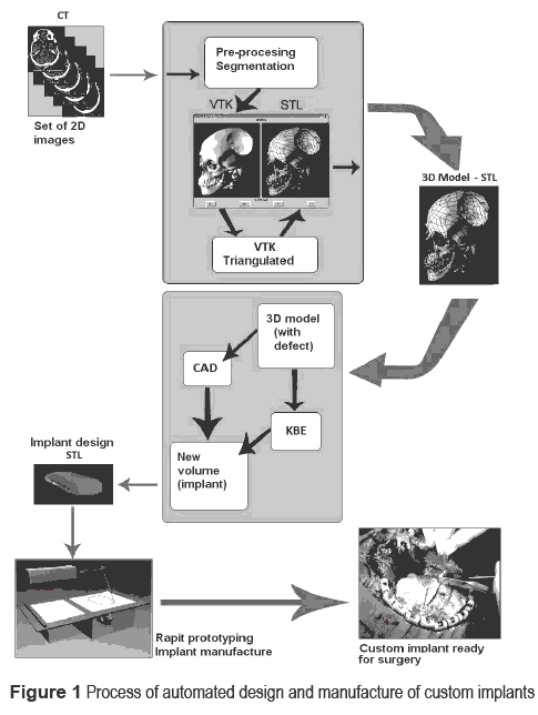
Although research on custom implants is narrowing its scope  to focus on specific types of implants, no studies have been found that deal  with specifically the automated design of cranio&shy;facial implants. This fact  shows the difficulty to create a system that considers all the steps of the  process. So, the aim of the present study is to deal with the technical design  and implementation of software, which accelerates the medical implants' design  process through the use of CAD (Computer Aided Design) and KBE-based tools  (Knowledge Based Engineering). The purpose is to obtain a remarkable  improvement both for patients and for service suppliers. The experimental  results presented in this article show two computer applications, the first of  them is aimed to create an STL (Stereolithography) format file from a CT scan  file, and the second one develops an automated model of a volume to cover  existing holes on a surface, beginning with a surface that could be the  previous STL file.</p>        ]]></body>
<body><![CDATA[<p>&nbsp;</p>     <p><font size="3"><b>Materials and methods</b></font></p>        <p>This research was based on the previous study of existing  softwares (such as MIMICS, 3D Slicer, ImLib3D, MITK, OsiriX and Visualization  Toolkit) and a study about materials used for implant manufacture and the  existing techniques to carry it out.</p>       <p>Medical imaging is based on projection technologies. CT  consists in an X-ray beam that crosses the analysed body and its projection is  detected varying the attenuation in function of the density of the tissues gone  through. Soft tissues will appear dark and bones will appear light. The result  is a transversal section, a 2D projection of tissues along X-rays direction.  MRI is a similar technique, but it uses a magnetic field instead of an X-ray  beam. The result is also a transversal section 2D image. Nevertheless, one  medical image has no sense on its own, but different images of the same patient  can be related to each other and, moreover, have associated a set of patient  data. Current image formats (TIFF, JPEG or GIF) are not enough. For that  reason, it was proposed that the image processing of the present research  follows the DICOM (Digital Imaging and Communications in Medicine) standard.  This standard describes the files format and the specification of patient  primordial data in an image, just as the required header, showing a common  language to the different medical systems. Images come enclosed with  measurements, calculations and other relevant descriptive information, as  patient's name, birth date, data acquisition equipment, image size, pixel data,  contrast used, etc.</p>       <p>The first computer application was defined as a prototype  for reconstruction of a 3D image from a set of 2D DICOM images and conversion  of VTK (Visualization Toolkit) format to an STL, which is recognizable for CAD  programs. 3D Slicer is the most well known open source application found for  visualization, medical data segmentation and 3D medical images reconstruction from  DICOM files, and it has been developed over the graphic library OpenGL, using  VTK for image processing. MIMICS (Materialised Interactive Medical Image</p>       <p>Control System) is an interactive commercial tool for the  visualization and segmentation of CT images as well as MRI images and 3D  rendering of objects. VTK is an open source library for computer visualization  of images and graphics, at a higher level of abstraction than other rendering  libraries, OpenGL or PEX. VTK applications can be directly written in C++, Tcl,  Java or Python. Design and implementation of the library have been strongly  influenced by oriented-to-objects programming principles. Operations on two or  three dimensions and model generation could be carried out by this library with  a few lines of code.</p>       <p>The technologies needed for the development ofthe second  computer application, which must be able to realize an automated design of an  implant, are KBS and CAD. There exist software applications based jointly in  KBE and CAD. Their respective KBS modules only use the systems capacities  associated to control of geometry variables, reutilization of geometry  information and control and management of rules and restrictions. This feature  provides the advantage of no excessive expert knowledge in KBS required for  application development in an efficient manner. The present application was  developed using the CATIA software suite, which has a module structure with  specialized functions groups. It allows development applications based on both  KBS and CAD, integrating the modules through APIs (Application Programming  Interface) program libraries. In CATIA, the ''Knowledgeware'' module  groups the functions related with knowledge (e.g., parameterize elements,  create formulas or rules, check, etc.), and allows it to expand design features  with knowledge elements. CATIA disposes of an FEM module (in the CAE section),  which gives the opportunity to analyse the response and evolution of a physical  system under simulated conditions of load, initial and boundary conditions,  assuring the integration with the KBS. In the particular case of implants, FEM  is used for strain an effort analysis from a given force field and evaluation  of bone growth. Furthermore, there is the chance to predict the facial  appearance after surgery through an FEM model applied to soft tissue.</p>       <p>The output of CATIA is presented in STL format, which  allows to be directly transferred into an RP (Rapid Prototiping) machine. RP is  the name given to the technology set used for three-dimension manufacture, directly  from CAD data source. This system drives to a design improvement, as well as  time manufacture reduction, better material use and excellent tolerances. A  review of RP techniques applicable to implant manufacture can be seen in past  research &#91;18&#93;.</p>        <p>&nbsp;</p>      <p><font size="3"><b>Results (development of the model)</b></font></p>        ]]></body>
<body><![CDATA[<p><a href="#Figura1">Figure 1</a> illustrates the global process of automated design  and manufacture of customized implants, as has been considered in the present work.  Processes enclosed inside the shaded boxes represent the two software  prototypes designed during the project development. The procedure of customized  craniofacial implant design began with medical data acquisition with the  already mentioned technology of CT. This process provided a set of  two-dimension images (transversal slices). In order to reconstruct the  three-dimension model, medical images must be treated. There exist two main  steps in order to obtain the desired results, that we named pre&shy;processing and  segmentation.</p>      <p align="center"><a name="Figura1"></a><img src="/img/revistas/rfiua/n68/n68a10i01.gif"></p>      <p>During  the first step of the pre-processing, there was an attempt to improve the image  as much as possible, since images from CT have a considerable quantity of  ''noise''. Transverse slices are frequently performed in all organs and  tissues within the exploration range. A previous process, segmentation, is  needed to select the objects of the organ or tissue considered whose  transformation to 3D is desired. Tissues (bone, skin, encephalic mass, etc.)  can usually be differentiated by the grey scale represented in the medical  image, in the segmentation process it is usual to select a point in a region to  be transformed, and its value in the grey scale is used as a reference value. </p>       <p>Once the area to be treated was selected, the procedure  continued with the 3D model building. Each transversal slice image is composed  by a bidimensional matrix of pixels, each one having a (x, y) position. If  individual images are overlaid, the pixels in every image for given (x, y)  coordinates will be aligned. Thus, pixels can be labelled with three  dimensional coordinates. They maintain their original (x, y) coordinates and  take a new z coordinate. The z coordinate is simply the slice number to which  pixels are associated. The concept of slices is eliminated and all pixels of  all the images are represented in a unique three-dimensional image. The new  volume unit so created is called voxel (which is a portmanteau word from  volumetric and pixel), which dimensions are 1 pixel wide x 1 pixel height x 1  pixel length.</p>       <p>Since most of the applications in open source capable of  treating medical images and convert those into a 3D geometry are based on VTK,  the next step was to develop a module to convert the VTK file into an STL file,  that CAD programs could recognise it. This development began with the  installation of the VTK library, which requires the compilation of the VTK  source code (in order to generate C++ libraries) and VTK wrapping code (to  generate executables in Java, Tcl and Python). TKinter library - DLL  (Dynamic-link library) - for Python, which is an open source library used to  develop user interfaces, has been used to develop the interface.</p>       <p>The first action carried out by the algorithm is to create  a vtkTriangleFilter type object. This object has a method that allows  triangulating a VTK model. Previous to the creation of the STL model, the  triangulation of VTK object must be done since STL format only accepts  triangular faced models. Subsequently, a vtkSTLWriter type object was created,  which has a function that allows converting a triangulated VTK model to STL  format.</p>       <p>
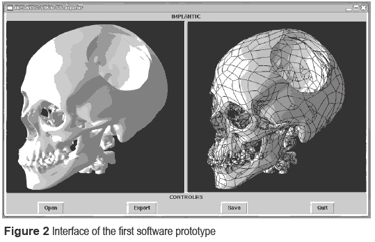
The first computerprogram developmentincludes the steps of  pre-processing, segmentation, 3D building and file format conversion. This is,  in general lines, a prototype to recognize data and convert them in a  recognizable format for CAD software. As shown in the <a href="#Figura2">figure 2</a>, the interface  of the prototype has two graphic windows, where the model can be visualized  both in VTK format and STL (triangulated) format. Once the STL model appears in  the corresponding graphic window, it could be saved in that format and be  retrieved in any CAD software.</p>      <p align="center"><a name="Figura2"></a><img src="/img/revistas/rfiua/n68/n68a10i02.gif"></p>        <p>
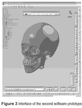
The next step consisted in developing a second computer  program prototype through CATIA software, which models automatically a volume  to cover a hole in an object. The use of the 3D STL model from the first  computer program prototype showed the possibility to create a custom implant  design adapted to the patient's anatomy, improving structural, functional and  aesthetic biocompatibility. The modelling of the prototype requires, firstly,  the manual definition and testing of all the steps of the modelling in order to  ensure that is working correctly. These steps are: locate the hole(s) on the  surface by means of object geometrical boundaries analysis; set the gap from  the hole boundaries that will be the outline of a new area; create the area  that will become the outer surface of the implant, considering the curvature  radius of the hole's zone; and extrude a volume from the surface. Once the  manual process is done, the automation of these steps is done through the  knowledge management module. Most of the instructions used by the program are  developed through APIs. Once the modelling process had been defined,  instructions are recorded into a macro in a code recognised by CATIA by means  of language VBScript. In order to conclude the customized design for final  user, some geometrical rules and restrictions can be included in the prototype  by means of databases, like ''gap between hole (cranium defect) and  modelled volume (implant)'' and ''thickness''. The result of  running the program is shown in <a href="#Figura3">figure 3</a>, as it is viewed in CATIA's interface.  Finally, the resulting implant must be exported into a format compatible with  the manufacturing process, rapid prototyping in our case. The format used in  this case is STL, an instruction has been added to the algorithm in order to  save the implant in a file ready to be transferred to an RP machine.</p>      <p align="center"><a name="Figura3"></a><img src="/img/revistas/rfiua/n68/n68a10i03.gif"></p>      ]]></body>
<body><![CDATA[<p>&nbsp;</p>     <p><font size="3"><b>Discussions</b> </font></p>      <p>The model, as shown in figure 1, has been built from two  software prototypes, which have been linked for the final result. The first  software prototype was to convert medical images to a 3D model in STL format. This  application improves those existing in the market since it not only  reconstructs a 3D image from a set of 2D image, but also prepares it for being  used in next design steps. Therefore, the advantage of the first software  prototype consists in time reduction and less human manipulation. The second  software prototype comprises the automated implant design process. Initial  tests show that the creation of a cover for a hole (this is, an implant) over a  cranium is possible in an automatic mode by means of KBS. However, medical  knowledge should be broadened in order to optimize the KBS database, which  should make it possible to differentiate natural skull holes from defects,  recognize cranial zone affected, set implant width and mechanical properties in  function of cranial zone, etc.</p>       <p>
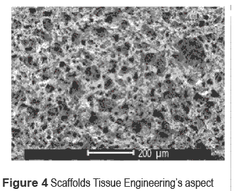
Along with the advantages mentioned above, RP allows  raising the proposal of using the technology of tissue engineering scaffolds,  in which an implant is used as a ''scaffold'' that will work as a base  to regenerate the tissue &#91;19&#93;. This ''scaffold'' form can be achieved  with RP techniques, which allow to control independently the porosity and the  pores' size by varying the working parameters of the machine. <a href="#Figura4">Figure 4</a> shows  the aspect of an application of this kind of technique. Within this same  proposal, the use of biomaterials can be taken into consideration, creating  implants combining bio-stable polymers with bio-degradable polymers, that at  the same time the regenerated tissue fills the existing pores in the implant  and new pores are created in order that the tissue can continue its expansion.  The material proposed as a bio-stable polymer is PEEK  (Poli-ether-ether-ketone), which is used at present in implant manufacturing  and has been tested in RP manufacturing, and the material proposed as  bio-degradable is HA (Hidroxiapatite), natural bone component that has been  experimentally tested together with PEEK using scaffolds technology &#91;20&#93;.</p>      <p align="center"><a name="Figura4"></a><img src="/img/revistas/rfiua/n68/n68a10i04.gif"></p>          <p>&nbsp;</p>       <p><font size="3"><b>Conclusions</b> </font></p>        <p>The present work presents a process to improve the  automated design and manufacture of customized cranial implants. The results  showed that a computer application for automated design of customized cranial  implants will provide numerous advantages, both for customers as well as for  surgeons, manufacturers and designers. Custom implants will improve functional  and structural biocompatibility, aesthetic, functionality, quality of life, and  they can also provide a less invasive surgery. Automating the process would  lead, ostensibly, to reduction in design and manufacture time and consequently  to lower costs, which provides new market opportunities.</p>       <p>Society in general demands those advantages, and they will  become a serious necessity in a near future. The computer software prototypes  presented in this article, as well as the proposals developed during the  research, establish the essential bases for the final ideal application.</p>      <p>&nbsp;</p>     ]]></body>
<body><![CDATA[<p><font size="3"><b>Acknowledgments</b> </font></p>      <p>The research presented in this paper was partially funded  by the Ministerio de Industria, Turismo y Comercio of Spain (project ref.  FIT-300100- 2006-64) and by the Ministerio de Educaci&oacute;n y Ciencia of Spain  (project ref. DPI2006- 15570-C02).</p>       <p>The authors also gratefully acknowledge the cooperation of  the staff of Plastiasite, S.A., Neos Surgery, S.L. and Fundaci&oacute;n ASCAMM.</p>        <p>&nbsp;</p>       <p><font size="3"><b>References</b> </font></p>      <!-- ref --><p>1. M. Cabrera, J. Burgelmen,  M. Boden, O da Costa, C. Rodr&iacute;guez. <i>eHealth in 2010:  Realising a Knowledge- based Approach to Healthcare in the EU</i>. European Commission  Directorate-General Joint Research Centre Technical Report EUR 21486. Seville,  Spain. 2004.    &nbsp;&nbsp;&nbsp;&nbsp;&nbsp;&nbsp;&nbsp;&nbsp;[&#160;<a href="javascript:void(0);" onclick="javascript: window.open('/scielo.php?script=sci_nlinks&ref=000068&pid=S0120-6230201300030001000001&lng=','','width=640,height=500,resizable=yes,scrollbars=1,menubar=yes,');">Links</a>&#160;]<!-- end-ref --></p>       <!-- ref --><p>2. P. D'Urso, W. Earwaker, T.  Barker, M. Redmond, R. Thompson, D. Effeney, F. Tomlinson. ''Custom  cranioplasty using stereolithography and acrylic''. <i>British Journal of  Plastic Surgery</i>.  Vol. 53. 2000. pp. 200-204.    &nbsp;&nbsp;&nbsp;&nbsp;&nbsp;&nbsp;&nbsp;&nbsp;[&#160;<a href="javascript:void(0);" onclick="javascript: window.open('/scielo.php?script=sci_nlinks&ref=000070&pid=S0120-6230201300030001000002&lng=','','width=640,height=500,resizable=yes,scrollbars=1,menubar=yes,');">Links</a>&#160;]<!-- end-ref --></p>       <!-- ref --><p>3. H.  Eufinger, C. Rasche, M. Wehmoller, K. Schmiederc, M. Scholzc, S. Weihed, P.  Scherera. ''CAD/CAM  titanium implants for cranioplasty--an evaluation of success and quality of  life of 169 consecutive implants with regard to size and location''. <i>Computer Assisted  Radiology and Surgery</i>. Vol. 1281. 2005. pp. 827-31.    &nbsp;&nbsp;&nbsp;&nbsp;&nbsp;&nbsp;&nbsp;&nbsp;[&#160;<a href="javascript:void(0);" onclick="javascript: window.open('/scielo.php?script=sci_nlinks&ref=000072&pid=S0120-6230201300030001000003&lng=','','width=640,height=500,resizable=yes,scrollbars=1,menubar=yes,');">Links</a>&#160;]<!-- end-ref --></p>       <!-- ref --><p>4. R. Dooley, G. Heimke, A. Dingankar, E. Berg,  E. Kimbrough. ''Automated design and analysis system for design of custom  orthopedic implants''. <i>Proceedings of the first international conference on  Industrial and engineering applications of artificial intelligence and expert  systems</i>.  Vol. 1. 1988. pp. 405-12.    &nbsp;&nbsp;&nbsp;&nbsp;&nbsp;&nbsp;&nbsp;&nbsp;[&#160;<a href="javascript:void(0);" onclick="javascript: window.open('/scielo.php?script=sci_nlinks&ref=000074&pid=S0120-6230201300030001000004&lng=','','width=640,height=500,resizable=yes,scrollbars=1,menubar=yes,');">Links</a>&#160;]<!-- end-ref --></p>       <!-- ref --><p>5. D. Whittaker.  ''Custom-designed hip implants through knowledge-based engineering''. <i>Materials &amp;  Design</i>. Vol.  12. 1991. pp. 103-4.    &nbsp;&nbsp;&nbsp;&nbsp;&nbsp;&nbsp;&nbsp;&nbsp;[&#160;<a href="javascript:void(0);" onclick="javascript: window.open('/scielo.php?script=sci_nlinks&ref=000076&pid=S0120-6230201300030001000005&lng=','','width=640,height=500,resizable=yes,scrollbars=1,menubar=yes,');">Links</a>&#160;]<!-- end-ref --></p>       <!-- ref --><p>6. O. Tziovaras. <i>Customised  implant design for the knee joint</i>. X Congreso Internacional de Ingenier&iacute;a de Proyectos. Valencia, Espa&ntilde;a. 2006. pp. 630-635.    &nbsp;&nbsp;&nbsp;&nbsp;&nbsp;&nbsp;&nbsp;&nbsp;[&#160;<a href="javascript:void(0);" onclick="javascript: window.open('/scielo.php?script=sci_nlinks&ref=000078&pid=S0120-6230201300030001000006&lng=','','width=640,height=500,resizable=yes,scrollbars=1,menubar=yes,');">Links</a>&#160;]<!-- end-ref --></p>       <!-- ref --><p>7. T. Wu, L.  Fietena, M. Engelhardt. ''Knowledge-based individual implant design based on  reference models for craniofacial reconstruction''. <i>International  Journal of Computer Assisted Radiology and Surgery</i>. Vol. 1. 2006. pp. 259-61.    &nbsp;&nbsp;&nbsp;&nbsp;&nbsp;&nbsp;&nbsp;&nbsp;[&#160;<a href="javascript:void(0);" onclick="javascript: window.open('/scielo.php?script=sci_nlinks&ref=000080&pid=S0120-6230201300030001000007&lng=','','width=640,height=500,resizable=yes,scrollbars=1,menubar=yes,');">Links</a>&#160;]<!-- end-ref --></p>       <!-- ref --><p>8. A. Rose, M.  Klein, T. Krueger. <i>Individual bone implant modeling using planned resection  lines for facial and cranial tumor resection</i>. 4<sup>th</sup> European  Conference of the International Federation for Medical and Biological  Engineering. Ed. Springer Berlin - Heidelberg. Berlin, Germany. Vol. 22. 2009.  pp. 944-947.    &nbsp;&nbsp;&nbsp;&nbsp;&nbsp;&nbsp;&nbsp;&nbsp;[&#160;<a href="javascript:void(0);" onclick="javascript: window.open('/scielo.php?script=sci_nlinks&ref=000082&pid=S0120-6230201300030001000008&lng=','','width=640,height=500,resizable=yes,scrollbars=1,menubar=yes,');">Links</a>&#160;]<!-- end-ref --></p>       <!-- ref --><p>9. J. Gao, W. Xu, Z. Ding.  ''3D finite element mesh generation of complicated tooth model based on CT  slices''.  <i>Computer Methods and Programs in Biomedicine</i>. Vol. 82. 2006. pp. 97-105.    &nbsp;&nbsp;&nbsp;&nbsp;&nbsp;&nbsp;&nbsp;&nbsp;[&#160;<a href="javascript:void(0);" onclick="javascript: window.open('/scielo.php?script=sci_nlinks&ref=000084&pid=S0120-6230201300030001000009&lng=','','width=640,height=500,resizable=yes,scrollbars=1,menubar=yes,');">Links</a>&#160;]<!-- end-ref --></p>       <!-- ref --><p>10. M. Truscott, D. de Beer, G. Booysen, L. Barnard. <i>Bone Development  through CT/CAD/CAM/RP</i>. 10&egrave;mes Assises Europ&eacute;ennes de Prototypage Rapide. AFPR - Association Fran&ccedil;aise  de Prototypage  Rapide. Paris,  France. 2004. pp.107-114&nbsp;&nbsp;&nbsp;&nbsp;&nbsp;&nbsp;&nbsp;&nbsp;[&#160;<a href="javascript:void(0);" onclick="javascript: window.open('/scielo.php?script=sci_nlinks&ref=000086&pid=S0120-6230201300030001000010&lng=','','width=640,height=500,resizable=yes,scrollbars=1,menubar=yes,');">Links</a>&#160;]<!-- end-ref --><!-- ref --><p>11. S.  Singare, L. Yaxiong, L. Dichen, L. Bingheng, H. Sanhu, L. Gang. ''Fabrication of  customised maxillo- facial prothesis using computer-aided design and rapid  prototyping techniques''. <i>Rapid Prototyping Journal</i>. Vol. 12. 2006. pp. 206-13.    &nbsp;&nbsp;&nbsp;&nbsp;&nbsp;&nbsp;&nbsp;&nbsp;[&#160;<a href="javascript:void(0);" onclick="javascript: window.open('/scielo.php?script=sci_nlinks&ref=000087&pid=S0120-6230201300030001000011&lng=','','width=640,height=500,resizable=yes,scrollbars=1,menubar=yes,');">Links</a>&#160;]<!-- end-ref --></p>       <!-- ref --><p>12. L. Hieu, N. Zlatov, J.  Vander, E. Bohez, L. Khanh, P. Binh, P. Oris, Y. Toshev. ''Medical  rapid prototyping applications and methods''. <i>Assembly  Automation</i>.  Vol. 25. 2005. pp. 284-292.    &nbsp;&nbsp;&nbsp;&nbsp;&nbsp;&nbsp;&nbsp;&nbsp;[&#160;<a href="javascript:void(0);" onclick="javascript: window.open('/scielo.php?script=sci_nlinks&ref=000089&pid=S0120-6230201300030001000012&lng=','','width=640,height=500,resizable=yes,scrollbars=1,menubar=yes,');">Links</a>&#160;]<!-- end-ref --></p>       <!-- ref --><p>13. J. Ho, S. Kleiven.  ''An automatic method to generate patient specific finite element head  model''.  <i>Journal of Biomechanics</i>. Vol. 39. 2006. pp. S428-S.    &nbsp;&nbsp;&nbsp;&nbsp;&nbsp;&nbsp;&nbsp;&nbsp;[&#160;<a href="javascript:void(0);" onclick="javascript: window.open('/scielo.php?script=sci_nlinks&ref=000091&pid=S0120-6230201300030001000013&lng=','','width=640,height=500,resizable=yes,scrollbars=1,menubar=yes,');">Links</a>&#160;]<!-- end-ref --></p>       ]]></body>
<body><![CDATA[<!-- ref --><p>14. B. Starly. <i>Biomimetic design and  fabrication of tissue engineered scaffolds using computer aided tissue  engineering</i>. PhD Thesis. Drexel University. Philadelphia, Pennsylvania,  USA. 2006.    &nbsp;&nbsp;&nbsp;&nbsp;&nbsp;&nbsp;&nbsp;&nbsp;[&#160;<a href="javascript:void(0);" onclick="javascript: window.open('/scielo.php?script=sci_nlinks&ref=000093&pid=S0120-6230201300030001000014&lng=','','width=640,height=500,resizable=yes,scrollbars=1,menubar=yes,');">Links</a>&#160;]<!-- end-ref --></p>       <!-- ref --><p>15. S. Lohfeld, V. Barron, P.  McHugh. ''Biomodels of bone: a review''. <i>Annals of  Biomedical Engineering</i>. Vol. 33. 2005. pp. 1295-1311.    &nbsp;&nbsp;&nbsp;&nbsp;&nbsp;&nbsp;&nbsp;&nbsp;[&#160;<a href="javascript:void(0);" onclick="javascript: window.open('/scielo.php?script=sci_nlinks&ref=000095&pid=S0120-6230201300030001000015&lng=','','width=640,height=500,resizable=yes,scrollbars=1,menubar=yes,');">Links</a>&#160;]<!-- end-ref --></p>       <!-- ref --><p>16. I. Gibson, L. Cheung, S.  Chow, W. Cheung, S. Beh, M. Savalani, S. Lee. ''The use of rapid  prototyping to assist medical applications''. <i>Rapid Prototyping  Journal</i>.  Vol. 12. 2006. pp. 53-58.    &nbsp;&nbsp;&nbsp;&nbsp;&nbsp;&nbsp;&nbsp;&nbsp;[&#160;<a href="javascript:void(0);" onclick="javascript: window.open('/scielo.php?script=sci_nlinks&ref=000097&pid=S0120-6230201300030001000016&lng=','','width=640,height=500,resizable=yes,scrollbars=1,menubar=yes,');">Links</a>&#160;]<!-- end-ref --></p>       <!-- ref --><p>17. M. Naing, C. Chua, K.  Leong, Y. Wang. ''Fabrication of customised scaffolds using computer-aided  design and rapid prototyping  techniques''. <i>Rapid Prototyping Journal</i>. Vol. 249.  2005. pp. 259.    &nbsp;&nbsp;&nbsp;&nbsp;&nbsp;&nbsp;&nbsp;&nbsp;[&#160;<a href="javascript:void(0);" onclick="javascript: window.open('/scielo.php?script=sci_nlinks&ref=000099&pid=S0120-6230201300030001000017&lng=','','width=640,height=500,resizable=yes,scrollbars=1,menubar=yes,');">Links</a>&#160;]<!-- end-ref --> </p>       <!-- ref --><p>18. V. Chulvi,  C. Mu&ntilde;oz. <i>Prototipado r&aacute;pido  + PEEK = Andamios para huesos</i>. XI Congreso Internacional de  Ingenier&iacute;a de Proyectos (AEIPRO). Lugo, Espa&ntilde;a. 2007. pp. 602-612.    &nbsp;&nbsp;&nbsp;&nbsp;&nbsp;&nbsp;&nbsp;&nbsp;[&#160;<a href="javascript:void(0);" onclick="javascript: window.open('/scielo.php?script=sci_nlinks&ref=000101&pid=S0120-6230201300030001000018&lng=','','width=640,height=500,resizable=yes,scrollbars=1,menubar=yes,');">Links</a>&#160;]<!-- end-ref --></p>       ]]></body>
<body><![CDATA[<!-- ref --><p>19. A. Lipowicz.  ''Advanced methods of hard tissue scaffold and implant fabrication''. <i>Journal of Biomechanics</i>. Vol. 39. 2006. pp. S216&nbsp;&nbsp;&nbsp;&nbsp;&nbsp;&nbsp;&nbsp;&nbsp;[&#160;<a href="javascript:void(0);" onclick="javascript: window.open('/scielo.php?script=sci_nlinks&ref=000103&pid=S0120-6230201300030001000019&lng=','','width=640,height=500,resizable=yes,scrollbars=1,menubar=yes,');">Links</a>&#160;]<!-- end-ref --><!-- ref --><p>20. G. Converse, W. Yue, R.  Roeder. ''Processing  and tensile properties of hydroxyapatite-whisker- reinforced  polyetheretherketone''. <i>Biomaterials</i>. Vol. 28. 2007. pp. 927-935.    &nbsp;&nbsp;&nbsp;&nbsp;&nbsp;&nbsp;&nbsp;&nbsp;[&#160;<a href="javascript:void(0);" onclick="javascript: window.open('/scielo.php?script=sci_nlinks&ref=000104&pid=S0120-6230201300030001000020&lng=','','width=640,height=500,resizable=yes,scrollbars=1,menubar=yes,');">Links</a>&#160;]<!-- end-ref --></p>        <p>&nbsp;</p>     <p>&nbsp;</p>     <p>&nbsp;</p>     <p>&nbsp;</p>    </font>     ]]></body><back>
<ref-list>
<ref id="B1">
<label>1</label><nlm-citation citation-type="book">
<person-group person-group-type="author">
<name>
<surname><![CDATA[Cabrera]]></surname>
<given-names><![CDATA[M]]></given-names>
</name>
<name>
<surname><![CDATA[Burgelmen]]></surname>
<given-names><![CDATA[J]]></given-names>
</name>
<name>
<surname><![CDATA[Boden]]></surname>
<given-names><![CDATA[M]]></given-names>
</name>
<name>
<surname><![CDATA[da Costa]]></surname>
<given-names><![CDATA[O]]></given-names>
</name>
<name>
<surname><![CDATA[Rodríguez]]></surname>
<given-names><![CDATA[C]]></given-names>
</name>
</person-group>
<source><![CDATA[eHealth in 2010: Realising a Knowledge- based Approach to Healthcare in the EU]]></source>
<year>2004</year>
<publisher-loc><![CDATA[Seville ]]></publisher-loc>
<publisher-name><![CDATA[European Commission Directorate-General Joint Research Centre Technical Report EUR 21486]]></publisher-name>
</nlm-citation>
</ref>
<ref id="B2">
<label>2</label><nlm-citation citation-type="journal">
<person-group person-group-type="author">
<name>
<surname><![CDATA[D'Urso]]></surname>
<given-names><![CDATA[P]]></given-names>
</name>
<name>
<surname><![CDATA[Earwaker]]></surname>
<given-names><![CDATA[W]]></given-names>
</name>
<name>
<surname><![CDATA[Barker]]></surname>
<given-names><![CDATA[T]]></given-names>
</name>
<name>
<surname><![CDATA[Redmond]]></surname>
<given-names><![CDATA[M]]></given-names>
</name>
<name>
<surname><![CDATA[Thompson]]></surname>
<given-names><![CDATA[R]]></given-names>
</name>
<name>
<surname><![CDATA[Effeney]]></surname>
<given-names><![CDATA[D]]></given-names>
</name>
<name>
<surname><![CDATA[Tomlinson]]></surname>
<given-names><![CDATA[F]]></given-names>
</name>
</person-group>
<article-title xml:lang="en"><![CDATA[Custom cranioplasty using stereolithography and acrylic]]></article-title>
<source><![CDATA[British Journal of Plastic Surgery]]></source>
<year>2000</year>
<volume>53</volume>
<page-range>200-204</page-range></nlm-citation>
</ref>
<ref id="B3">
<label>3</label><nlm-citation citation-type="journal">
<person-group person-group-type="author">
<name>
<surname><![CDATA[Eufinger]]></surname>
<given-names><![CDATA[H]]></given-names>
</name>
<name>
<surname><![CDATA[Rasche]]></surname>
<given-names><![CDATA[C]]></given-names>
</name>
<name>
<surname><![CDATA[Wehmoller]]></surname>
<given-names><![CDATA[M]]></given-names>
</name>
<name>
<surname><![CDATA[Schmiederc]]></surname>
<given-names><![CDATA[K]]></given-names>
</name>
<name>
<surname><![CDATA[Scholzc]]></surname>
<given-names><![CDATA[M]]></given-names>
</name>
<name>
<surname><![CDATA[Weihed]]></surname>
<given-names><![CDATA[S]]></given-names>
</name>
<name>
<surname><![CDATA[Scherera]]></surname>
<given-names><![CDATA[P]]></given-names>
</name>
</person-group>
<article-title xml:lang="en"><![CDATA[CAD/CAM titanium implants for cranioplasty--an evaluation of success and quality of life of 169 consecutive implants with regard to size and location]]></article-title>
<source><![CDATA[Computer Assisted Radiology and Surgery]]></source>
<year>2005</year>
<volume>1281</volume>
<page-range>827-31</page-range></nlm-citation>
</ref>
<ref id="B4">
<label>4.</label><nlm-citation citation-type="journal">
<person-group person-group-type="author">
<name>
<surname><![CDATA[Dooley]]></surname>
<given-names><![CDATA[R]]></given-names>
</name>
<name>
<surname><![CDATA[Heimke]]></surname>
<given-names><![CDATA[G]]></given-names>
</name>
<name>
<surname><![CDATA[Dingankar]]></surname>
<given-names><![CDATA[A]]></given-names>
</name>
<name>
<surname><![CDATA[Berg]]></surname>
<given-names><![CDATA[E]]></given-names>
</name>
<name>
<surname><![CDATA[Kimbrough]]></surname>
<given-names><![CDATA[E]]></given-names>
</name>
</person-group>
<article-title xml:lang="en"><![CDATA[Automated design and analysis system for design of custom orthopedic implants]]></article-title>
<source><![CDATA[Proceedings of the first international conference on Industrial and engineering applications of artificial intelligence and expert systems]]></source>
<year>1988</year>
<volume>1</volume>
<page-range>405-12</page-range></nlm-citation>
</ref>
<ref id="B5">
<label>5</label><nlm-citation citation-type="journal">
<person-group person-group-type="author">
<name>
<surname><![CDATA[Whittaker]]></surname>
<given-names><![CDATA[D]]></given-names>
</name>
</person-group>
<article-title xml:lang="en"><![CDATA[Custom-designed hip implants through knowledge-based engineering]]></article-title>
<source><![CDATA[Materials & Design]]></source>
<year>1991</year>
<volume>12</volume>
<page-range>103-4</page-range></nlm-citation>
</ref>
<ref id="B6">
<label>6</label><nlm-citation citation-type="confpro">
<person-group person-group-type="author">
<name>
<surname><![CDATA[Tziovaras]]></surname>
<given-names><![CDATA[O]]></given-names>
</name>
</person-group>
<source><![CDATA[Customised implant design for the knee joint]]></source>
<year></year>
<conf-name><![CDATA[X Congreso Internacional de Ingeniería de Proyectos]]></conf-name>
<conf-date>2006</conf-date>
<conf-loc>Valencia </conf-loc>
<page-range>630-635</page-range></nlm-citation>
</ref>
<ref id="B7">
<label>7</label><nlm-citation citation-type="journal">
<person-group person-group-type="author">
<name>
<surname><![CDATA[Wu]]></surname>
<given-names><![CDATA[T]]></given-names>
</name>
<name>
<surname><![CDATA[Fietena]]></surname>
<given-names><![CDATA[L]]></given-names>
</name>
<name>
<surname><![CDATA[Engelhardt]]></surname>
<given-names><![CDATA[M]]></given-names>
</name>
</person-group>
<article-title xml:lang="en"><![CDATA[Knowledge-based individual implant design based on reference models for craniofacial reconstruction]]></article-title>
<source><![CDATA[International Journal of Computer Assisted Radiology and Surgery]]></source>
<year>2006</year>
<volume>1</volume>
<page-range>259-61</page-range></nlm-citation>
</ref>
<ref id="B8">
<label>8</label><nlm-citation citation-type="confpro">
<person-group person-group-type="author">
<name>
<surname><![CDATA[Rose]]></surname>
<given-names><![CDATA[A]]></given-names>
</name>
<name>
<surname><![CDATA[Klein]]></surname>
<given-names><![CDATA[M]]></given-names>
</name>
<name>
<surname><![CDATA[Krueger]]></surname>
<given-names><![CDATA[T]]></given-names>
</name>
</person-group>
<source><![CDATA[Individual bone implant modeling using planned resection lines for facial and cranial tumor resection]]></source>
<year>2009</year>
<volume>22</volume>
<conf-name><![CDATA[4th European Conference of the International Federation for Medical and Biological Engineering]]></conf-name>
<conf-loc> </conf-loc>
<page-range>944-947</page-range><publisher-loc><![CDATA[Berlin ]]></publisher-loc>
<publisher-name><![CDATA[Ed. Springer Berlin - Heidelberg]]></publisher-name>
</nlm-citation>
</ref>
<ref id="B9">
<label>9</label><nlm-citation citation-type="journal">
<person-group person-group-type="author">
<name>
<surname><![CDATA[Gao]]></surname>
<given-names><![CDATA[J]]></given-names>
</name>
<name>
<surname><![CDATA[Xu]]></surname>
<given-names><![CDATA[W]]></given-names>
</name>
<name>
<surname><![CDATA[Ding]]></surname>
<given-names><![CDATA[Z]]></given-names>
</name>
</person-group>
<article-title xml:lang="en"><![CDATA[3D finite element mesh generation of complicated tooth model based on CT slices]]></article-title>
<source><![CDATA[Computer Methods and Programs in Biomedicine]]></source>
<year>2006</year>
<volume>82</volume>
<page-range>97-105</page-range></nlm-citation>
</ref>
<ref id="B10">
<label>10</label><nlm-citation citation-type="confpro">
<person-group person-group-type="author">
<name>
<surname><![CDATA[Truscott]]></surname>
<given-names><![CDATA[M]]></given-names>
</name>
<name>
<surname><![CDATA[de Beer]]></surname>
<given-names><![CDATA[D]]></given-names>
</name>
<name>
<surname><![CDATA[Booysen]]></surname>
<given-names><![CDATA[G]]></given-names>
</name>
<name>
<surname><![CDATA[Barnard]]></surname>
<given-names><![CDATA[L]]></given-names>
</name>
</person-group>
<source><![CDATA[Bone Development through CT/CAD/CAM/RP]]></source>
<year>2004</year>
<conf-name><![CDATA[10èmes Européennes de Prototypage Rapide. AFPR - Association Française de Prototypage Rapide]]></conf-name>
<conf-loc>Paris </conf-loc>
<page-range>107-114</page-range></nlm-citation>
</ref>
<ref id="B11">
<label>11</label><nlm-citation citation-type="journal">
<person-group person-group-type="author">
<name>
<surname><![CDATA[Singare]]></surname>
<given-names><![CDATA[S]]></given-names>
</name>
<name>
<surname><![CDATA[Yaxiong]]></surname>
<given-names><![CDATA[L]]></given-names>
</name>
<name>
<surname><![CDATA[Dichen]]></surname>
<given-names><![CDATA[L]]></given-names>
</name>
<name>
<surname><![CDATA[Bingheng]]></surname>
<given-names><![CDATA[L]]></given-names>
</name>
<name>
<surname><![CDATA[Sanhu]]></surname>
<given-names><![CDATA[H]]></given-names>
</name>
<name>
<surname><![CDATA[Gang]]></surname>
<given-names><![CDATA[L]]></given-names>
</name>
</person-group>
<article-title xml:lang="en"><![CDATA[Fabrication of customised maxillo- facial prothesis using computer-aided design and rapid prototyping techniques]]></article-title>
<source><![CDATA[Rapid Prototyping Journal]]></source>
<year>2006</year>
<volume>12</volume>
<page-range>206-13</page-range></nlm-citation>
</ref>
<ref id="B12">
<label>12</label><nlm-citation citation-type="journal">
<person-group person-group-type="author">
<name>
<surname><![CDATA[Hieu]]></surname>
<given-names><![CDATA[L]]></given-names>
</name>
<name>
<surname><![CDATA[Zlatov]]></surname>
<given-names><![CDATA[N]]></given-names>
</name>
<name>
<surname><![CDATA[Vander]]></surname>
<given-names><![CDATA[J]]></given-names>
</name>
<name>
<surname><![CDATA[Bohez]]></surname>
<given-names><![CDATA[E]]></given-names>
</name>
<name>
<surname><![CDATA[Khanh]]></surname>
<given-names><![CDATA[L]]></given-names>
</name>
<name>
<surname><![CDATA[Binh]]></surname>
<given-names><![CDATA[P]]></given-names>
</name>
<name>
<surname><![CDATA[Oris]]></surname>
<given-names><![CDATA[P]]></given-names>
</name>
<name>
<surname><![CDATA[Toshev]]></surname>
<given-names><![CDATA[Y]]></given-names>
</name>
</person-group>
<article-title xml:lang="en"><![CDATA[Medical rapid prototyping applications and methods]]></article-title>
<source><![CDATA[Assembly Automation]]></source>
<year>2005</year>
<volume>25</volume>
<page-range>284-292</page-range></nlm-citation>
</ref>
<ref id="B13">
<label>13</label><nlm-citation citation-type="journal">
<person-group person-group-type="author">
<name>
<surname><![CDATA[Ho]]></surname>
<given-names><![CDATA[J]]></given-names>
</name>
<name>
<surname><![CDATA[Kleiven]]></surname>
<given-names><![CDATA[S]]></given-names>
</name>
</person-group>
<article-title xml:lang="en"><![CDATA[An automatic method to generate patient specific finite element head model]]></article-title>
<source><![CDATA[Journal of Biomechanics]]></source>
<year>2006</year>
<volume>39</volume>
<page-range>S428-S</page-range></nlm-citation>
</ref>
<ref id="B14">
<label>14</label><nlm-citation citation-type="">
<person-group person-group-type="author">
<name>
<surname><![CDATA[Starly]]></surname>
<given-names><![CDATA[B]]></given-names>
</name>
</person-group>
<source><![CDATA[Biomimetic design and fabrication of tissue engineered scaffolds using computer aided tissue engineering]]></source>
<year></year>
</nlm-citation>
</ref>
<ref id="B15">
<label>15</label><nlm-citation citation-type="journal">
<person-group person-group-type="author">
<name>
<surname><![CDATA[Lohfeld]]></surname>
<given-names><![CDATA[S]]></given-names>
</name>
<name>
<surname><![CDATA[Barron]]></surname>
<given-names><![CDATA[V]]></given-names>
</name>
<name>
<surname><![CDATA[McHugh]]></surname>
<given-names><![CDATA[P]]></given-names>
</name>
</person-group>
<article-title xml:lang="en"><![CDATA[Biomodels of bone: a review]]></article-title>
<source><![CDATA[Annals of Biomedical Engineering]]></source>
<year>2005</year>
<volume>33</volume>
<page-range>1295-1311</page-range></nlm-citation>
</ref>
<ref id="B16">
<label>16</label><nlm-citation citation-type="journal">
<person-group person-group-type="author">
<name>
<surname><![CDATA[Gibson]]></surname>
<given-names><![CDATA[I]]></given-names>
</name>
<name>
<surname><![CDATA[Cheung]]></surname>
<given-names><![CDATA[L]]></given-names>
</name>
<name>
<surname><![CDATA[Chow]]></surname>
<given-names><![CDATA[S]]></given-names>
</name>
<name>
<surname><![CDATA[Cheung]]></surname>
<given-names><![CDATA[W]]></given-names>
</name>
<name>
<surname><![CDATA[Beh]]></surname>
<given-names><![CDATA[S]]></given-names>
</name>
<name>
<surname><![CDATA[Savalani]]></surname>
<given-names><![CDATA[M]]></given-names>
</name>
<name>
<surname><![CDATA[Lee]]></surname>
<given-names><![CDATA[S]]></given-names>
</name>
</person-group>
<article-title xml:lang="en"><![CDATA[The use of rapid prototyping to assist medical applications]]></article-title>
<source><![CDATA[Rapid Prototyping Journal]]></source>
<year>2006</year>
<volume>12</volume>
<page-range>53-58</page-range></nlm-citation>
</ref>
<ref id="B17">
<label>17</label><nlm-citation citation-type="journal">
<person-group person-group-type="author">
<name>
<surname><![CDATA[Naing]]></surname>
<given-names><![CDATA[M]]></given-names>
</name>
<name>
<surname><![CDATA[Chua]]></surname>
<given-names><![CDATA[C]]></given-names>
</name>
<name>
<surname><![CDATA[Leong]]></surname>
<given-names><![CDATA[K]]></given-names>
</name>
<name>
<surname><![CDATA[Wang]]></surname>
<given-names><![CDATA[Y]]></given-names>
</name>
</person-group>
<article-title xml:lang="en"><![CDATA[Fabrication of customised scaffolds using computer-aided design and rapid prototyping techniques]]></article-title>
<source><![CDATA[Rapid Prototyping Journal]]></source>
<year>2005</year>
<volume>249</volume>
<page-range>259</page-range></nlm-citation>
</ref>
<ref id="B18">
<label>18</label><nlm-citation citation-type="confpro">
<person-group person-group-type="author">
<name>
<surname><![CDATA[Chulvi]]></surname>
<given-names><![CDATA[V]]></given-names>
</name>
<name>
<surname><![CDATA[Muñoz]]></surname>
<given-names><![CDATA[C]]></given-names>
</name>
</person-group>
<source><![CDATA[Prototipado rápido + PEEK = Andamios para huesos]]></source>
<year></year>
<conf-name><![CDATA[XI Congreso Internacional de Ingeniería de Proyectos (AEIPRO)]]></conf-name>
<conf-date>2007</conf-date>
<conf-loc>Lugo </conf-loc>
<page-range>602-612</page-range></nlm-citation>
</ref>
<ref id="B19">
<label>19</label><nlm-citation citation-type="journal">
<person-group person-group-type="author">
<name>
<surname><![CDATA[Lipowicz]]></surname>
<given-names><![CDATA[A]]></given-names>
</name>
</person-group>
<article-title xml:lang="en"><![CDATA[Advanced methods of hard tissue scaffold and implant fabrication]]></article-title>
<source><![CDATA[Journal of Biomechanics]]></source>
<year>2006</year>
<volume>39</volume>
<page-range>S216</page-range></nlm-citation>
</ref>
<ref id="B20">
<label>20</label><nlm-citation citation-type="journal">
<person-group person-group-type="author">
<name>
<surname><![CDATA[Converse]]></surname>
<given-names><![CDATA[G]]></given-names>
</name>
<name>
<surname><![CDATA[Yue]]></surname>
<given-names><![CDATA[W]]></given-names>
</name>
<name>
<surname><![CDATA[Roeder]]></surname>
<given-names><![CDATA[R]]></given-names>
</name>
</person-group>
<article-title xml:lang="en"><![CDATA[Processing and tensile properties of hydroxyapatite-whisker- reinforced polyetheretherketone]]></article-title>
<source><![CDATA[Biomaterials]]></source>
<year>2007</year>
<volume>28</volume>
<page-range>927-935</page-range></nlm-citation>
</ref>
</ref-list>
</back>
</article>
