Enzymatic dissociation of long muscles from mice: a model for the study of skeletal muscle fiber types
Disociación enzimática de músculos largos de ratón: un modelo para el estudio de los tipos de fibras del músculo esquelético
Juan Camilo Calderón*
]]> * Médico y cirujano. Doctor en Ciencias, mención Fisiología y Biofísica. Grupo de Investigación en Fisiología y Bioquímica – Physis. Profesor, Facultad de Medicina, Universidad de Antioquia, Medellín, Colombia. jcalderonv00@yahoo.com
Recibido: febrero 22 de 2012
Aceptado: octubre 22 de 2012
SUMMARY
The enzymatic dissociation of short muscles from mice, such as flexor digitorum brevis, has allowed a great accumulation of physiological, pharmacological and biochemical knowledge about skeletal muscle. However, this body of knowledge has been restricted to the types of fibers present in these muscles. Information about the other fiber types has been limited and has been primarily obtained by the manual isolation of fibers from other species, typically rats, via a difficult and time-consuming procedure. In this report, the author describes a technique for the enzymatic dissociation of long muscles, such as soleus or extensor digitorum longus (EDL), which can be applied to study a wider spectrum of fiber types and larger quantities of cells. Additionally, the kinetics of Ca2+ transients obtained in soleus and EDL fibers are compared in this report. The usefulness of this methodology for other physiological, biochemical and molecular biology experiments is also discussed. This methodology introduces the possibility of using the whole spectrum of fiber types to study normal muscle biology and the pathophysiology of muscle diseases.
KEY WORDS
]]> Calcium Signaling, Muscle, Skeletal, Myosin Heavy Chains, Soleus MuscleRESUMEN
La disociación enzimática de músculos cortos de ratón, como el flexor digitorum brevis, ha permitido la acumulación de gran cantidad de información fisiológica, farmacológica y bioquímica sobre el músculo esquelético, la cual, sin embargo, ha estado limitada a los tipos de fibras presentes en estos músculos. La acumulación de información sobre los demás tipos de fibras ha sido escasa y se ha logrado mediante el aislamiento manual de fibras en otras especies, clásicamente en ratas: un procedimiento difícil y que toma mucho tiempo. En este trabajo se presenta una descripción de la técnica que permite la disociación enzimática de músculos largos de ratón como el soleus o el extensor digitorum longus (EDL), lo cual aumenta el espectro de tipos de fibras y el número de ellas disponibles para el estudio de los fenómenos biológicos en el músculo esquelético. Además se compara la cinética de los transitorios de Ca2+ en fibras de soleus y EDL y se prueba la utilidad del modelo en otros experimentos que utilizan técnicas de fisiología, bioquímica y biología celular. Esto abre la posibilidad de utilizar todo el espectro de fibras musculares para estudiar la biología muscular y la fisiopatología de las enfermedades musculares.
PALABRAS CLAVE
Cadenas Pesadas de Miosina, Músculo Esquelético, Músculo Soleus, Señalización del Calcio
INTRODUCTION
To study skeletal muscle physiology, single muscle fibers can be obtained from mice by either manual isolation or enzymatic dissociation. However, long muscle fibers from mice are difficult to isolate by both techniques. Accordingly, many published reports are based solely on the enzymatic dissociation of short muscles (less than 1 mm long), such as the flexor digitorum brevis (FDB) or the interossei (1-13).
Fibers obtained from dissociated short muscles have been used to study the recovery of the inactivation of Ca2+ release (1,2), the sarcoplasmic reticulum (SR) Ca2+ content and emptying, and the storeoperated Ca2+-entry (SOCE) phenomenon (3,6,12). Furthermore, researchers have utilized short fibers to evaluate the effects of multiple compounds and treatments on excitation-contraction coupling (ECC), measure sarcomere size, analyze the distribution and function of mitochondria and study transgenically modified forms of ECC proteins (4,5,7-11). However, the use of short muscles has been accompanied by some limitations: the fibers are difficult to classify using polyacrylamide gel electrophoresis (SDS-PAGE), the muscles that are commonly used for experiments lack some fiber types, and much of the information available on muscle physiology and biochemistry was obtained using whole muscles, such as the soleus and extensor digitorum longus (EDL).
]]> The enzymatic dissociation of longer, bulkier mouse muscles (longer than 1 mm), such as the soleus and EDL, was previously used in experiments that addressed only a few physiological variables in a small number of cells with unspecified fiber types (14,15). Only recently, the technique was adapted to evaluate the kinetics of Ca2+ transients in different fiber types from soleus and EDL muscles (8,16) as well as to study other physiological phenomena (17-19). Regarding the kinetics of Ca2+ transients, the different amplitudes and half-widths of the slow and fast-twitch single Ca2+ transients found by different authors (16,20) have sparked some controversy about both the kinetics of Ca2+ release in these fibers and the authors' corresponding experimental models; this issue is worthy of further investigation.The aim of this work is to demonstrate that a standard methodology for dissociating long muscles of the murine hind leg will expand the use of enzymatic dissociation to the study of other muscle fibers types (via multiple physiological, biochemical, cellular and molecular techniques). This is possible because long muscles such as the soleus and EDL are composed of different fiber types than those of the FDB and other short muscles. This document has three major components: i) a detailed methodology for the enzymatic dissociation of long muscles, ii) the results from our analysis of Ca2+ transient kinetics in adult mouse soleus and EDL fibers, using the largest number of fibers known to date, and iii) a discussion of the applicability of our model to the study of multiple phenomena with various experimental techniques.
METHODS
Ethical approval
All manipulations and procedures performed on mice during the development of this work were approved by the local Bioethics Committee on Animal Research (COBIANIM) at the Venezuelan Institute for Scientific Research (IVIC).
Preparation of the fibers
The method of enzymatic dissociation was based on a protocol for rat muscle that was originally described by Bekoff & Betz (21) and modified by Capote et al. (2). NMRI and C57BL/6 mice were housed at room temperature with free access to food and water. They were maintained on light-darkness cycles of 12-12 h. At an age between 42-49 days and weight between 30 to 40 g, the mice were sacrificed by cervical dislocation. Muscles were immediately dissected (the viability diminishes with time following dissection) and immersed for 1 hour in various quantities of collagenase type II (Worthington, USA), at a temperature between 36.6 and 36.8 °C. The amount of enzyme used and incubation time should be standardized for each muscle according to laboratory conditions and the collagenase batch. Table 1 shows the conditions under which the best yield was obtained in the dissociation of the soleus and EDL muscles for this work; the (FDB) is also shown for comparison. The muscles were then thoroughly washed in Tyrode solution (and occasionally maintained in culture medium) and dissociated with fire-polished glass Pasteur pipettes.
Experimental procedures with fibers obtained by enzymatic dissociation
A total of 208 fibers, between 1 and 12 from each mouse, were used for all experimental procedures. The dissociated fibers were transferred to an experimental chamber for passive loading with several fluorescent indicators. For obtaining Ca2+ transients, the cells were incubated for 30 to 45 min in 8-10 µM Magfluo-4 AM (Life Technologies, USA), a low-affinity Ca2+ and Mg2+ dye. Once loaded, cells were washed before the chamber was mounted on the stage of a fluorescence microscope. The cells were then electrically stimulated to generate single and tetanic Ca2+ transients. Recordings were obtained, kept and analyzed using pClamp 6.0 (Molecular Devices, USA). A square stimulation that was 1 ms in duration was used to elicit single twitches (2,8). To reduce muscle fiber movement, which can alter the morphology of Ca2+ transients, and to prevent the fibers from exiting the light excitation field (e.g., during tetani or long protocols of stimulation), they were placed on laminin-coated slides (1 mg/mL, Sigma-Aldrich, USA; see also ref 8). Alternatively, other compounds, such as butanedionemonoxime (BDM) (2) and N-benzyl p-toluene sulphonamide (BTS) (8), can be used.
]]> For structural studies, fibers were loaded with MitotrackerGreen-FM (Life Technologies, USA) to stain mitochondria and Di-8Anepps (Life Technologies, USA) to stain the plasma membrane and T-tubules. Fibers were visualized using a Nikon Eclipse TE2000 confocal microscope.Soleus and EDL fiber typing
A major advantage of using long muscle fibers is that typing via immunostaining or SDS-PAGE is possible. These techniques identify isoforms of myosin heavy chain (MHC), the protein that classifies a fiber, present in the cells. For immunostaining, the fibers were fixed with 4% paraformaldehyde, permeabilized with 1% Triton X-100, blocked with 1% bovine serum albumin, incubated with an anti-myosin II (My-32, Sigma) antibody and then incubated with a secondary antibody coupled to the fluorescent compound AlexaFluor-488 (Life Technologies, USA). The fibers were visualized using a Nikon Eclipse TE2000 confocal microscope.
For MHC determination by SDS-PAGE, isolated fibers were incubated in 30 µl of loading solution (62.5 mM Tris, 1% sodium dodecylsulfate, 0.01 of bromophenol blue, 5% mercaptoethanol, 15.2% glycerol), sonicated for 40-60 s (Fisher Sonic Dismembrator Model 550) and frozen at -80 °C until the SDS-PAGE was carried out. The SDS-PAGE protocol was developed by Talmadge and Roy (22) and modified by Calderón et al. (8). In summary, the stacking gel was a 4% acrylamide/polyacrylamide mixture with 30% glycerol and 4 mM EDTA, and the separating gel was 8% acrylamide/polyacrylamide with 30% glycerol. Either Mini-Protean II or Mini-Protean III can be used for SDSPAGE. The internal buffer, which is in contact with the gel, and external buffer solutions were different (8,16) and were not mixed. To improve the resolution of the bands, 2-mercaptoethanol was added to the internal solution at a final concentration of 10 mM (23). The electrophoresis was run at 70 V at a temperature of 4-6 °C for 26-28 hours. The gels were stained with silver nitrate or Coomassie blue.
Statistics
Values are reported as the mean ± standard error (SEM). Comparisons were performed with Origin software, version 7.5 (OriginLab Corporation, USA). Differences were considered statistically significant at P<0.05. ImageJ software, version 1.45 (http://rsbweb. nih.gov/ij/) was used for image analysis.
RESULTS
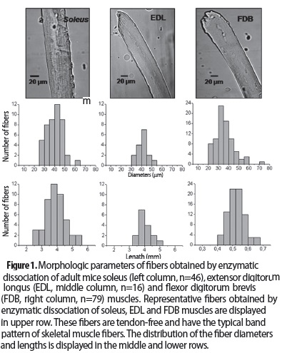
Preparation of the fibers Figure 1 shows the fibers typically obtained with the dissociation protocol and also compares the sizes of soleus (3.9±0.1 mm, n=46) and EDL (4.0±0.1 mm, n=16) fibers with those of the shorter FDB fibers (0.52±0.006 mm, n=79). The diameters of the soleus (39.5±1.1 µm) and EDL (40.8±1.5 µm) fibers are also greater than those from the FDB (35.1±1 µm). FDB fibers were significantly different from soleus and EDL ones.
]]> Ca2+ transients in soleus and EDL fibers obtained by enzymatic dissociationIn figure 2, representative single Ca2+ transients obtained from one soleus (A) and one EDL (B) fiber are shown. Table 2 summarizes the kinetics of single Ca2+ transients obtained in 45 soleus and 26 EDL fibers. A double exponential function was fitted to calculate the decay kinetics (8). Soleus fibers were significantly slower for all variables and had smaller amplitudes than the EDL fibers.
Experimental procedures with fibers obtained by enzymatic dissociation
Figure 3 is a confocal image of a fiber labeled with MitotrackerGreen-FM and Di-8Anepps. The picture demonstrates that the procedure renders structurally normal cells and also allows us to know that the distribution of labeled mitochondria (figure 3C) is similar to that reported for fast FDB cells (5). The sarcomere length is also comparable to that of fast fibers, despite the fact that it differs in some structural protein isoforms (5). The sarcomere size based on the non-fitted profile plot of the Di-8Anepps image is preferred (figure 3B) because of potential bias when the calculation is based on a fitted mitochondrial array (Calderón J, unpublished results).
Soleus and EDL fiber typing
Figure 4A is a confocal image of a type II fiber isolated from the soleus. The fiber was labeled with an anti-myosin II antibody prior to incubation with a secondary antibody coupled to AlexaFluor-488, a fluorescent compound. In figure 4B, separate gels were loaded with either an isolated soleus or EDL fiber. Several MHC bands may be present in each fiber. In mice, the observed pattern of MHC electrophoretic mobility is IIA, IIX/D, IIB and I (24,25). Table 3 lists the proportion of pure and hybrid fibers present in 54 soleus and EDL fibers, as analyzed by SDS-PAGE. The soleus is comprised of a greater proportion of hybrid fibers than the EDL.
DISCUSSION
Although enzymatic dissociation of mammalian muscles was first described decades ago (21), dissociation and physiological experimentation on muscle cells longer than 1 mm are seldom performed. The many publications using the rat FDB, mouse FDB and mouse interossei (1-13,21) have produced a large amount of physiological information on a subset of fiber types, leading to an unbalanced understanding of muscle fiber physiology.
In mammalian skeletal muscle, there are four major types of fibers: I, IIA, IIX/D and IIB (26). Multiple contractile properties have been studied in all types, including the maximum speed of contraction, the characteristics of the force-speed curve, the maximal power, the timing of contraction and relaxation, the maximal tension, and ATP consumption. (27,28). However, other functional properties have only been studied in a limited number of fiber types. These properties include the kinetics of the Ca2+ transients and Ca2+ flow in both developing and mature fibers, the characteristics and factors that affect SOCE, the role of mitochondria in Ca2+ regulation within muscle fibers, and alterations that occur during muscle fatigue (1-4,6,29). In these publications, the fiber type used in the experiments was not specified; however, the fibers can be assumed to be type IIX/D because they are the predominant type in the FDB (8,30), which is the most frequently studied muscle. The dissociation of muscles such as the soleus or the EDL introduces the possibility of using the whole spectrum of fiber types. The results of this study show that the soleus of mature NMRI mice is composed of fiber types I, IIA and hybrid fiber types IC and IIC. The EDL is composed of fiber types IIX/D, IIB and hybrid fibers with different proportions of IIX/D and IIB.
]]> Although not yet confirmed by other researchers (20), each fiber type uses different proteins for Ca2+ handling and has unique ECC properties (16). This work presents the mean values of kinetic variables of Ca2+ transients obtained from the largest number of soleus and EDL fibers so far published in the literature. The results include some fibers that were already published and others obtained during the last two years. The results are very similar to those previously reported (8,16), suggesting that the data are reproducible and the experiments were performed well. Some differences among earlier studies (8,16,20) may have arisen from several factors: dissociated fibers are shortened approximately 20% compared to stretched fibers mounted in a transducer (5,8), different loading methods may result in differences in intracellular dye concentrations, and manually isolated cells (20) may be damaged because of imperceptible stretches during the isolation procedure.One main advantage of using long muscle fibers in physiological experiments is the ability to determine MHC isoforms of the fiber via SDS-PAGE. In short muscles fibers, the bands are difficult to distinguish, and there is occasionally too little protein for sufficient staining with silver nitrate (Calderón J, unpublished results), likely leading to the underestimation of some MHC isoforms in hybrid fibers. As the soleus and EDL fibers are approximately eight times longer than those of FDB (figure 2), the amount of myosin (and other proteins) present in the soleus and EDL fibers can be considered eight times larger than the amount in the FDB fibers. Therefore, long muscles generate cleaner bands.
One use for models of dissociated long muscles is to study hybrid fibers. Hybrid fibers were first observed in the early 1990s in rats and named by the Pette group (31-33). Their physiological significance began to be recognized approximately a decade later (34,35). While they have been studied in rats (33-36), little is known about hybrid fibers in mice; this shortcoming is not trivial because the mouse is the preferred model for muscle physiological studies. Gorza described the frequency of hybrid fibers in the tibialis anterior of mouse using antibodies against different myosin types (37); however, because there are no antibodies against myosin type IIX/D, the hybrid fibers expressing this myosin type could only be detected by indirect means (38). Hence, the study of hybrid fibers in mouse muscles and the correlation of physiological phenomena with the content of MHC in these fibers have been hindered by technical difficulties related with the dissociation or isolation of the mouse fibers, the decreased amount of myosin present in short muscles in comparison with rat muscle fibers and the lack of specific antibodies to some myosin isoforms. The case of the study of the Ca2+ transients according to the fiber type (pure and hybrid) seems to be illustrative: the rat fibers that yield good bands via SDS-PAGE are very long and difficult to be successfully dissociated; short mouse muscles (i.e., FDB), easier to dissociate, are very small to give good bands via SDS-PAGE, but those of long muscles of mouse are susceptible to dissociation and give nice bands in the SDS-PAGE. Our results corroborate the findings by Gorza (31) that fast muscles are composed of fewer hybrid fibers than slow muscles. This difference could be related to the decreased variability among type II fibers (which predominate in fast muscles) than among fibers of types I and II (both of which are present in slow muscles).
Increasing the knowledge about hybrid fibers may help answer many questions in muscle physiology and biochemistry, especially in regard to the structure and function of the proteins involved in the ECC and genetic regulation (34). This knowledge may have important applications to sports medicine, musculoskeletal rehabilitation and muscle disease therapies.
There are several advantages to the described methodology: i) experiments on larger muscles are possible, ii) a greater number of cells can be studied, iii) the methodology is a more reliable way to classify muscle fibers, and iv) the methodology widens the spectrum of fibers to be studied. However, some limitations of the methodology exist: i) it requires more training than dissociating short muscles, and ii) in some experiments, more laminin is required to attach the fibers to the cover slide.
In summary, a methodology to enzymatically dissociate long mouse muscles was presented. Additionally, the usefulness of the methodology was demonstrated for studying phenomena such as the kinetics of the Ca2+ transients, evaluating different structures and reliably determining the fiber type used in immunostaining or SDS-PAGE experiments. Moreover, the greatest number of experiments on Ca2+ transients kinetics in the soleus and EDL was presented, showing that those of the soleus are lower and slower than those of EDL. This methodology is expected to be applied to other types of experiments (not described here, such as inactivation of Ca2+ release, SOCE regulation and fatigue) (17), increasing the knowledge of muscle fiber biology and pathophysiology, especially that related to pure and hybrid fiber types.
ACKNOWLEDGEMENTS
I would like to thank Dr. Carlo Caputo and Pura Bolaños from Cellular Physiology Laboratory, Venezuelan Institute for Scientific Research. I would also like to thank doctor Raul Narvaez-Sanchez and doctor Marco Giraldo from University of Antioquia for comments to this manuscript.
]]> BIBLIOGRAPHIC REFERENCES
1. Caputo C, Bolaños P, Gonzalez A. Inactivation of Ca2+ transients in amphibian and mammalian muscle fibres. J Muscle Res Cell Motil. 2004 Jan;25(4- 5):315–28. [ Links ]
2. Capote J, Bolaños P, Schuhmeier RP, Melzer W, Caputo C. Calcium transients in developing mouse skeletal muscle fibres. J Physiol. 2005 Apr 15;564(Pt 2):451–64. [ Links ]
3. González Narváez AA, Castillo A. Ca2+ store determines gating of store operated calcium entry in mammalian skeletal muscle. J Muscle Res Cell Motil. 2007 Jan;28(2-3):105–13. [ Links ]
4. Caputo C, Bolaños P. Effect of mitochondria poisoning by FCCP on Ca2+ signaling in mouse skeletal muscle fibers. Pflugers Arch. 2008 Jan;455(4):733–43. [ Links ]
5. Bolaños P, Guillen A, Rojas H, Boncompagni S, Caputo C. The use of CalciumOrange-5N as a specific marker of mitochondrial Ca2+ in mouse skeletal muscle fibers. Pflugers Arch. 2008 Jan;455(4):721–31. [ Links ]
6. Bolaños P, Guillén A, DiPolo R, Caputo C. Factors affecting SOCE activation in mammalian skeletal muscle fibers. J Physiol Sci. 2009 Jul;59(4):317–28. [ Links ]
7. Legrand C, Giacomello E, Berthier C, Allard B, Sorrentino V, Jacquemond V. Spontaneous and voltage-activated Ca2+ release in adult mouse skeletal muscle fibres expressing the type 3 ryanodine receptor. J Physiol. 2008 Jan 15;586(2):441–57. [ Links ]
8. Calderón JC, Bolaños P, Torres SH, Rodríguez-Arroyo G, Caputo C. Different fibre populations distinguished by their calcium transient characteristics in enzymatically dissociated murine flexor digitorum brevis and soleus muscles. J Muscle Res Cell Motil. 2009 Jan;30(3-4):125–37. [ Links ]
9. Apostol S, Ursu D, Lehmann-Horn F, Melzer W. Local calcium signals induced by hyper-osmotic stress in mammalian skeletal muscle cells. J Muscle Res Cell Motil. 2009 Jan;30(3-4):97–109. [ Links ]
10. DiFranco M, Quinonez M, Capote J, Vergara J. DNA transfection of mammalian skeletal muscles using in vivo electroporation. J Vis Exp. 2009 Jan;(32) [ Links ].
11. DiFranco M, Tran P, Quiñonez M, Vergara JL. Functional expression of transgenic a 1sDHPR channels in adult mammalian skeletal muscle fibres. J Physiol. 2011 Mar 15;589(Pt 6):1421–42. [ Links ]
12. Sztretye M, Yi J, Figueroa L, Zhou J, Royer L, Ríos E. D4cpv-calsequestrin: a sensitive ratiometric biosensor accurately targeted to the calcium store of skeletal muscle. J Gen Physiol. 2011 Aug;138(2):211–29. [ Links ]
13. Figueroa L, Shkryl VM, Zhou J, Manno C, Momotake A, Brum G, et al. Synthetic localized calcium transients directly probe signalling mechanisms in skeletal muscle. J Physiol. 2012 Mar 15;590(Pt 6):1389–411. [ Links ]
14. Williams DA, Head SI, Bakker AJ, Stephenson DG. Resting calcium concentrations in isolated skeletal muscle fibres of dystrophic mice. J Physiol. 1990 Sep;428:243–56. [ Links ]
15. Szentesi P, Jacquemond V, Kovács L, Csernoch L. Intramembrane charge movement and sarcoplasmic calcium release in enzymatically isolated mammalian skeletal muscle fibres. J Physiol. 1997 Dec 1;505 (Pt 2):371-84. [ Links ]
16. Calderón JC, Bolaños P, Caputo C. Myosin heavy chain isoform composition and Ca2+ transients in fibres from enzymatically dissociated murine soleus and extensor digitorum longus muscles. J Physiol. 2010 Jan 1;588(Pt 1):267-79. [ Links ]
17. Calderón JC, Bolaños P, Caputo C. Kinetic changes in tetanic Ca2+ transients in enzymatically dissociated muscle fibres under repetitive stimulation. J Physiol. 2011 Nov 1;589(Pt 21):5269-83. [ Links ]
18. Chemello F, Bean C, Cancellara P, Laveder P, Reggiani C, Lanfranchi G. Microgenomic analysis in skeletal muscle: expression signatures of individual fast and slow myofibers. PLoS One. 2011 Jan;6(2):e16807. [ Links ]
19. Head SI. Branched fibres in old dystrophic mdx muscle are associated with mechanical weakening of the sarcolemma, abnormal Ca2+ transients and a breakdown of Ca2+ homeostasis during fatigue. Exp Physiol. 2010 May;95(5):641–56. [ Links ]
20. Hollingworth S, Kim MM, Baylor SM. Measurement and simulation of myoplasmic calcium transients in mouse slow-twitch muscle fibres. J Physiol. 2012 Feb 1;590(Pt 3):575–94. [ Links ]
21. Bekoff A, Betz WJ. Physiological properties of dissociated muscle fibres obtained from innervated and denervated adult rat muscle. J Physiol. 1977 Sep;271(1):25–40. [ Links ]
22. Talmadge RJ, Roy RR. Electrophoretic separation of rat skeletal muscle myosin heavy-chain isoforms. J Appl Physiol. 1993 Nov;75(5):2337–40. [ Links ]
23. Blough ER, Rennie ER, Zhang F, Reiser PJ. Enhanced electrophoretic separation and resolution of myosin heavy chains in mammalian and avian skeletal muscles. Anal Biochem. 1996 Jan 1;233(1):31–5. [ Links ]
24. Hämäläinen N, Pette D. The histochemical profiles of fast fiber types IIB, IID, and IIA in skeletal muscles of mouse, rat, and rabbit. J Histochem Cytochem. 1993 May;41(5):733–43. [ Links ]
25. Agbulut O, Li Z, Mouly V, Butler-Browne GS. Analysis of skeletal and cardiac muscle from desmin knockout and normal mice by high resolution separation of myosin heavy-chain isoforms. Biol Cell. 1996 Jan;88(3):131–5. [ Links ]
26. Schiaffino S, Reggiani C. Myosin isoforms in mammalian skeletal muscle. J Appl Physiol. 1994 Aug;77(2):493–501. [ Links ]
27. Bottinelli R. Functional heterogeneity of mammalian single muscle fibres: do myosin isoforms tell the whole story? Pflugers Arch. 2001 Oct;443(1):6–17. [ Links ]
28. Bottinelli R, Reggiani C. Human skeletal muscle fibres: molecular and functional diversity. Prog Biophys Mol Biol. 2000 Jan;73(2-4):195–262. [ Links ]
29. Westerblad H, Allen DG. Changes of myoplasmic calcium concentration during fatigue in single mouse muscle fibers. J Gen Physiol. 1991 Sep;98(3):615–35. [ Links ]
30. Gonzalez E, Messi ML, Zheng Z, Delbono O. Insulinlike growth factor-1 prevents age-related decrease in specific force and intracellular Ca2+ in single intact muscle fibres from transgenic mice. J Physiol. 2003 Nov 1;552(Pt 3):833–44. [ Links ]
31. Termin A, Staron RS, Pette D. Myosin heavy chain isoforms in histochemically defined fiber types of rat muscle. Histochemistry. 1989 Jan;92(6):453–7. [ Links ]
32. Termin A, Staron RS, Pette D. Changes in myosin heavy chain isoforms during chronic low-frequency stimulation of rat fast hindlimb muscles. A single-fiber study. Eur J Biochem. 1989 Dec 22;186(3):749–54. [ Links ]
33. Staron RS, Pette D. The continuum of pure and hybrid myosin heavy chain-based fibre types in rat skeletal muscle. Histochemistry. 1993 Aug;100(2):149–53. [ Links ]
34. Stephenson G. Hybrid skeletal muscle fibres: a rare or common phenomenon? Proc Austr Physiol Pharmacol Soc. 2001;32(1):69–87. [ Links ]
35. Caiozzo VJ, Haddad F, Baker M, McCue S, Baldwin KM. MHC polymorphism in rodent plantaris muscle: effects of mechanical overload and hypothyroidism. Am J Physiol Cell Physiol. 2000 Apr;278(4):C709–17. [ Links ]
36. Staron RS, Kraemer WJ, Hikida RS, Fry AC, Murray JD, Campos GE. Fiber type composition of four hindlimb muscles of adult Fisher 344 rats. Histochem Cell Biol. 1999 Feb;111(2):117–23. [ Links ]
37. Gorza L. Identification of a novel type 2 fiber population in mammalian skeletal muscle by combined use of histochemical myosin ATPase and anti-myosin monoclonal antibodies. J Histochem Cytochem. 1990 Feb;38(2):257–65. [ Links ]
38. Bottinelli R, Schiaffino S, Reggiani C. Force-velocity relations and myosin heavy chain isoform compositions of skinned fibres from rat skeletal muscle. J Physiol. 1991 Jun;437:655–72. [ Links ]
]]>