<?xml version="1.0" encoding="ISO-8859-1"?><article xmlns:mml="http://www.w3.org/1998/Math/MathML" xmlns:xlink="http://www.w3.org/1999/xlink" xmlns:xsi="http://www.w3.org/2001/XMLSchema-instance">
<front>
<journal-meta>
<journal-id>0121-4004</journal-id>
<journal-title><![CDATA[Vitae]]></journal-title>
<abbrev-journal-title><![CDATA[Vitae]]></abbrev-journal-title>
<issn>0121-4004</issn>
<publisher>
<publisher-name><![CDATA[Facultad de Química Farmacéutica, Universidad de Antioquia]]></publisher-name>
</publisher>
</journal-meta>
<article-meta>
<article-id>S0121-40042013000200006</article-id>
<title-group>
<article-title xml:lang="en"><![CDATA[HEPATOPROTECTIVE EFFECT OF CALYCES EXTRACT OF Physalis peruviana ON HEPATOTOXICITY INDUCED BY CCl4 IN WISTAR RATS]]></article-title>
<article-title xml:lang="es"><![CDATA[EFECTO HEPATOPROTECTOR DE UN EXTRACTO DE CÁLICES DE Physalis peruviana EN HEPATOTOXICIDAD INDUCIDA POR CCl4 EN RATAS WISTAR]]></article-title>
</title-group>
<contrib-group>
<contrib contrib-type="author">
<name>
<surname><![CDATA[TORO A.]]></surname>
<given-names><![CDATA[Reina Marcela]]></given-names>
</name>
<xref ref-type="aff" rid="A01"/>
</contrib>
<contrib contrib-type="author">
<name>
<surname><![CDATA[ARAGÓN N.]]></surname>
<given-names><![CDATA[Diana Marcela]]></given-names>
</name>
<xref ref-type="aff" rid="A01"/>
</contrib>
<contrib contrib-type="author">
<name>
<surname><![CDATA[OSPINA G.]]></surname>
<given-names><![CDATA[Luis Fernando]]></given-names>
</name>
<xref ref-type="aff" rid="A01"/>
</contrib>
</contrib-group>
<aff id="A01">
<institution><![CDATA[,Universidad Nacional de Colombia Departamento de Farmacia de Investigación ''Principios Bioactivos de Plantas'']]></institution>
<addr-line><![CDATA[Bogotá D.C.]]></addr-line>
<country>Colombia</country>
</aff>
<pub-date pub-type="pub">
<day>00</day>
<month>08</month>
<year>2013</year>
</pub-date>
<pub-date pub-type="epub">
<day>00</day>
<month>08</month>
<year>2013</year>
</pub-date>
<volume>20</volume>
<numero>2</numero>
<fpage>125</fpage>
<lpage>132</lpage>
<copyright-statement/>
<copyright-year/>
<self-uri xlink:href="http://www.scielo.org.co/scielo.php?script=sci_arttext&amp;pid=S0121-40042013000200006&amp;lng=en&amp;nrm=iso"></self-uri><self-uri xlink:href="http://www.scielo.org.co/scielo.php?script=sci_abstract&amp;pid=S0121-40042013000200006&amp;lng=en&amp;nrm=iso"></self-uri><self-uri xlink:href="http://www.scielo.org.co/scielo.php?script=sci_pdf&amp;pid=S0121-40042013000200006&amp;lng=en&amp;nrm=iso"></self-uri><abstract abstract-type="short" xml:lang="en"><p><![CDATA[Background: Physalis peruviana (''uchuva'', Solanaceae) is a widespread species of the South American Andes and widely used in traditional medicine. Its fruits are consumed as food and for the treatment of diabetes. The juice of Physalis peruviana fruits is topically applied in the eyes for pterigyum treatment. Previous works reported that the fruit extracts has modulating activity of oxidative stress in experimental diabetes models induced by streptozotocin. It has been attributed antipyretic, antimicrobial, analgesic and anti-inflammatory properties to the calyces enveloping the fruit. Reported literature demonstrates in vivo and in vitro that different calyx's extracts have antioxidant and anti-inflammatory activities. Objectives: To evaluate the in vivo hepatoprotective effect of the extract of Physalis peruviana calyces, involving inflammation and oxidative stress models at hepatic level. Methods: Hepatotoxicity was induced by single oral administration of CCl4 (2 mL / Kg in olive oil) in Wistar rats. Physalis peruviana extract (250 mg/Kg) and silymarin (200 mg/Kg), used as control drug, were administrated twice a day for five days. At the end of the experiment, animals were euthanized and the liver enzymes alanine aminotransferase, aspartate aminotransferase, and alkaline phosphatase were measured as well as some parameters of hepatic antioxidant status like superoxide dismutase and catalase activities, protein oxidation and lipid peroxidation. Results: Extract of Physalis peruviana calyces inhibited significantly (p < 0.001) liver oxidative stress caused by CCl4, maintaining superoxide dismutase and catalase activities close to normal. Studied extract also reduced significantly liver enzymes levels increased by CCl4 administration. Conclusion: It was suggested that the extract of Physalis peruviana calyces presents a hepatoprotective effect related to its antioxidant activity, especially regarding to lipid peroxidation inhibition.]]></p></abstract>
<abstract abstract-type="short" xml:lang="es"><p><![CDATA[Antecedentes: Physalis peruviana (''uchuva'', Solanaceae) es una especie distribuida en los Andes suramericanos y de amplio uso a nivel etnofarmacológico. Sus frutos, además de ser usados como alimento, también son consumidos para el tratamiento de la diabetes. Por su parte, el zumo del fruto es aplicado localmente para el tratamiento de pterigios. En modelos de diabetes experimental inducidos por estreptozotocina, trabajos previos han reportado que los extractos de los frutos poseen actividad moduladora del estrés oxidativo. A los cálices que envuelven el fruto, se les atribuyen propiedades antipiréticas, antimicrobianas, analgésicas y antiinflamatorias, entre otras. Estudios previos han demostrado en modelos in vivo e in vitro que diferentes extractos de los cálices presentan actividad antioxidante y antiinflamatoria. Objetivo: Evaluar el posible efecto hepatoprotector de un extracto de cálices de Physalis peruviana en un modelo experimental in vivo que involucra procesos de inflamación y de estrés oxidativo a nivel hepático. Métodos: Se empleó un modelo de hepatotoxicidad inducida por CCl4 en ratas Wistar, mediante una única administración oral de CCl4. Tanto el extracto de Physalis peruviana (250 mg/Kg) como la silimarina (200 mg/Kg), empleada como patrón de referencia, fueron administradas dos veces al día durante cinco días. Al final del experimento, los animales fueron sacrificados y se evaluaron los niveles hepáticos de las enzimas alanina-aminotranferasa, aspartato-transaminasa y fosfatasa alcalina, al igual que algunos parámetros del estado antioxidante hepático como actividad de superóxido dismutasa y catalasa, oxidación proteica y peroxidación lipídica. Resultados: El extracto evaluado de Physalis peruviana inhibió significativamente (p < 0,001) el estrés oxidativo causado a nivel hepático por el CCl4, manteniendo las actividades de superóxido dismutasa y catalasa con valores cercanos a los normales. El extracto también redujo significativamente los niveles de las enzimas hepáticas que fueron incrementados después de la administración de CCl4. Conclusión: Estos resultados sugieren que el extracto de cálices de Physalis peruviana posee un efecto hepatoprotector relacionado con su actividad antioxidante, especialmente en lo referente a la inhibición de la peroxidación lipídica.]]></p></abstract>
<kwd-group>
<kwd lng="en"><![CDATA[Physalys peruviana]]></kwd>
<kwd lng="en"><![CDATA[hepatotoxicity]]></kwd>
<kwd lng="en"><![CDATA[oxidative stress]]></kwd>
<kwd lng="en"><![CDATA[antioxidant]]></kwd>
<kwd lng="es"><![CDATA[Physalys peruviana]]></kwd>
<kwd lng="es"><![CDATA[hepatotoxicidad]]></kwd>
<kwd lng="es"><![CDATA[estrés oxidativo]]></kwd>
<kwd lng="es"><![CDATA[antioxidante]]></kwd>
</kwd-group>
</article-meta>
</front><body><![CDATA[  <font face="Verdana, Arial, Helvetica, sans-serif" size="2">     <p align="right"> <b>PHARMACOLOGY AND TOXICOLOGY</b></p>     <p>&nbsp;</p>     <p align="center"><b><font size="4">HEPATOPROTECTIVE EFFECT OF CALYCES EXTRACT OF       <i><i>Physalis peruviana</i></i> ON HEPATOTOXICITY INDUCED BY CCl<sub>4</sub> IN WISTAR RATS</font></b></p>     <p>&nbsp;</p>     <p align="center"><b><font size="3"> EFECTO HEPATOPROTECTOR DE UN EXTRACTO DE C&Aacute;LICES DE <i><i>Physalis peruviana</i></i> EN HEPATOTOXICIDAD INDUCIDA POR CCl<sub>4</sub> EN RATAS WISTAR</font></b></p>     <p>&nbsp;</p>     <p>&nbsp;</p>     <p><b> Reina Marcela TORO A. QF<sup>1</sup>; Diana Marcela ARAG&Oacute;N N. PhD<sup>1</sup>; Luis Fernando OSPINA G. PhD<sup>1</sup>*</b></p>     <p>1 Grupo de Investigaci&oacute;n ''Principios Bioactivos de Plantas''; Departamento de Farmacia, Universidad Nacional de Colombia, A.A. 14490, Cra 30 No. 45-03. Bogot&aacute; D.C.,Colombia.</p>     ]]></body>
<body><![CDATA[<p> * Autor a quien se debe dirigir la correspondencia: <a href="mailto:lfospinag@unal.edu.co">lfospinag@unal.edu.co</a>.</p>     <p>&nbsp;</p>     <p>Recibido: Noviembre 06 de 2013     <br> Aceptado: Octubre 15 de 2013</p>     <p>&nbsp;</p> <hr noshade size="1">     <p><b> ABSTRACT</b></p>     <p><b>Background</b>: <i>Physalis peruviana</i> (''uchuva'', Solanaceae) is a widespread species of the South American   Andes and widely used in traditional medicine. Its fruits are consumed as food and for the treatment of   diabetes. The juice of <i>Physalis peruviana</i> fruits is topically applied in the eyes for pterigyum treatment.   Previous works reported that the fruit extracts has modulating activity of oxidative stress in experimental   diabetes models induced by streptozotocin. It has been attributed antipyretic, antimicrobial, analgesic   and anti-inflammatory properties to the calyces enveloping the fruit. Reported literature demonstrates <i>in   vivo</i> and<i> in vitro</i> that different calyx's extracts have antioxidant and anti-inflammatory activities. <b>Objectives</b>:   To evaluate the in vivo hepatoprotective effect of the extract of <i>Physalis peruviana</i> calyces, involving   inflammation and oxidative stress models at hepatic level. <b>Methods</b>: Hepatotoxicity was induced by   single oral administration of CCl<sub>4</sub> (2 mL / Kg in olive oil) in Wistar rats. <i>Physalis peruviana</i> extract (250   mg/Kg) and silymarin (200 mg/Kg), used as control drug, were administrated twice a day for five days.   At the end of the experiment, animals were euthanized and the liver enzymes alanine aminotransferase,   aspartate aminotransferase, and alkaline phosphatase were measured as well as some parameters of hepatic   antioxidant status like superoxide dismutase and catalase activities, protein oxidation and lipid peroxidation.   <b>Results</b>: Extract of <i>Physalis peruviana</i> calyces inhibited significantly (p &lt; 0.001) liver oxidative   stress caused by CCl4, maintaining superoxide dismutase and catalase activities close to normal. Studied   extract also reduced significantly liver enzymes levels increased by CCl<sub>4</sub> administration. <b>Conclusion</b>:   It was suggested that the extract of <i>Physalis peruviana</i> calyces presents a hepatoprotective effect related to its antioxidant activity, especially regarding to lipid peroxidation inhibition.</p>     <p> <b>Keywords</b>: <i>Physalys peruviana</i>, hepatotoxicity, oxidative stress, antioxidant.</p> <hr noshade size="1">     <p> <b>RESUMEN</b></p>     <p><b>Antecedentes</b>: <i>Physalis peruviana</i> (''uchuva'', Solanaceae) es una especie distribuida en los Andes suramericanos   y de amplio uso a nivel etnofarmacol&oacute;gico. Sus frutos, adem&aacute;s de ser usados como alimento,   tambi&eacute;n son consumidos para el tratamiento de la diabetes. Por su parte, el zumo del fruto es aplicado   localmente para el tratamiento de pterigios. En modelos de diabetes experimental inducidos por estreptozotocina,   trabajos previos han reportado que los extractos de los frutos poseen actividad moduladora   del estr&eacute;s oxidativo. A los c&aacute;lices que envuelven el fruto, se les atribuyen propiedades antipir&eacute;ticas, antimicrobianas, analg&eacute;sicas y antiinflamatorias, entre otras. Estudios previos han demostrado en modelos   <i>in vivo</i> e<i> in vitro</i> que diferentes extractos de los c&aacute;lices presentan actividad antioxidante y antiinflamatoria.   <b>Objetivo</b>: Evaluar el posible efecto hepatoprotector de un extracto de c&aacute;lices de <i>Physalis peruviana</i> en un   modelo experimental<i> in vivo </i>que involucra procesos de inflamaci&oacute;n y de estr&eacute;s oxidativo a nivel hep&aacute;tico.   <b>M&eacute;todos</b>: Se emple&oacute; un modelo de hepatotoxicidad inducida por CCl4 en ratas Wistar, mediante una   &uacute;nica administraci&oacute;n oral de CCl<sub>4</sub>. Tanto el extracto de <i>Physalis peruviana</i> (250 mg/Kg) como la silimarina   (200 mg/Kg), empleada como patr&oacute;n de referencia, fueron administradas dos veces al d&iacute;a durante cinco   d&iacute;as. Al final del experimento, los animales fueron sacrificados y se evaluaron los niveles hep&aacute;ticos de   las enzimas alanina-aminotranferasa, aspartato-transaminasa y fosfatasa alcalina, al igual que algunos   par&aacute;metros del estado antioxidante hep&aacute;tico como actividad de super&oacute;xido dismutasa y catalasa, oxidaci&oacute;n   proteica y peroxidaci&oacute;n lip&iacute;dica. <b>Resultados</b>: El extracto evaluado de <i>Physalis peruviana</i> inhibi&oacute;   significativamente (p &lt; 0,001) el estr&eacute;s oxidativo causado a nivel hep&aacute;tico por el CCl<sub>4</sub>, manteniendo las   actividades de super&oacute;xido dismutasa y catalasa con valores cercanos a los normales. El extracto tambi&eacute;n   redujo significativamente los niveles de las enzimas hep&aacute;ticas que fueron incrementados despu&eacute;s de la   administraci&oacute;n de CCl4. Conclusi&oacute;n: Estos resultados sugieren que el extracto de c&aacute;lices de <i>Physalis   peruviana</i> posee un efecto hepatoprotector relacionado con su actividad antioxidante, especialmente en lo referente a la inhibici&oacute;n de la peroxidaci&oacute;n lip&iacute;dica.</p>     ]]></body>
<body><![CDATA[<p> <b>Palabras clave:</b> <i>Physalys peruviana</i>, hepatotoxicidad, estr&eacute;s oxidativo, antioxidante.  </p> <hr noshade size="1">     <p>&nbsp;</p>     <p>&nbsp;</p>     <p><font size="3"><b>INTRODUCTION</b></font></p>     <p>Oxidative stress is employed to indicate the   oxidative damage produced to biological molecules   such as nucleic acids, proteins, lipids, and carbohydrates   (1). This phenomenon occurs when reactive   oxygen species (ROS), including superoxide anion,   hydrogen peroxide (H<sub>2</sub>O<sub>2</sub>) and hydroxyl radical   (&bull;OH) exceed the cellular antioxidant defense. This   imbalance produces adverse effects on the tissues   and eventually cardiovascular or liver diseases, inflammatory   processes and other chronic diseases such as hepatic fibrosis (2, 3).</p>     <p> The administration of carbon tetrachloride   (CCl4) in rodents is the most studied experimental   model of liver inflammation. The CCl<sub>4</sub> is biotransformed   in the liver by cytochrome P450 and   produces trichloromethyl radical (CCl<sub>3</sub>&bull;) and ROS,   initiating a lipid peroxidation leading to hepatocyte   death and subsequent inflammation of the organ   (4). These changes modify the normal performance   of liver and alter serum levels of liver alanine aminotransferase   (ALT), aspartate aminotransferase   (AST) and alkaline phosphatase (AP).</p>     <p> Cape gooseberry (<i>Physalis peruviana</i> L., Solanaceae)   is an abundant specie of the South American   Andes that has been widely used and studied for   medicinal purposes; this specie has been used for its   anti-mycobacterial, antipyretic, analgesic, diuretic,   and anti-inflammatory properties (5, 6). Extracts   from the calyces of <i>Physalis peruviana</i> have demonstrated   anti-inflammatory (7), immunomodulatory   (8) and anti-nociceptive activities (9). Furthermore,   it has been reported antioxidant and hypoglycemic   activity in fruit extracts (10), and effectiveness in   the treatment of pterygium, due to the inhibition   of fibroblasts proliferation (11). Therefore, the aim   of the present study was to evaluate the possible   hepatoprotective effect of calyces extract of <i>P. peruviana</i>  in CCl<sub>4</sub> hepatotoxicity model.</p>     <p>&nbsp;</p>     <p><font size="3"> <b>METHODS AND MATERIALS</b></font></p>     <p><b> Plant Material and extraction</b></p>     ]]></body>
<body><![CDATA[<p>  <i>Physalis peruviana</i> calyces were obtained from a   local market of Bogota city (voucher specimen was   deposited at The Herbario Nacional Colombiano   -COL- 51200, identified by the Botanic C.I. Orozco-).   Fresh calyces were selected and dried in air   circulating oven at 50&deg;C for 36 h. Dried material   was milled and then was extracted by exhaustive   percolation with petroleum ether according to   other works (7). The extract in petroleum ether   was fractionated by a liquid - liquid partition with   methanol-water (9:1). The methanol-water fraction   was dried in a rotary evaporator and used for this   work.</p>     <p> <b>Animals</b></p>     <p> Male Wistar rats (220-250 g) were bred and   housed under standard conditions (12 h light/12 h   dark cycles, temperature 22 &plusmn; 2 &deg;C). Food and water   were available <i>ad libitum</i>. The protocol was approved   by the Ethics Committee of Science Faculty of the   National University of Colombia. The animals   were cared for in accordance with the international   ethical guidelines (12).</p>     <p> <b>Treatment</b></p>     <p> 
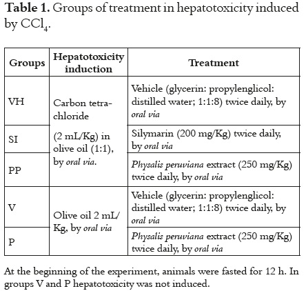
The rats were randomly divided into five groups   of 10 animals each one. <a href="#t1">Table 1</a> describe the treatments.</p>       <p align="center"><a name="t1"></a><img src="/img/revistas/vitae/v20n2/v20n2a6t1.jpg"></p>     <p>&nbsp;</p>     <p>  At the end of the experiment, all animals were   anesthetized for cardiac puncture in order to obtain   blood for ferric reduction ability of plasma   (FRAP) and biochemical determinations (alanine   aminotransferase-ALT, alanine aminotransferase-   AST, and alkaline phosphatase-AP). Finally, the   rats were sacrificed by cervical dislocation and   the livers were quickly removed, place in ice cold,   rinsed, cut and homogenized (Polytron<sup>&reg;</sup> PT-2100)   in cold Tris&#8211;HCl (5 mM, pH 7.4). Homogenates   were centrifuged (10000 rpm, 4&deg;C, 10 min), and   supernatants were used to evaluate the oxidative   stress parameters. The protein concentration was   determined using Bradford reagent.</p>     <p> <b>Histopathological studies</b></p>     <p> Sections of liver samples were washed and fixed   with 10% neutralized formalin (pH 7.4) and then   stained with hematoxylin and eosin, and were kept   under observation for pathological changes.</p>     ]]></body>
<body><![CDATA[<p> <b>Ferric reduction ability of plasma (FRAP)</b></p>     <p> This method measure the antioxidants ability in   biological sample by reducing the 2,4,6-tripyridyls-   triazine (Fe<sup>3+</sup>-TPTZ) to the ferrous form (Fe<sup>2+</sup>)   (13). The FRAP was expressed as mg FeSO<sub>4</sub>/mg   protein.</p>     <p> <b>Index of lipid peroxidation</b></p>     <p> This parameter was evaluated by the measure   of thiobarbituric acid reactive substances (TBARs).   Briefly, 450&mu;L of homogenated were added to 50&mu;L   of phosphate buffers (50 mM, pH 7,4) and 1 mL of   trichloroacetic acid (10%). The mix was centrifuged   (2000 rpm, 10 min, 4&deg;C) and 1 mL of supernatant   was added to 1 mL of thiobarbithuric acid (0.67%).   This mixture was boiled for 30 min, with the   subsequent absorbance measurement at 532 nm.   Concentration of the TBARs were expressed as   (mmol/mL)/mg protein, using the expression &epsilon; =   A/c*h, where &epsilon; = 153000 M<sup>&#8211;1</sup>cm<sup>&#8211;1</sup>.</p>     <p> <b>Protein carbonyl content (COP)</b></p>     <p> This parameter was measured using 2,4-dinotrophenylhydrazine   (DNPH) according to methods   previously described (14). Briefly, 50 &mu;L of homogenate   was added to 250 &mu;L of 2,4-dinitrophenylhydrazine   (DNPH, 10 mM in HCl 2M) and left to   stand at room temperature for 1h stirring every 15   min. Thereafter, 500 &mu;L of cold trichloroacetic acid   (TCA, 20%) were added and left to stand for 15 min   at 4&deg;C. Then, the mixture was centrifuged at 11000   rpm for 5 min, the supernatant was removed and   the pellet was rinsed three times with ethanol: ethyl   acetate (1:1), and centrifuged after each rinse for 7   min at 3000 rpm. Finally, the pellet was dissolved   in 250 &mu;L of Guanidine 6 M and incubated at 37&deg;C   for 10 min. The absorbance was measured at 360   nm. The carbonyl content was calculated using the   expression &epsilon; = A/c*h, where &epsilon; = 22000 M<sup>&#8211;1</sup>cm<sup>&#8211;1</sup>   and expressed as nmol/mg protein.</p>     <p> <b>Superoxide dismutase activity (SOD)</b></p>     <p> The Cu-SOD and Mn-SOD were determined   by the riboflavin-o-dianisidine test (13). 4 &mu;L of   homogenate was added to 200 &mu;L of the reaction   mixture (0,1 mL of o-dianisidine 6 mM; 0,1 mL of   riboflavin 0.29 mM and phosphate buffer, 50 mM,   pH 7.8). The mixture was incubated at dark for 5   min at 25&deg;C and then exposed for 8 min to fluorescent   light; the absorbance was measured at 415   nm before and after the light exposition. The SOD   concentration was extrapolated from a calibration   curve and expressed as U/mg protein.</p>     <p> <b>Catalase activity (CAT)</b></p>     <p> The method is based on the H<sub>2</sub>O<sub>2</sub> decomposition   and the rate constant determination k (s<sup>&#8211;1</sup>) (15).   Briefly, 250 &mu;L of H<sub>2</sub>O<sub>2</sub> (10 mM) were added to 5 &mu;L of homogenate and 45 &mu;L of phosphate buffer (50 mM, pH 7.0). Immediately, the change in the absorbance was measured at 240 nm for 30 sec. The enzyme activity was expressed as k (s mg protein)<sup>&#8211;1</sup>.</p>     ]]></body>
<body><![CDATA[<p> <b>Statistical Analysis</b></p>     <p> The results were expressed as means &plusmn; SD and   were analyzed by one-way analysis of variance   (ANOVA), followed by Dunnett's test. Statistical   differences were considered (*) when p &lt; 0.05.</p>     <p>&nbsp;</p>     <p><font size="3"> <b>RESULTS</b></font></p>     <p> Significant increases of enzymes AST, ALT   and ALP (more than 5 times baseline levels) were   found in the control group (VH) (<a href="/img/revistas/vitae/v20n2/v20n2a6f1.jpg" target="_blank">figure 1</a>). These   results are similar to those reported by other authors   and were consistent to the mode of action of   CCl<sub>4</sub>, which induces a large lipid peroxidation in   the endoplasmic reticulum of hepatocytes (16, 17).</p>     <p> Histopathological studies showed that rats   without CCl4 had a normal hepatic architecture,   indicating that the vehicle and the extract of <i>Physalis   peruviana</i> do not have hepatotoxic effect (<a href="/img/revistas/vitae/v20n2/v20n2a6f2.jpg" target="_blank">figure 2</a>).    The administration of CCl<sub>4</sub> induced sub-massive   hepatic necrosis and a pronounced hepatic microvacuolar   steatosis (<a href="/img/revistas/vitae/v20n2/v20n2a6f2.jpg" target="_blank">figure 2</a>).</p>     <p>  Regarding to oxidative stress parameters, a   significant increase (around 50%) in SOD and   CAT activities &#91;1.87 &plusmn; 0.13 units/ mg protein and   3.16 &plusmn; 0.04 k/(s* mg protein), respectively&#93; in the   group VH respect to group V was found &#91;0.60&plusmn;   0.16 units/ mg protein and 1.15 &plusmn; 0.17 k/(s* mg   protein), respectively&#93;, while silymarin and <i>P. peruviana</i>   maintain enzyme activities near to normal   (<a href="/img/revistas/vitae/v20n2/v20n2a6f3.jpg" target="_blank">figures 3A</a> y <a href="/img/revistas/vitae/v20n2/v20n2a6f3.jpg" target="_blank">3B</a>).</p>     <p> Furthermore, a high level of carbonyl (<a href="/img/revistas/vitae/v20n2/v20n2a6f3.jpg" target="_blank">figure   3C</a>) was found in group VH (5.78 &plusmn; 0.94 nmol/mg   protein) being three times more than in group V   (1.45 &plusmn; 0.06 nmol/mg protein). Similar to the other   parameters evaluated, silymarin and the extract of<i> P.   peruviana</i> showed a decrease in the levels of carbonyl   groups. Lipid peroxidation, expressed as nmoles of   malondialdehyde (MDA), were increased almost   twice in the group VH (9.29 &plusmn; 0.19 nmol MDA/mg   protein) (<a href="/img/revistas/vitae/v20n2/v20n2a6f3.jpg" target="_blank">figure 3D</a>). In this case, silymarin prevented   lipid peroxidation produced in the liver by CCl4 in a   50%, while the extract of <i>P. peruviana</i> reduced significantly   this damage, in a 30% respect to group VH.</p>     <p>&nbsp;</p>     <p><font size="3">  <b>DISCUSSION</b></font></p>     ]]></body>
<body><![CDATA[<p> Serum levels of liver enzymes such as ALT, AST   and ALP, and other compounds such as urea, are   important markers of liver function and the possible   damage to hepatocytes. These enzymes are released   from the cytosol and mitochondria, therefore lesions   that alter the permeability of hepatocytes   elevate the normal levels of ALT, AST and ALP   more than three times, allowing the passage of these   enzymes to peripheral blood (16, 18).</p>     <p> Treatment with silymarin (SI) and <i>P. peruviana</i>   (PP) prevent a significant increase in the levels of   ALT, AST and ALP. This could be due to the antioxidant   activity of these treatments. Silymarin is a   mixture of flavonolignans extracted from<i> Silybum   marianum</i>, which had reported protective effect   against various liver diseases (19), including those   induced by oxidative stress like cirrhosis, diabetes   and liver fibrosis (20-22). Its mechanism of action is   still unknown; however, two possible mechanisms   proposed are through free radical scavenging and   regulation of immune functions modulated by   cytokines (23, 24). The decrease in enzyme levels   (AST, ALT and ALP) in silymarin group (SI) is   probably due to the antioxidant activity of silymarin,   demonstrated against DPPH, ROS such   as &bull;OH, HOCl and against lipid peroxidation. A   similar effect can be attributed to the extract of <i>P.   peruviana</i> which had demonstrated superoxide radical   scavenging activity, inhibition of lipid peroxidation   <i>in vitro</i> and scavenging of ROS like H<sub>2</sub>O<sub>2</sub> (6).</p>     <p> Microvacuolar steatosis found in histopalogical   studies is associated with a state of oxidative stress   generated, since the trichloromethyl radical (&bull;CCl<sub>3</sub>)   covalently binds to cellular components such as   glutathione reductase, inhibiting the secretion of lipoproteins   causing the subsequent steatosis. On the   other hand, after the &bull;CCl<sub>3</sub> reaction with molecular   oxygen forms the peroxyl radical (&bull;OOCCl<sub>3</sub>), the   process of lipid peroxidation initiates generating   disruption of membrane cell of the hepatocytes   (25, 26). Furthermore, histopathological findings in   liver tissues of rats with silymarin and <i>P. peruviana</i>  presented a slight attenuation of liver steatosis, and   absence of hepatic necrosis in the treatment with   <i>P. peruviana</i> (<a href="/img/revistas/vitae/v20n2/v20n2a6f2.jpg" target="_blank">figure 2</a>).</p>     <p> As mentioned before, after CCl<sub>4</sub> administration   diverse reactive oxygen species (ROS) are generated,   which must be neutralized by the antioxidant defense   system of the organism like the enzymes SOD and   CAT. The role of SOD is the dismutation of the   superoxide radical to hydrogen peroxide, which is   converted in water by CAT . For this reason, in most   events of oxidative stress these two enzymes are   affected (27). Previous studies have fully described   the alteration; generally, increase in the activity of   antioxidant enzymes such as SOD and CAT after   administration of CCl<sub>4</sub> to Wistar rats (28). Results   found in this investigation with <i>P. peruviana</i> were   similar to those obtained from silymarin and other   plants such as <i>Solanum tuberosum</i> (29), <i>Phyllanthus amarus</i> (30) and <i>Hippophae rhamnoides </i>(31).</p>     <p> Recently, it was studied the possibility that free   radicals induced not only lipid peroxidation but   also oxidation of carbonyl groups of proteins. In   this way, it was established that &bull;OH radicals can   extract an H of any carbon of amino acids, forming   a carbonyl radical which subsequently can be   converted to an alkyl peroxyl radical. The radical   formed can break peptide bonds, generating hydrolysis   and oxidation of other proteins (18). Finally,   free radicals can cause significant alterations of cell   function, (including death) causing oxidation and   inactivation of enzymes and accumulation of other   oxidized proteins. The effect of CCl<sub>4</sub> on protein   oxidation is not fully elucidated, but recent studies   demonstrated that in the presence of this toxic the   liver shows an increase (about 50%) in the levels   of carbonyl groups (32). The effect of silymarin   and the extract of <i>P. peruviana</i> in the decreasing of   carbonyl groups levels could be associated with the   antioxidant activity previously described.</p>     <p> Lipid peroxidation results are similar to those   reported for other antioxidants such as carotenoids   and tocopherols (33) in models of hepatotoxicity   induced by CCl<sub>4</sub>. Clearly, there is a strong relationship   between in vivo antioxidant activity and <i>in vitro</i>   inhibition of lipid peroxidation reported for the   extracts of <i>Physalis peruviana</i> (6).</p>     <p>&nbsp;</p>     <p><font size="3"> <b>CONCLUSIONS</b></font></p>     <p> In conclusion, the evaluated calyces extract of   <i>Physalis peruviana</i> has a significant hepatoprotective   activity that could be related to its antioxidant capacity,   especially by the inhibition of lipid peroxidation.</p>     <p>&nbsp;</p>     ]]></body>
<body><![CDATA[<p> <font size="3"><b>ACKNOWLEDGEMENTS</b></font></p>     <p> This work was supported by the National University   of Colombia, CIAT (International Center   of Tropical Agriculture) and the Ministerio de   Agricultura y Desarrollo Rural of Colombian government.</p>     <p> <b>Conflict of interest:</b> The author reports no   declaration of interest.</p>     <p>&nbsp;</p>     <p><font size="3"> <b>REFERENCES</b></font></p>     <!-- ref --><p> 1. Cadenas E. Biochemistry of oxygen toxicity. Ann Rev Biochem.   1989 Jun; 58 (1): 79-110.    &nbsp;&nbsp;&nbsp;&nbsp;&nbsp;&nbsp;&nbsp;&nbsp;[&#160;<a href="javascript:void(0);" onclick="javascript: window.open('/scielo.php?script=sci_nlinks&ref=000078&pid=S0121-4004201300020000600001&lng=','','width=640,height=500,resizable=yes,scrollbars=1,menubar=yes,');">Links</a>&#160;]<!-- end-ref --></p>     <!-- ref --><p> 2. Halliwell B. Antioxidants and human disease: A general introduction.   Nutrition Rev. 1997 Jan; 55 (1): 44-52.    &nbsp;&nbsp;&nbsp;&nbsp;&nbsp;&nbsp;&nbsp;&nbsp;[&#160;<a href="javascript:void(0);" onclick="javascript: window.open('/scielo.php?script=sci_nlinks&ref=000080&pid=S0121-4004201300020000600002&lng=','','width=640,height=500,resizable=yes,scrollbars=1,menubar=yes,');">Links</a>&#160;]<!-- end-ref --></p>     <!-- ref --><p> 3. Parola M, Robino G. Oxidative stress-related molecules and liver   fibrosis. J Hepatol. 2001 Aug; 35 (2): 297-306.    &nbsp;&nbsp;&nbsp;&nbsp;&nbsp;&nbsp;&nbsp;&nbsp;[&#160;<a href="javascript:void(0);" onclick="javascript: window.open('/scielo.php?script=sci_nlinks&ref=000082&pid=S0121-4004201300020000600003&lng=','','width=640,height=500,resizable=yes,scrollbars=1,menubar=yes,');">Links</a>&#160;]<!-- end-ref --></p>     <!-- ref --><p> 4. Recknagel RO, Glende EA, Dolak JA, Waller RL. Mechanisms   of carbon tetrachloride toxicity. Pharmacol Ther. 1989 Jan; 43   (1): 139-154.    &nbsp;&nbsp;&nbsp;&nbsp;&nbsp;&nbsp;&nbsp;&nbsp;[&#160;<a href="javascript:void(0);" onclick="javascript: window.open('/scielo.php?script=sci_nlinks&ref=000084&pid=S0121-4004201300020000600004&lng=','','width=640,height=500,resizable=yes,scrollbars=1,menubar=yes,');">Links</a>&#160;]<!-- end-ref --></p>     <!-- ref --><p> 5. Fischer G, Miranda D, Piedrahita W, Romero J. Avances en   cultivo poscosecha y exportaci&oacute;n de la uchuva, <i>Physalis peruviana</i>   L., en Colombia. Bogot&aacute;, Colombia: Unibiblios; 2008. 325p.    &nbsp;&nbsp;&nbsp;&nbsp;&nbsp;&nbsp;&nbsp;&nbsp;[&#160;<a href="javascript:void(0);" onclick="javascript: window.open('/scielo.php?script=sci_nlinks&ref=000086&pid=S0121-4004201300020000600005&lng=','','width=640,height=500,resizable=yes,scrollbars=1,menubar=yes,');">Links</a>&#160;]<!-- end-ref --></p>     <!-- ref --><p> 6. Wu SJ, Ng LT, Huang YM, Lin DL, Wang SS, Huang S, <i>et al.</i>   Antioxidant activities of <i>Physalis peruviana</i>. Biol Pharm Bull. 2005   Jun; 28 (6): 963-966.    &nbsp;&nbsp;&nbsp;&nbsp;&nbsp;&nbsp;&nbsp;&nbsp;[&#160;<a href="javascript:void(0);" onclick="javascript: window.open('/scielo.php?script=sci_nlinks&ref=000088&pid=S0121-4004201300020000600006&lng=','','width=640,height=500,resizable=yes,scrollbars=1,menubar=yes,');">Links</a>&#160;]<!-- end-ref --></p>     <!-- ref --><p> 7. Franco LA, Matiz GE, Calle J, Pinz&oacute;n R, Ospina LF. Actividad   antiinflamatoria de extractos y fracciones obtenidas de c&aacute;lices   de <i>Physalis peruviana</i> L. Biomedica. 2007 Mar; 27 (1): 110-115.    &nbsp;&nbsp;&nbsp;&nbsp;&nbsp;&nbsp;&nbsp;&nbsp;[&#160;<a href="javascript:void(0);" onclick="javascript: window.open('/scielo.php?script=sci_nlinks&ref=000090&pid=S0121-4004201300020000600007&lng=','','width=640,height=500,resizable=yes,scrollbars=1,menubar=yes,');">Links</a>&#160;]<!-- end-ref --></p>     <!-- ref --><p> 8. Mart&iacute;nez W, Ospina LF, Granados D, Delgado G. In vitro studies   on the relationship between the anti-inflammatory activity of   <i>Physalis peruviana</i> extracts and the phagocytic process. Immunopharmamacol   Immunotoxicol. 2010 Mar; 32 (1): 63-73.    &nbsp;&nbsp;&nbsp;&nbsp;&nbsp;&nbsp;&nbsp;&nbsp;[&#160;<a href="javascript:void(0);" onclick="javascript: window.open('/scielo.php?script=sci_nlinks&ref=000092&pid=S0121-4004201300020000600008&lng=','','width=640,height=500,resizable=yes,scrollbars=1,menubar=yes,');">Links</a>&#160;]<!-- end-ref --></p>     <!-- ref --><p> 9. Mu&ntilde;oz CE, Vergel NE, Arag&oacute;n DM, Ospina LF. Efecto antinociceptivo   de <i>Critoniella acuminata,</i> <i>Physalis peruviana</i> y <i>Salvia rubescens</i>.   Rev Col Cienc Qu&iacute;m Farm. 2009 Jan- Jun; 38 (1): 31-41.    &nbsp;&nbsp;&nbsp;&nbsp;&nbsp;&nbsp;&nbsp;&nbsp;[&#160;<a href="javascript:void(0);" onclick="javascript: window.open('/scielo.php?script=sci_nlinks&ref=000094&pid=S0121-4004201300020000600009&lng=','','width=640,height=500,resizable=yes,scrollbars=1,menubar=yes,');">Links</a>&#160;]<!-- end-ref --></p>     <!-- ref --><p> 10. Mora AC, Arag&oacute;n DM, Ospina LF. Effects of <i>Physalis peruviana</i>   fruit extract on stress oxidative parameters in streptozotocindiabetic   rats. Lat Am J Pharm. 2010 Jul; 29 (7): 1132-1136.    &nbsp;&nbsp;&nbsp;&nbsp;&nbsp;&nbsp;&nbsp;&nbsp;[&#160;<a href="javascript:void(0);" onclick="javascript: window.open('/scielo.php?script=sci_nlinks&ref=000096&pid=S0121-4004201300020000600010&lng=','','width=640,height=500,resizable=yes,scrollbars=1,menubar=yes,');">Links</a>&#160;]<!-- end-ref --></p>     <!-- ref --><p> 11. Pardo JM, Fontanilla MR, Ospina LF, Espinosa L. Determining   the pharmacological activity of <i>Physalis peruviana</i> fruit juice on   rabbit eyes and fibroblast primary cultures. Invest Ophtalmol   Vis Sci. 2008 Jul; 49 (7): 3074-3079.    &nbsp;&nbsp;&nbsp;&nbsp;&nbsp;&nbsp;&nbsp;&nbsp;[&#160;<a href="javascript:void(0);" onclick="javascript: window.open('/scielo.php?script=sci_nlinks&ref=000098&pid=S0121-4004201300020000600011&lng=','','width=640,height=500,resizable=yes,scrollbars=1,menubar=yes,');">Links</a>&#160;]<!-- end-ref --></p>     <!-- ref --><p> 12. National Research Council. Guide for the Care and Use of   Laboratory Animals. Washington, D.C: National Academy Press;   1996. 98 p.    &nbsp;&nbsp;&nbsp;&nbsp;&nbsp;&nbsp;&nbsp;&nbsp;[&#160;<a href="javascript:void(0);" onclick="javascript: window.open('/scielo.php?script=sci_nlinks&ref=000100&pid=S0121-4004201300020000600012&lng=','','width=640,height=500,resizable=yes,scrollbars=1,menubar=yes,');">Links</a>&#160;]<!-- end-ref --></p>     <!-- ref --><p> 13. Paynter J. Changes in activity of the manganese superoxide   dismutase enzyme in tissues of the rat with changes in dietary   manganese. J Nutr. 1980 Mar; 110 (3): 437-447.    &nbsp;&nbsp;&nbsp;&nbsp;&nbsp;&nbsp;&nbsp;&nbsp;[&#160;<a href="javascript:void(0);" onclick="javascript: window.open('/scielo.php?script=sci_nlinks&ref=000102&pid=S0121-4004201300020000600013&lng=','','width=640,height=500,resizable=yes,scrollbars=1,menubar=yes,');">Links</a>&#160;]<!-- end-ref --></p>     <!-- ref --><p> 14. Baltacio&#287;lu F, Akalin E, Alver A, De&#287;er O, Karabulut E. Protein   carbonyl levels in serum and gingival crevicular fluid in patients   with chronic periodontitis. Arch Oral Biol. 2008 Aug; 53 (8):   716-722.    &nbsp;&nbsp;&nbsp;&nbsp;&nbsp;&nbsp;&nbsp;&nbsp;[&#160;<a href="javascript:void(0);" onclick="javascript: window.open('/scielo.php?script=sci_nlinks&ref=000104&pid=S0121-4004201300020000600014&lng=','','width=640,height=500,resizable=yes,scrollbars=1,menubar=yes,');">Links</a>&#160;]<!-- end-ref --></p>     <!-- ref --><p> 15. Aebi H. Catalase in vitro. Meth Enzymol. 1981 Jan; 105 (1):   121-126.    &nbsp;&nbsp;&nbsp;&nbsp;&nbsp;&nbsp;&nbsp;&nbsp;[&#160;<a href="javascript:void(0);" onclick="javascript: window.open('/scielo.php?script=sci_nlinks&ref=000106&pid=S0121-4004201300020000600015&lng=','','width=640,height=500,resizable=yes,scrollbars=1,menubar=yes,');">Links</a>&#160;]<!-- end-ref --></p>     <!-- ref --><p> 16. Wang PY, Kaneko T, Tsukada H, Nakano M, Nakajima T, Sato   A. Time courses of hepatic injuries induced by chloroform and   by carbon tetrachloride: comparison of biochemical and histopathological   changes. Arch Toxicol. 1997 Oct; 71 (10): 638-645.    &nbsp;&nbsp;&nbsp;&nbsp;&nbsp;&nbsp;&nbsp;&nbsp;[&#160;<a href="javascript:void(0);" onclick="javascript: window.open('/scielo.php?script=sci_nlinks&ref=000108&pid=S0121-4004201300020000600016&lng=','','width=640,height=500,resizable=yes,scrollbars=1,menubar=yes,');">Links</a>&#160;]<!-- end-ref --></p>     <!-- ref --><p> 17. Basu S. Carbon tetrachloride-induced lipid peroxidation: eicosanoid   formation and their regulation by antioxidant nutrients.   Toxicology. 2003 Jul; 189 (1-2): 113-127.    &nbsp;&nbsp;&nbsp;&nbsp;&nbsp;&nbsp;&nbsp;&nbsp;[&#160;<a href="javascript:void(0);" onclick="javascript: window.open('/scielo.php?script=sci_nlinks&ref=000110&pid=S0121-4004201300020000600017&lng=','','width=640,height=500,resizable=yes,scrollbars=1,menubar=yes,');">Links</a>&#160;]<!-- end-ref --></p>     <!-- ref --><p> 18. Smart R, Hodgson E. Molecular and biochemical toxicology.   3rd ed. New York, USA: John Wiley and Sons; 2008, 535 p.    &nbsp;&nbsp;&nbsp;&nbsp;&nbsp;&nbsp;&nbsp;&nbsp;[&#160;<a href="javascript:void(0);" onclick="javascript: window.open('/scielo.php?script=sci_nlinks&ref=000112&pid=S0121-4004201300020000600018&lng=','','width=640,height=500,resizable=yes,scrollbars=1,menubar=yes,');">Links</a>&#160;]<!-- end-ref --></p>     <!-- ref --><p> 19. Shaker E, Mahmoud H, Mnaa S. Silymarin, the antioxidant   component and <i>Silybum marianum</i> extracts prevent liver damage.   Food Chem Toxicol. 2010 Mar; 48 (3): 803-806.    &nbsp;&nbsp;&nbsp;&nbsp;&nbsp;&nbsp;&nbsp;&nbsp;[&#160;<a href="javascript:void(0);" onclick="javascript: window.open('/scielo.php?script=sci_nlinks&ref=000114&pid=S0121-4004201300020000600019&lng=','','width=640,height=500,resizable=yes,scrollbars=1,menubar=yes,');">Links</a>&#160;]<!-- end-ref --></p>     <!-- ref --><p> 20. Lieber CS, Leo MA, Cao Q, Ren C, DeCarli LM. Silymarin   retards the progression of alcohol-induced hepatic fibrosis in   baboons. J Clinic Gastroenterol. 2003 Oct; 37 (4): 336-339.    &nbsp;&nbsp;&nbsp;&nbsp;&nbsp;&nbsp;&nbsp;&nbsp;[&#160;<a href="javascript:void(0);" onclick="javascript: window.open('/scielo.php?script=sci_nlinks&ref=000116&pid=S0121-4004201300020000600020&lng=','','width=640,height=500,resizable=yes,scrollbars=1,menubar=yes,');">Links</a>&#160;]<!-- end-ref --></p>     <!-- ref --><p> 21. Soto C, P&eacute;rez J, Garc&iacute;a V, Ur&iacute;a E, Vadillo M, Raya L. Effect of   silymarin on kidneys of rats suffering from alloxan-induced   diabetes mellitus. Phytomedicine 2010 Dec; 17 (14): 1090-1094.    &nbsp;&nbsp;&nbsp;&nbsp;&nbsp;&nbsp;&nbsp;&nbsp;[&#160;<a href="javascript:void(0);" onclick="javascript: window.open('/scielo.php?script=sci_nlinks&ref=000118&pid=S0121-4004201300020000600021&lng=','','width=640,height=500,resizable=yes,scrollbars=1,menubar=yes,');">Links</a>&#160;]<!-- end-ref --></p>     <!-- ref --><p> 22. Shaker ME, Zalata KR, Mehal WZ, Shiha GE, Ibrahim TM.   Comparison of imatinib, nilotinib and silymarin in the treatment   of carbon tetrachloride-induced hepatic oxidative stress, injury   and fibrosis. Toxicol Appl Pharmacol. 2011 Apr; 252 (2): 165-175.    &nbsp;&nbsp;&nbsp;&nbsp;&nbsp;&nbsp;&nbsp;&nbsp;[&#160;<a href="javascript:void(0);" onclick="javascript: window.open('/scielo.php?script=sci_nlinks&ref=000120&pid=S0121-4004201300020000600022&lng=','','width=640,height=500,resizable=yes,scrollbars=1,menubar=yes,');">Links</a>&#160;]<!-- end-ref --></p>     <!-- ref --><p> 23. Ferenci P, Dragosics B, Dittrich H, Frank H, Benda L, Lochs   H, <i>et al.</i> Randomized controlled trial of silymarin treatment   in patients with cirrhosis of the liver. J Hepatol. 1989 Jul; 9 (1):   105-113.    &nbsp;&nbsp;&nbsp;&nbsp;&nbsp;&nbsp;&nbsp;&nbsp;[&#160;<a href="javascript:void(0);" onclick="javascript: window.open('/scielo.php?script=sci_nlinks&ref=000122&pid=S0121-4004201300020000600023&lng=','','width=640,height=500,resizable=yes,scrollbars=1,menubar=yes,');">Links</a>&#160;]<!-- end-ref --></p>     <!-- ref --><p> 24. Luper S. A review of plants used in the treatment of liver disease:   Part 1. Altern Med Rev. 1989 Dec; 3 (6): 410-421.    &nbsp;&nbsp;&nbsp;&nbsp;&nbsp;&nbsp;&nbsp;&nbsp;[&#160;<a href="javascript:void(0);" onclick="javascript: window.open('/scielo.php?script=sci_nlinks&ref=000124&pid=S0121-4004201300020000600024&lng=','','width=640,height=500,resizable=yes,scrollbars=1,menubar=yes,');">Links</a>&#160;]<!-- end-ref --></p>     <!-- ref --><p> 25. Boll M, Weber LW, Becker E, Stampfl A. Mechanism of carbon   tetrachloride induced hepatotoxicity. Hepatocellular damage by   reactive carbon tetrachloride metabolites. Z Naturforsch C. 2001   Jul-Aug; 56 (7-8): 649-659.    &nbsp;&nbsp;&nbsp;&nbsp;&nbsp;&nbsp;&nbsp;&nbsp;[&#160;<a href="javascript:void(0);" onclick="javascript: window.open('/scielo.php?script=sci_nlinks&ref=000126&pid=S0121-4004201300020000600025&lng=','','width=640,height=500,resizable=yes,scrollbars=1,menubar=yes,');">Links</a>&#160;]<!-- end-ref --></p>     <!-- ref --><p> 26. Weber LW, Boll M, Stampfl A. Hepatotoxicity and mechanism   of action of haloalkanes: carbon tetrachloride as a toxicological   model. Crit Rev Toxicol, 2003 Mar; 33 (2): 105-136.    &nbsp;&nbsp;&nbsp;&nbsp;&nbsp;&nbsp;&nbsp;&nbsp;[&#160;<a href="javascript:void(0);" onclick="javascript: window.open('/scielo.php?script=sci_nlinks&ref=000128&pid=S0121-4004201300020000600026&lng=','','width=640,height=500,resizable=yes,scrollbars=1,menubar=yes,');">Links</a>&#160;]<!-- end-ref --></p>     <!-- ref --><p> 27. Halliwell B, Gutteridge J. Free radical in biology and medicine.   4 ed. New York, USA: Oxford University Press; 2007. 851 p.    &nbsp;&nbsp;&nbsp;&nbsp;&nbsp;&nbsp;&nbsp;&nbsp;[&#160;<a href="javascript:void(0);" onclick="javascript: window.open('/scielo.php?script=sci_nlinks&ref=000130&pid=S0121-4004201300020000600027&lng=','','width=640,height=500,resizable=yes,scrollbars=1,menubar=yes,');">Links</a>&#160;]<!-- end-ref --></p>     <!-- ref --><p> 28. Szymonik-Lesiuk S, Czechowska G, Stryjecka-Zimmer M,   Siomka M, Madro A, Celinski K, <i>et al.</i> The <i>in vitro</i> and <i>in vivo</i>   antioxidant properties of seabuckthorn (<i>Hippophae rhamnoides L.</i>)   seed oil. Food Chem. 2011 Mar; 125 (2): 652-659.    &nbsp;&nbsp;&nbsp;&nbsp;&nbsp;&nbsp;&nbsp;&nbsp;[&#160;<a href="javascript:void(0);" onclick="javascript: window.open('/scielo.php?script=sci_nlinks&ref=000132&pid=S0121-4004201300020000600028&lng=','','width=640,height=500,resizable=yes,scrollbars=1,menubar=yes,');">Links</a>&#160;]<!-- end-ref --></p>     <!-- ref --><p> 29. Singha N, Kamatha V, Narasimhamurthy M, Rajini PS. Protective   effect of potato peel extract against carbon tetrachlorideinduced   liver injury in rats. Environ Toxicol Pharmacol. 2008   Aug; 26 (8): 241-246.    &nbsp;&nbsp;&nbsp;&nbsp;&nbsp;&nbsp;&nbsp;&nbsp;[&#160;<a href="javascript:void(0);" onclick="javascript: window.open('/scielo.php?script=sci_nlinks&ref=000134&pid=S0121-4004201300020000600029&lng=','','width=640,height=500,resizable=yes,scrollbars=1,menubar=yes,');">Links</a>&#160;]<!-- end-ref --></p>     <!-- ref --><p> 30. Bhattacharjee R, Sil PC. Protein isolate from the herb,<i> Phyllanthus   niruri L.</i> (Euphorbiaceae), plays hepatoprotective role against   carbon tetrachloride e induced liver damage via its antioxidant   properties. Food Chem Toxicol. 2007 May; 45 (5): 817-826.    &nbsp;&nbsp;&nbsp;&nbsp;&nbsp;&nbsp;&nbsp;&nbsp;[&#160;<a href="javascript:void(0);" onclick="javascript: window.open('/scielo.php?script=sci_nlinks&ref=000136&pid=S0121-4004201300020000600030&lng=','','width=640,height=500,resizable=yes,scrollbars=1,menubar=yes,');">Links</a>&#160;]<!-- end-ref --></p>     <!-- ref --><p> 31. Maheshwari DT, Yogendra Kumar MS, Verma SK, Singh VK,   Singh SN. Antioxidant and hepatoprotective activities of phenolic   rich fraction of Seabuckthorn (<i>Hippophae rhamnoides </i>L.) leaves.   Food Chem Toxicol. 2011 Sept; 49 (9): 2422-2428.    &nbsp;&nbsp;&nbsp;&nbsp;&nbsp;&nbsp;&nbsp;&nbsp;[&#160;<a href="javascript:void(0);" onclick="javascript: window.open('/scielo.php?script=sci_nlinks&ref=000138&pid=S0121-4004201300020000600031&lng=','','width=640,height=500,resizable=yes,scrollbars=1,menubar=yes,');">Links</a>&#160;]<!-- end-ref --></p>     <!-- ref --><p> 32. Sundari P, Banumath W. Does oxidative protein damage play a   role in the pathogenesis of carbon tetrachloride-induced liver   injury in the rat?. Biochim Biophys Acta. 1997 Dec; 162 (2-3):   169-176.    &nbsp;&nbsp;&nbsp;&nbsp;&nbsp;&nbsp;&nbsp;&nbsp;[&#160;<a href="javascript:void(0);" onclick="javascript: window.open('/scielo.php?script=sci_nlinks&ref=000140&pid=S0121-4004201300020000600032&lng=','','width=640,height=500,resizable=yes,scrollbars=1,menubar=yes,');">Links</a>&#160;]<!-- end-ref --></p>     <!-- ref --><p> 33. Krinsky NI. Antioxidant functions of carotenoids. Free Radic   Biol Med. 1989 Jun; 7 (6): 617-635.    &nbsp;&nbsp;&nbsp;&nbsp;&nbsp;&nbsp;&nbsp;&nbsp;[&#160;<a href="javascript:void(0);" onclick="javascript: window.open('/scielo.php?script=sci_nlinks&ref=000142&pid=S0121-4004201300020000600033&lng=','','width=640,height=500,resizable=yes,scrollbars=1,menubar=yes,');">Links</a>&#160;]<!-- end-ref --></p>     <p>&nbsp;</p> </font>      ]]></body><back>
<ref-list>
<ref id="B1">
<label>1</label><nlm-citation citation-type="journal">
<person-group person-group-type="author">
<name>
<surname><![CDATA[Cadenas]]></surname>
<given-names><![CDATA[E.]]></given-names>
</name>
</person-group>
<article-title xml:lang="en"><![CDATA[Biochemistry of oxygen toxicity]]></article-title>
<source><![CDATA[Ann Rev Biochem]]></source>
<year>1989</year>
<month> J</month>
<day>un</day>
<volume>58</volume>
<numero>1</numero>
<issue>1</issue>
<page-range>79-110</page-range></nlm-citation>
</ref>
<ref id="B2">
<label>2</label><nlm-citation citation-type="journal">
<person-group person-group-type="author">
<name>
<surname><![CDATA[Halliwell]]></surname>
<given-names><![CDATA[B.]]></given-names>
</name>
</person-group>
<article-title xml:lang="en"><![CDATA[Antioxidants and human disease: A general introduction]]></article-title>
<source><![CDATA[Nutrition Rev]]></source>
<year>1997</year>
<month> J</month>
<day>an</day>
<volume>55</volume>
<numero>1</numero>
<issue>1</issue>
<page-range>44-52</page-range></nlm-citation>
</ref>
<ref id="B3">
<label>3</label><nlm-citation citation-type="journal">
<person-group person-group-type="author">
<name>
<surname><![CDATA[Parola]]></surname>
<given-names><![CDATA[M]]></given-names>
</name>
<name>
<surname><![CDATA[Robino]]></surname>
<given-names><![CDATA[G.]]></given-names>
</name>
</person-group>
<article-title xml:lang="en"><![CDATA[Oxidative stress-related molecules and liver fibrosis]]></article-title>
<source><![CDATA[J Hepatol]]></source>
<year>2001</year>
<month> A</month>
<day>ug</day>
<volume>35</volume>
<numero>2</numero>
<issue>2</issue>
<page-range>297-306</page-range></nlm-citation>
</ref>
<ref id="B4">
<label>4</label><nlm-citation citation-type="journal">
<person-group person-group-type="author">
<name>
<surname><![CDATA[Recknagel]]></surname>
<given-names><![CDATA[RO]]></given-names>
</name>
<name>
<surname><![CDATA[Glende]]></surname>
<given-names><![CDATA[EA]]></given-names>
</name>
<name>
<surname><![CDATA[Dolak]]></surname>
<given-names><![CDATA[JA]]></given-names>
</name>
<name>
<surname><![CDATA[Waller]]></surname>
<given-names><![CDATA[RL.]]></given-names>
</name>
</person-group>
<article-title xml:lang="en"><![CDATA[Mechanisms of carbon tetrachloride toxicity]]></article-title>
<source><![CDATA[Pharmacol Ther]]></source>
<year>1989</year>
<month> J</month>
<day>an</day>
<volume>43</volume>
<numero>1</numero>
<issue>1</issue>
<page-range>139-154</page-range></nlm-citation>
</ref>
<ref id="B5">
<label>5</label><nlm-citation citation-type="book">
<person-group person-group-type="author">
<name>
<surname><![CDATA[Fischer]]></surname>
<given-names><![CDATA[G]]></given-names>
</name>
<name>
<surname><![CDATA[Miranda]]></surname>
<given-names><![CDATA[D]]></given-names>
</name>
<name>
<surname><![CDATA[Piedrahita]]></surname>
<given-names><![CDATA[W]]></given-names>
</name>
<name>
<surname><![CDATA[Romero]]></surname>
<given-names><![CDATA[J]]></given-names>
</name>
</person-group>
<source><![CDATA[Avances en cultivo poscosecha y exportación de la uchuva, Physalis peruviana L., en Colombia]]></source>
<year>2008</year>
<page-range>325</page-range><publisher-loc><![CDATA[Bogotá ]]></publisher-loc>
<publisher-name><![CDATA[Unibiblios]]></publisher-name>
</nlm-citation>
</ref>
<ref id="B6">
<label>6</label><nlm-citation citation-type="journal">
<person-group person-group-type="author">
<name>
<surname><![CDATA[Wu]]></surname>
<given-names><![CDATA[SJ]]></given-names>
</name>
<name>
<surname><![CDATA[Ng]]></surname>
<given-names><![CDATA[LT]]></given-names>
</name>
<name>
<surname><![CDATA[Huang]]></surname>
<given-names><![CDATA[YM]]></given-names>
</name>
<name>
<surname><![CDATA[Lin]]></surname>
<given-names><![CDATA[DL]]></given-names>
</name>
<name>
<surname><![CDATA[Wang]]></surname>
<given-names><![CDATA[SS]]></given-names>
</name>
<name>
<surname><![CDATA[Huang]]></surname>
<given-names><![CDATA[S]]></given-names>
</name>
</person-group>
<article-title xml:lang="en"><![CDATA[Antioxidant activities of Physalis peruviana]]></article-title>
<source><![CDATA[Biol Pharm Bull]]></source>
<year>2005</year>
<month> J</month>
<day>un</day>
<volume>28</volume>
<numero>6</numero>
<issue>6</issue>
<page-range>963-966</page-range></nlm-citation>
</ref>
<ref id="B7">
<label>7</label><nlm-citation citation-type="journal">
<person-group person-group-type="author">
<name>
<surname><![CDATA[Franco]]></surname>
<given-names><![CDATA[LA]]></given-names>
</name>
<name>
<surname><![CDATA[Matiz]]></surname>
<given-names><![CDATA[GE]]></given-names>
</name>
<name>
<surname><![CDATA[Calle]]></surname>
<given-names><![CDATA[J]]></given-names>
</name>
<name>
<surname><![CDATA[Pinzón]]></surname>
<given-names><![CDATA[R]]></given-names>
</name>
<name>
<surname><![CDATA[Ospina]]></surname>
<given-names><![CDATA[LF.]]></given-names>
</name>
</person-group>
<article-title xml:lang="es"><![CDATA[Actividad antiinflamatoria de extractos y fracciones obtenidas de cálices de Physalis peruviana L]]></article-title>
<source><![CDATA[Biomedica]]></source>
<year>2007</year>
<month> M</month>
<day>ar</day>
<volume>27</volume>
<numero>1</numero>
<issue>1</issue>
<page-range>110-115</page-range></nlm-citation>
</ref>
<ref id="B8">
<label>8</label><nlm-citation citation-type="journal">
<person-group person-group-type="author">
<name>
<surname><![CDATA[Martínez]]></surname>
<given-names><![CDATA[W]]></given-names>
</name>
<name>
<surname><![CDATA[Ospina]]></surname>
<given-names><![CDATA[LF]]></given-names>
</name>
<name>
<surname><![CDATA[Granados]]></surname>
<given-names><![CDATA[D]]></given-names>
</name>
<name>
<surname><![CDATA[Delgado]]></surname>
<given-names><![CDATA[G.]]></given-names>
</name>
</person-group>
<article-title xml:lang="en"><![CDATA[In vitro studies on the relationship between the anti-inflammatory activity of Physalis peruviana extracts and the phagocytic process]]></article-title>
<source><![CDATA[Immunopharmamacol Immunotoxicol]]></source>
<year>2010</year>
<month> M</month>
<day>ar</day>
<volume>32</volume>
<numero>1</numero>
<issue>1</issue>
<page-range>63-73</page-range></nlm-citation>
</ref>
<ref id="B9">
<label>9</label><nlm-citation citation-type="journal">
<person-group person-group-type="author">
<name>
<surname><![CDATA[Muñoz]]></surname>
<given-names><![CDATA[CE]]></given-names>
</name>
<name>
<surname><![CDATA[Vergel]]></surname>
<given-names><![CDATA[NE]]></given-names>
</name>
<name>
<surname><![CDATA[Aragón]]></surname>
<given-names><![CDATA[DM]]></given-names>
</name>
<name>
<surname><![CDATA[Ospina]]></surname>
<given-names><![CDATA[LF.]]></given-names>
</name>
</person-group>
<article-title xml:lang="es"><![CDATA[Efecto antinociceptivo de Critoniella acuminata, Physalis peruviana y Salvia rubescens]]></article-title>
<source><![CDATA[Rev Col Cienc Quím Farm]]></source>
<year>2009</year>
<month> J</month>
<day>an</day>
<volume>38</volume>
<numero>1</numero>
<issue>1</issue>
<page-range>31-41</page-range></nlm-citation>
</ref>
<ref id="B10">
<label>10</label><nlm-citation citation-type="journal">
<person-group person-group-type="author">
<name>
<surname><![CDATA[Mora]]></surname>
<given-names><![CDATA[AC]]></given-names>
</name>
<name>
<surname><![CDATA[Aragón]]></surname>
<given-names><![CDATA[DM]]></given-names>
</name>
<name>
<surname><![CDATA[Ospina]]></surname>
<given-names><![CDATA[LF.]]></given-names>
</name>
</person-group>
<article-title xml:lang="en"><![CDATA[Effects of Physalis peruviana fruit extract on stress oxidative parameters in streptozotocindiabetic rats]]></article-title>
<source><![CDATA[Lat Am J Pharm]]></source>
<year>2010</year>
<month> J</month>
<day>ul</day>
<volume>29</volume>
<numero>7</numero>
<issue>7</issue>
<page-range>1132-1136</page-range></nlm-citation>
</ref>
<ref id="B11">
<label>11</label><nlm-citation citation-type="journal">
<person-group person-group-type="author">
<name>
<surname><![CDATA[Pardo]]></surname>
<given-names><![CDATA[JM]]></given-names>
</name>
<name>
<surname><![CDATA[Fontanilla]]></surname>
<given-names><![CDATA[MR]]></given-names>
</name>
<name>
<surname><![CDATA[Ospina]]></surname>
<given-names><![CDATA[LF]]></given-names>
</name>
<name>
<surname><![CDATA[Espinosa]]></surname>
<given-names><![CDATA[L.]]></given-names>
</name>
</person-group>
<article-title xml:lang="en"><![CDATA[Determining the pharmacological activity of Physalis peruviana fruit juice on rabbit eyes and fibroblast primary cultures]]></article-title>
<source><![CDATA[Invest Ophtalmol Vis Sci]]></source>
<year>2008</year>
<month> J</month>
<day>ul</day>
<volume>49</volume>
<numero>7</numero>
<issue>7</issue>
<page-range>3074-3079</page-range></nlm-citation>
</ref>
<ref id="B12">
<label>12</label><nlm-citation citation-type="book">
<collab>National Research Council</collab>
<source><![CDATA[Guide for the Care and Use of Laboratory Animals]]></source>
<year>1996</year>
<page-range>98</page-range><publisher-loc><![CDATA[Washington^eD.C D.C]]></publisher-loc>
<publisher-name><![CDATA[National Academy Press]]></publisher-name>
</nlm-citation>
</ref>
<ref id="B13">
<label>13</label><nlm-citation citation-type="journal">
<person-group person-group-type="author">
<name>
<surname><![CDATA[Paynter]]></surname>
<given-names><![CDATA[J.]]></given-names>
</name>
</person-group>
<article-title xml:lang="en"><![CDATA[Changes in activity of the manganese superoxide dismutase enzyme in tissues of the rat with changes in dietary manganese]]></article-title>
<source><![CDATA[J Nutr]]></source>
<year>1980</year>
<month> M</month>
<day>ar</day>
<volume>110</volume>
<numero>3</numero>
<issue>3</issue>
<page-range>437-447</page-range></nlm-citation>
</ref>
<ref id="B14">
<label>14</label><nlm-citation citation-type="journal">
<person-group person-group-type="author">
<name>
<surname><![CDATA[Baltacio&#287;lu]]></surname>
<given-names><![CDATA[F]]></given-names>
</name>
<name>
<surname><![CDATA[Akalin]]></surname>
<given-names><![CDATA[E]]></given-names>
</name>
<name>
<surname><![CDATA[Alver]]></surname>
<given-names><![CDATA[A]]></given-names>
</name>
<name>
<surname><![CDATA[De&#287;er]]></surname>
<given-names><![CDATA[O]]></given-names>
</name>
<name>
<surname><![CDATA[Karabulut]]></surname>
<given-names><![CDATA[E.]]></given-names>
</name>
</person-group>
<article-title xml:lang="en"><![CDATA[Protein carbonyl levels in serum and gingival crevicular fluid in patients with chronic periodontitis]]></article-title>
<source><![CDATA[Arch Oral Biol]]></source>
<year>2008</year>
<month> A</month>
<day>ug</day>
<volume>53</volume>
<numero>8</numero>
<issue>8</issue>
<page-range>716-722</page-range></nlm-citation>
</ref>
<ref id="B15">
<label>15</label><nlm-citation citation-type="journal">
<person-group person-group-type="author">
<name>
<surname><![CDATA[Aebi]]></surname>
<given-names><![CDATA[H.]]></given-names>
</name>
</person-group>
<article-title xml:lang="en"><![CDATA[Catalase in vitro]]></article-title>
<source><![CDATA[Meth Enzymol]]></source>
<year>1981</year>
<month> J</month>
<day>an</day>
<volume>105</volume>
<numero>1</numero>
<issue>1</issue>
<page-range>121-126</page-range></nlm-citation>
</ref>
<ref id="B16">
<label>16</label><nlm-citation citation-type="journal">
<person-group person-group-type="author">
<name>
<surname><![CDATA[Wang]]></surname>
<given-names><![CDATA[PY]]></given-names>
</name>
<name>
<surname><![CDATA[Kaneko]]></surname>
<given-names><![CDATA[T]]></given-names>
</name>
<name>
<surname><![CDATA[Tsukada]]></surname>
<given-names><![CDATA[H]]></given-names>
</name>
<name>
<surname><![CDATA[Nakano]]></surname>
<given-names><![CDATA[M]]></given-names>
</name>
<name>
<surname><![CDATA[Nakajima]]></surname>
<given-names><![CDATA[T]]></given-names>
</name>
<name>
<surname><![CDATA[Sato]]></surname>
<given-names><![CDATA[A.]]></given-names>
</name>
</person-group>
<article-title xml:lang="en"><![CDATA[Time courses of hepatic injuries induced by chloroform and by carbon tetrachloride: comparison of biochemical and histopathological changes]]></article-title>
<source><![CDATA[Arch Toxicol]]></source>
<year>1997</year>
<month> O</month>
<day>ct</day>
<volume>71</volume>
<numero>10</numero>
<issue>10</issue>
<page-range>638-645</page-range></nlm-citation>
</ref>
<ref id="B17">
<label>17</label><nlm-citation citation-type="journal">
<person-group person-group-type="author">
<name>
<surname><![CDATA[Basu]]></surname>
<given-names><![CDATA[S.]]></given-names>
</name>
</person-group>
<article-title xml:lang="en"><![CDATA[Carbon tetrachloride-induced lipid peroxidation: eicosanoid formation and their regulation by antioxidant nutrients]]></article-title>
<source><![CDATA[Toxicology]]></source>
<year>2003</year>
<month> J</month>
<day>ul</day>
<volume>189</volume>
<numero>1-2</numero>
<issue>1-2</issue>
<page-range>113-127</page-range></nlm-citation>
</ref>
<ref id="B18">
<label>18</label><nlm-citation citation-type="book">
<person-group person-group-type="author">
<name>
<surname><![CDATA[Smart]]></surname>
<given-names><![CDATA[R]]></given-names>
</name>
<name>
<surname><![CDATA[Hodgson]]></surname>
<given-names><![CDATA[E]]></given-names>
</name>
</person-group>
<source><![CDATA[Molecular and biochemical toxicology]]></source>
<year>2008</year>
<edition>3rd</edition>
<page-range>535</page-range><publisher-loc><![CDATA[New York ]]></publisher-loc>
<publisher-name><![CDATA[John Wiley and Sons]]></publisher-name>
</nlm-citation>
</ref>
<ref id="B19">
<label>19</label><nlm-citation citation-type="journal">
<person-group person-group-type="author">
<name>
<surname><![CDATA[Shaker]]></surname>
<given-names><![CDATA[E]]></given-names>
</name>
<name>
<surname><![CDATA[Mahmoud]]></surname>
<given-names><![CDATA[H]]></given-names>
</name>
<name>
<surname><![CDATA[Mnaa]]></surname>
<given-names><![CDATA[S.]]></given-names>
</name>
</person-group>
<article-title xml:lang="en"><![CDATA[Silymarin, the antioxidant component and Silybum marianum extracts prevent liver damage]]></article-title>
<source><![CDATA[Food Chem Toxicol]]></source>
<year>2010</year>
<month> M</month>
<day>ar</day>
<volume>48</volume>
<numero>3</numero>
<issue>3</issue>
<page-range>803-806</page-range></nlm-citation>
</ref>
<ref id="B20">
<label>20</label><nlm-citation citation-type="journal">
<person-group person-group-type="author">
<name>
<surname><![CDATA[Lieber]]></surname>
<given-names><![CDATA[CS]]></given-names>
</name>
<name>
<surname><![CDATA[Leo]]></surname>
<given-names><![CDATA[MA]]></given-names>
</name>
<name>
<surname><![CDATA[Cao]]></surname>
<given-names><![CDATA[Q]]></given-names>
</name>
<name>
<surname><![CDATA[Ren]]></surname>
<given-names><![CDATA[C]]></given-names>
</name>
<name>
<surname><![CDATA[DeCarli]]></surname>
<given-names><![CDATA[LM.]]></given-names>
</name>
</person-group>
<article-title xml:lang="en"><![CDATA[Silymarin retards the progression of alcohol-induced hepatic fibrosis in baboons]]></article-title>
<source><![CDATA[J Clinic Gastroenterol]]></source>
<year>2003</year>
<month> O</month>
<day>ct</day>
<volume>37</volume>
<numero>4</numero>
<issue>4</issue>
<page-range>336-339</page-range></nlm-citation>
</ref>
<ref id="B21">
<label>21</label><nlm-citation citation-type="journal">
<person-group person-group-type="author">
<name>
<surname><![CDATA[Soto]]></surname>
<given-names><![CDATA[C]]></given-names>
</name>
<name>
<surname><![CDATA[Pérez]]></surname>
<given-names><![CDATA[J]]></given-names>
</name>
<name>
<surname><![CDATA[García]]></surname>
<given-names><![CDATA[V]]></given-names>
</name>
<name>
<surname><![CDATA[Uría]]></surname>
<given-names><![CDATA[E]]></given-names>
</name>
<name>
<surname><![CDATA[Vadillo]]></surname>
<given-names><![CDATA[M]]></given-names>
</name>
<name>
<surname><![CDATA[Raya]]></surname>
<given-names><![CDATA[L.]]></given-names>
</name>
</person-group>
<article-title xml:lang="en"><![CDATA[Effect of silymarin on kidneys of rats suffering from alloxan-induced diabetes mellitus]]></article-title>
<source><![CDATA[Phytomedicine]]></source>
<year>2010</year>
<month> D</month>
<day>ec</day>
<volume>17</volume>
<numero>14</numero>
<issue>14</issue>
<page-range>1090-1094</page-range></nlm-citation>
</ref>
<ref id="B22">
<label>22</label><nlm-citation citation-type="journal">
<person-group person-group-type="author">
<name>
<surname><![CDATA[Shaker]]></surname>
<given-names><![CDATA[ME]]></given-names>
</name>
<name>
<surname><![CDATA[Zalata]]></surname>
<given-names><![CDATA[KR]]></given-names>
</name>
<name>
<surname><![CDATA[Mehal]]></surname>
<given-names><![CDATA[WZ]]></given-names>
</name>
<name>
<surname><![CDATA[Shiha]]></surname>
<given-names><![CDATA[GE]]></given-names>
</name>
<name>
<surname><![CDATA[Ibrahim]]></surname>
<given-names><![CDATA[TM.]]></given-names>
</name>
</person-group>
<article-title xml:lang="en"><![CDATA[Comparison of imatinib, nilotinib and silymarin in the treatment of carbon tetrachloride-induced hepatic oxidative stress, injury and fibrosis]]></article-title>
<source><![CDATA[Toxicol Appl Pharmacol]]></source>
<year>2011</year>
<month> A</month>
<day>pr</day>
<volume>252</volume>
<numero>2</numero>
<issue>2</issue>
<page-range>165-175</page-range></nlm-citation>
</ref>
<ref id="B23">
<label>23</label><nlm-citation citation-type="journal">
<person-group person-group-type="author">
<name>
<surname><![CDATA[Ferenci]]></surname>
<given-names><![CDATA[P]]></given-names>
</name>
<name>
<surname><![CDATA[Dragosics]]></surname>
<given-names><![CDATA[B]]></given-names>
</name>
<name>
<surname><![CDATA[Dittrich]]></surname>
<given-names><![CDATA[H]]></given-names>
</name>
<name>
<surname><![CDATA[Frank]]></surname>
<given-names><![CDATA[H]]></given-names>
</name>
<name>
<surname><![CDATA[Benda]]></surname>
<given-names><![CDATA[L]]></given-names>
</name>
<name>
<surname><![CDATA[Lochs]]></surname>
<given-names><![CDATA[H]]></given-names>
</name>
</person-group>
<article-title xml:lang="en"><![CDATA[Randomized controlled trial of silymarin treatment in patients with cirrhosis of the liver]]></article-title>
<source><![CDATA[J Hepatol]]></source>
<year>1989</year>
<month> J</month>
<day>ul</day>
<volume>9</volume>
<numero>1</numero>
<issue>1</issue>
<page-range>105-113</page-range></nlm-citation>
</ref>
<ref id="B24">
<label>24</label><nlm-citation citation-type="journal">
<person-group person-group-type="author">
<name>
<surname><![CDATA[Luper]]></surname>
<given-names><![CDATA[S.]]></given-names>
</name>
</person-group>
<article-title xml:lang="en"><![CDATA[A review of plants used in the treatment of liver disease: Part 1]]></article-title>
<source><![CDATA[Altern Med Rev]]></source>
<year>1989</year>
<month> D</month>
<day>ec</day>
<volume>3</volume>
<numero>6</numero>
<issue>6</issue>
<page-range>410-421</page-range></nlm-citation>
</ref>
<ref id="B25">
<label>25</label><nlm-citation citation-type="journal">
<person-group person-group-type="author">
<name>
<surname><![CDATA[Boll]]></surname>
<given-names><![CDATA[M]]></given-names>
</name>
<name>
<surname><![CDATA[Weber]]></surname>
<given-names><![CDATA[LW]]></given-names>
</name>
<name>
<surname><![CDATA[Becker]]></surname>
<given-names><![CDATA[E]]></given-names>
</name>
<name>
<surname><![CDATA[Stampfl]]></surname>
<given-names><![CDATA[A.]]></given-names>
</name>
</person-group>
<article-title xml:lang="en"><![CDATA[Mechanism of carbon tetrachloride induced hepatotoxicity. Hepatocellular damage by reactive carbon tetrachloride metabolites]]></article-title>
<source><![CDATA[Z Naturforsch C]]></source>
<year>2001</year>
<month> J</month>
<day>ul</day>
<volume>56</volume>
<numero>7-8</numero>
<issue>7-8</issue>
<page-range>649-659</page-range></nlm-citation>
</ref>
<ref id="B26">
<label>26</label><nlm-citation citation-type="journal">
<person-group person-group-type="author">
<name>
<surname><![CDATA[Weber]]></surname>
<given-names><![CDATA[LW]]></given-names>
</name>
<name>
<surname><![CDATA[Boll]]></surname>
<given-names><![CDATA[M]]></given-names>
</name>
<name>
<surname><![CDATA[Stampfl]]></surname>
<given-names><![CDATA[A.]]></given-names>
</name>
</person-group>
<article-title xml:lang="en"><![CDATA[Hepatotoxicity and mechanism of action of haloalkanes: carbon tetrachloride as a toxicological model]]></article-title>
<source><![CDATA[Crit Rev Toxicol,]]></source>
<year>2003</year>
<month> M</month>
<day>ar</day>
<volume>33</volume>
<numero>2</numero>
<issue>2</issue>
<page-range>105-136</page-range></nlm-citation>
</ref>
<ref id="B27">
<label>27</label><nlm-citation citation-type="book">
<person-group person-group-type="author">
<name>
<surname><![CDATA[Halliwell]]></surname>
<given-names><![CDATA[B]]></given-names>
</name>
<name>
<surname><![CDATA[Gutteridge]]></surname>
<given-names><![CDATA[J]]></given-names>
</name>
</person-group>
<source><![CDATA[Free radical in biology and medicine]]></source>
<year>2007</year>
<edition>4</edition>
<page-range>851</page-range><publisher-loc><![CDATA[New York ]]></publisher-loc>
<publisher-name><![CDATA[Oxford University Press]]></publisher-name>
</nlm-citation>
</ref>
<ref id="B28">
<label>28</label><nlm-citation citation-type="journal">
<person-group person-group-type="author">
<name>
<surname><![CDATA[Szymonik-Lesiuk]]></surname>
<given-names><![CDATA[S]]></given-names>
</name>
<name>
<surname><![CDATA[Czechowska]]></surname>
<given-names><![CDATA[G]]></given-names>
</name>
<name>
<surname><![CDATA[Stryjecka-Zimmer]]></surname>
<given-names><![CDATA[M]]></given-names>
</name>
<name>
<surname><![CDATA[Siomka]]></surname>
<given-names><![CDATA[M]]></given-names>
</name>
<name>
<surname><![CDATA[Madro]]></surname>
<given-names><![CDATA[A]]></given-names>
</name>
<name>
<surname><![CDATA[Celinski]]></surname>
<given-names><![CDATA[K]]></given-names>
</name>
</person-group>
<article-title xml:lang="en"><![CDATA[The in vitro and in vivo antioxidant properties of seabuckthorn (Hippophae rhamnoides L.) seed oil]]></article-title>
<source><![CDATA[Food Chem]]></source>
<year>2011</year>
<month> M</month>
<day>ar</day>
<volume>125</volume>
<numero>2</numero>
<issue>2</issue>
<page-range>652-659</page-range></nlm-citation>
</ref>
<ref id="B29">
<label>29</label><nlm-citation citation-type="journal">
<person-group person-group-type="author">
<name>
<surname><![CDATA[Singha]]></surname>
<given-names><![CDATA[N]]></given-names>
</name>
<name>
<surname><![CDATA[Kamatha]]></surname>
<given-names><![CDATA[V]]></given-names>
</name>
<name>
<surname><![CDATA[Narasimhamurthy]]></surname>
<given-names><![CDATA[M]]></given-names>
</name>
<name>
<surname><![CDATA[Rajini]]></surname>
<given-names><![CDATA[PS.]]></given-names>
</name>
</person-group>
<article-title xml:lang="en"><![CDATA[Protective effect of potato peel extract against carbon tetrachlorideinduced liver injury in rats]]></article-title>
<source><![CDATA[Environ Toxicol Pharmacol]]></source>
<year>2008</year>
<month> A</month>
<day>ug</day>
<volume>26</volume>
<numero>8</numero>
<issue>8</issue>
<page-range>241-246</page-range></nlm-citation>
</ref>
<ref id="B30">
<label>30</label><nlm-citation citation-type="journal">
<person-group person-group-type="author">
<name>
<surname><![CDATA[Bhattacharjee]]></surname>
<given-names><![CDATA[R]]></given-names>
</name>
<name>
<surname><![CDATA[Sil]]></surname>
<given-names><![CDATA[PC.]]></given-names>
</name>
</person-group>
<article-title xml:lang="en"><![CDATA[Protein isolate from the herb, Phyllanthus niruri L. (Euphorbiaceae), plays hepatoprotective role against carbon tetrachloride e induced liver damage via its antioxidant properties]]></article-title>
<source><![CDATA[Food Chem Toxicol]]></source>
<year>2007</year>
<month> M</month>
<day>ay</day>
<volume>45</volume>
<numero>5</numero>
<issue>5</issue>
<page-range>817-826</page-range></nlm-citation>
</ref>
<ref id="B31">
<label>31</label><nlm-citation citation-type="journal">
<person-group person-group-type="author">
<name>
<surname><![CDATA[Maheshwari]]></surname>
<given-names><![CDATA[DT]]></given-names>
</name>
<name>
<surname><![CDATA[Yogendra]]></surname>
<given-names><![CDATA[Kumar MS]]></given-names>
</name>
<name>
<surname><![CDATA[Verma]]></surname>
<given-names><![CDATA[SK]]></given-names>
</name>
<name>
<surname><![CDATA[Singh]]></surname>
<given-names><![CDATA[VK]]></given-names>
</name>
<name>
<surname><![CDATA[Singh]]></surname>
<given-names><![CDATA[SN.]]></given-names>
</name>
</person-group>
<article-title xml:lang="en"><![CDATA[Antioxidant and hepatoprotective activities of phenolic rich fraction of Seabuckthorn (Hippophae rhamnoides L.) leaves]]></article-title>
<source><![CDATA[Food Chem Toxicol]]></source>
<year>2011</year>
<month> S</month>
<day>ep</day>
<volume>49</volume>
<numero>9</numero>
<issue>9</issue>
<page-range>2422-2428</page-range></nlm-citation>
</ref>
<ref id="B32">
<label>32</label><nlm-citation citation-type="journal">
<person-group person-group-type="author">
<name>
<surname><![CDATA[Sundari]]></surname>
<given-names><![CDATA[P]]></given-names>
</name>
<name>
<surname><![CDATA[Banumath]]></surname>
<given-names><![CDATA[W.]]></given-names>
</name>
</person-group>
<article-title xml:lang="en"><![CDATA[Does oxidative protein damage play a role in the pathogenesis of carbon tetrachloride-induced liver injury in the rat?]]></article-title>
<source><![CDATA[Biochim Biophys Acta]]></source>
<year>1997</year>
<month> D</month>
<day>ec</day>
<volume>162</volume>
<numero>2-3</numero>
<issue>2-3</issue>
<page-range>169-176</page-range></nlm-citation>
</ref>
<ref id="B33">
<label>33</label><nlm-citation citation-type="journal">
<person-group person-group-type="author">
<name>
<surname><![CDATA[Krinsky]]></surname>
<given-names><![CDATA[NI.]]></given-names>
</name>
</person-group>
<article-title xml:lang="en"><![CDATA[Antioxidant functions of carotenoids]]></article-title>
<source><![CDATA[Free Radic Biol Med]]></source>
<year>1989</year>
<month> J</month>
<day>un</day>
<volume>7</volume>
<numero>6</numero>
<issue>6</issue>
<page-range>617-635</page-range></nlm-citation>
</ref>
</ref-list>
</back>
</article>
