


Services on Demand
Journal
Article
 text in
  English (pdf)
 Article in xml format
 Article references
 Send this article by e-mail
Indicators
-
 Cited by SciELO
 -
 Access statistics
Related links
-
 Cited by Google
 -
 Similars in
SciELO
Similars in
SciELO -
 Similars in Google
Share

 Permalink
Colombian Journal of Anestesiology
Print version ISSN 0120-3347
Rev. colomb. anestesiol. vol.39 no.1 Bogotá Jan./Mar. 2011
https://doi.org/10.5554/rca.v39i1.71
Reporte de Caso
Radiological Measurement of Cervical Angulation Comparing Direct Laryngoscopy with Miller Blade vs. lightwand
Maria Claudia Nino*, Francisco José Ramirez**, Andrea Carolina Perez Pradilla***
* Médica neuroanestesióloga. Hospital Universitario Fundación Santa Fe. Bogotá, Colombia, e-mail: gigi87@yahoo.com
** Médico anestesiólogo e intensivista. Jefe de la Unidad de Cuidado Intensivo de la Clínica Santa Ana, Cúcuta, Colombia.
*** Médica residente de tercer año de Anestesiología y Reanimación en la Universidad El Bosque. Hospital Universitario Fundación Santa Fe. Bogotá, Colombia.
Recibido: julio 29 de 2010. Enviado para modificaciones: agosto 28 de 2010. Aceptado: octubre 9 de 2010.
SUMMARY
Objective. To compare the cervical spine motion with direct laryngoscopy using the Miller blade with cervical protection, triple maneuver and intubation with lightwand in patients with no predictors of a difficult airway.
Methods. This is a series of 5 female patients who underwent elective embolization of cerebral arteriovenous malformations between January and March 2003, at the Fundación de Santa Fe University Hospital, Bogotá. Videofuoroscopic measurements were taken to determine the angular displacement of the cervical spine.
Results. Rhe study included five female patients with an average age of 43 years. C1 and C5 were the segments with greater displacement. Baseline measurements were taken with the head of the patient in neutral position to compare against subsequent measurements.
Less motion was observed at the segments with greater displacement (C1 y C5) with the use of the lightwand versus direct laryngoscopy with the Miller blade (8° vs. 13.2° at C1 and 6.4° vs. 15.6° at C5). The triple maneuver and the fixation of the endotracheal tube caused less angulation than the intubation maneuver.
Conclusions. Lightwand intubation could be a useful and safe alternative in patients with cervical spine disorders and movement limitations.
Keywords: Intubation, Laryngoscopy, Radiometry, Cervical Vertebrae, Fluoroscopy. (Source: MeSH, NLM).
INTRODUCTION
Tongue and epiglottis displacement occurs during direct laryngoscopy and orotracheal intubation, in addition to cranial-cervical extension to align the oral, pharyngeal and laryngeal axis and to enable the visualization of the glottis (1). Maintaining a safe and patent airway to ensure oxygenation and to prevent the aspiration of gastric contents is one of the key treatment objectives in patients with trauma injuries or other pathologies related to instability of the cervical spine (2).
Although direct laryngoscopy is a reliable and readily available technique, its use in cervical spine instability patients, even under cervical protection, may result in extension of the cervical spine and increase the risk of spinal cord injuries (3-7).
No cinefluoroscopy studies have been made to assess cervical displacement comparing the lightwand versus direct laryngoscopy with the Miller blade. Hence, the main objective of this study was to compare the cervical spine motion during direct laryngoscopy with Miller blade and cervical protection, versus the triple maneuver and intubation with the lightwand in patients with no difficult airway predictors, undergoing interventionist radiology procedures under general anesthesia.
METHODS
This was an observational study of a series of cases including five patients over 18 years of age, who signed the informed consent and were scheduled for embolization of cerebral arteriovenous malformation, with no difficult airway predictors. The exclusion criteria were patients who underwent emergency procedures with a history of coronary disease, high blood pressure, lung disease or coagulation disorders, full stomach and preexisting oral or pharyngeal pathology.
The patients included had met the inclusion and exclusion criteria for three months and agreed to be part of the trial. Venous access was previously secured and vital signs monitoring was in place: EKG, BP, pulse oximetry. The induction was achieved with midazolam 0.01 - 0.03 mg/ Kg, fentanyl 0.5 - 3.5 mg/Kg, sodium thiopental 2-6 mg/Kg and rocuronium 0.6 - 1 mg/Kg was administered as a musle relaxant.
When the patient reached the appropriate anesthetic level, an initial fluoroscopic lateral image of the cervical spine was obtained, with the head in neutral position (Figure 1a), using a facemask to maintain the patient ventilated; a second image was captured performing the triple maneuver (Figure 1b); and a third image using direct laryngoscopy with the Miller blade, until the vocal cords were visualized (Figure 1c). A fourth image visualized the light rhombus resulting from the transillumination of the lightwand (Figure 1d); and a final image of the patient intubated and with the head in the position obtained after fixing the tube (Figure 1e). The neuroradiologist obtained these images at the hemodynamics room, with an ANGIOMAT 6000 unit, a General Electric digital injection system that renders images using digital substraction techniques.
Mobility segments of the cervical vertebrae were defined for the measurements obtained, taking as a limit a line drawn between the lower cortical margin of the vertebra and the lower margin of the spinous process of the corresponding vertebra. The angles were measured using a line parallel to the longitudinal axis of the hemodynamics table.
A neuroradiologist made the readings of the various images and their respective measurements and angulations along each one of the cervical segments; central trend measurements were then obtained using descriptive statistics.
RESULTS
The sample studied belongs to 5 women, average age of 43 years. The angulations of the cervical segments were measured during the different positions as shown in Table 1.
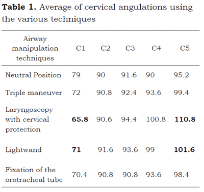
In C1 the minimum value measured with laryngoscopy was 60° and the maximum 72°. The minimum value with the lightwand was 66° and the maximum 82°. In C5 the minimum value measured with laryngoscopy was 100°, and the maximum 120°. With the lightwand, the minimum value obtained was 98°, and the maximum 112°.
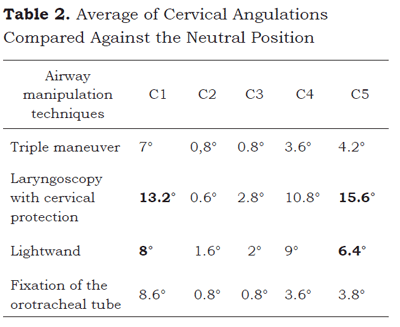
Later on, when comparing the angulations obtained using the maneuvers with the patient in neutral position, the degrees of motion in each cervical segment assessed were observed, as shown in Table 2.
DISCUSSION
Cervical trauma occurs in 1.5 % or every major trauma; it is usually caused by traffic accidents in young men 15 to 35 years old and by falls or other penetrating and sports injuries (8). Taking into account that the incidence of difficult airway may be higher in these patients, and that up to 28 % of the legal claims for anesthesia-related deaths and permanent brain injury are due to respiratory events -according to the analysis of Closed Claims of the American Society of Anesthesia (9)- it is mandatory for anesthesiologist to do an optimal management of the airway in these patients.
The current recommendations for cervical trauma indicate orotracheal intubation with immobilization or manual cervical protection (10). However, very often the immobilization maneuver hinders the visualization of the larynx and creates difficulties for intubation (11-13). Hence, the use of multiple devices has been described to facilitate the visualization of the glottis and orotracheal intubation.
Studies aimed at assessing the impact of using Macintosh blades showed that the longer extension occurs between the occiput and C1, and between C1 and C2; the extension is shorter in the sub-axial segments (1,14,15). Comparisons between direct laryngoscopy with Miller blades versus Macintosh blades have shown that despite a shorter cervical extension with the Miller blades (9.5+-3.8 vs. 12.1 +-4.9, p = 0.012), such shorter cervical extension is neglectable and probably unimportant for clinical practice (16).
The lightwand is a useful tool to facilitate orotracheal intubation in difficult airway patients (cervical trauma, micrognatia, macroglosia, mandibular immobility) (17-20). The lightwand can be used alone or in combination with other devices to facilitate airway access. Furthermore, in contrast with fiberoptic instruments, lightwands are easily cleaned and sterilized and are easy to transport, require minimal prep in emergency situations and are readily available (21).
No differences in mobility of the cervical spine have been found during orotracheal intubation with the lightwand vs. the flexible fiberscope (12° + -6° vs. 11° + -5°; p = 0.5). The segments with larger displacement are C0-1 and C1-2, exhibiting greater mobility during the introduction of both devices (22). Intubation time is shorter with the flexible fiberscope (34 + -17s vs. 60 + -15 s; p < 0.001) (22).
It has been shown that a shorter time is required for orotracheal intubation using the lightwand vs. Fastrach (23.9 + -9 s vs. 71 + -24 s) as well as a higher first attempt success rate (90,5 % vs. 79,8 %) (23).
The displacement of the cervical spine in the four segments assessed (Occiput- C1, C1-2, C2-5, C5-toracic segment) is 57 % less using the lightwand versus laryngoscopy with the Macintosh blade (p=0.03), with no significant difference in the time required for intubation using any of the two techniques. Although the intubation with the Glidescope is comparable to the lightwand in terms of cervical displacement, the time required using the Glidescope is 62 % longer that with the lightwand (24).
In our study, the critical points in terms of movement of the cervical spine were C1 and C5, which is comparable to the results of previous studies. When comparing both intubation techniques used in the study (laryngoscopy with cervical protection vs. lightwand intubation), the cervical angulation at the critical points -C1 and C5- is less when using the lightwand (8° vs. 13.2° in C1, and 6.4° vs. 15.6° in C5), with regards to the measurements taken with the head in neutral position. The triple maneuver and the fixation of the endotracheal tube result in smaller angulations as compared with the angulations obtained during the intubation maneuver.
We acknowledge the limitation of this study in terms of the size of the sample used which otherwise would enhance the validity of the results. However, the analysis of the results indicates that intubation with the lightwand not only reduces the cervical angulation as compared to intubation with the laryngoscope and cervical protection, but is also comparable to the Bullard laryngoscope that reports up to 4° angulations in C5, and to the fiberbroncoscope that reports up to 7° angulations in C1.
It is important to mention that only straight blades (Miller) were used in this study to assess cervical mobility, because there is a high percentage of routine utilization of Miller blades for direct laryngoscopy at our institution. However, the use of curved blades (Macintosh) is a common practice among anesthetists and one of the key reasons for choosing one against the other is the preference and experience of the practicing anesthetist, in addition to availability of the devices. Although there are no studies showing the preference of anesthetists in Colombia for any of these two devices, we believe that there could be a limitation for the applicability of the results of the study.
Lightwand intubation is a useful tool in cervical trauma patients with an empty stomach and should be considered an alternative airway management approach in these patients.
ACKNOWLEDGEMENTS
We would like to express our gratitude to Dr. Orlando Díaz and Dr. Sonia Bermúdez, of the Department of diagnostic Images of the Santa Fe Foundation, Bogotá and to Dr. Alexandra Chaves, for their collaboration with this study.
REFERENCES
1. Sawin PD, Todd MM, Traynelis VC, Farrell SB, Nader A, Sato Y, et al. Cervical spine motion with direct laryngoscopy and orotracheal intubation: an in vivo cinefluoroscopic study of subjects without cervical abnormality. Anesthesiology. 1996; 85(1):26-36.
2. Langeron O, Birenbaum A, Amour J. Airway management in trauma. Minerva Anestesiol. 2009;75(5): 307-11.
3. Suderman VS, Crosby ET, Lui A. Elective oral tracheal intubation in cervical spine- injured adults. Can J Anaesth. 1991;38(6):785-9.
4. Calder I, Calder J, Crockard HA. Difficult direct laryngoscopy in patients with cervical spine disease. Anaesthesia. 1995;50(9):756-63.
5. Hastings RH, Wood PR. Head extension and laryngeal view during laryngoscopy with cervical spine stabilization maneuvers. Anesthesiology. 1994;80(4):825-31.
6. Lennarson PJ, Smith D, Todd MM, Carras D, Sawin PD, Brayton J, et al. Segmental cervical spine motion during orotracheal intubation of the intact and injured spine with and without external stabilization. J Neurosurg. 2000;92(2Suppl):201-6.
7. McLeod ADM, Calder I. Spinal cord injury and direct laryngoscopy--the legend lives on. Br J Anaesth. 2000;84(6):705-9.
8. Bryson BL, Mulkey M, Mumford B. Cervical spine injury, incidence and diagnosis. J Trauma. 1986;26(7):669-74.
9. Cheney FW, Posner KL, Lee LA, Caplan RA, Domino KB. Trends in anesthesia-related death and brain damage: a closed claims analysis. Anesthesiology. 2006;105(6):1081-6.
10. American College of Surgeons Committee on Trauma. Advanced trauma life support course for doctors. 6th ed. Chicago: American College of Surgeons; 1997.
11. Nolan JP, Wilson ME. Orotracheal intubation in patients with potential cervical spine injuries. An indication for the gum elastic bougie. Anaesthesia. 1993;48(7):630-3.
12. Hastings RH, Wood PR. Head extension and laryngeal view during laryngoscopy with cervical spine stabilization maneuvers. Anesthesiology. 1994;80(4):825-31.
13. Heath KJ. The effect of laryngoscopy of different cervical spine immobilisation techniques. Anaesthesia. 1994;49(10):843-5.
14. Hastings RH, Vigil AC, Hanna R, Yang BY, Sartoris DJ. Cervical spine movement during laryngoscopy with the Bullard, Macintosh, and Miller laryngoscopes. Anesthesiology. 1995; 82(4):859-69.
15. Watts AD, Gelb AW, Bach DB, Pelz DM. Comparison of the Bullard and Macintosh laryngoscopes for endotracheal intubation of patients with a potential cervical spine injury. Anesthesiology. 1997; 87(6): 1335-42.
16. LeGrand SA, Hindman BJ, Dexter F, Weeks JB, Todd MM. Craniocervical motion during direct laryngoscopy and orotracheal intubation with the Macintosh and Miller blades: an in vivo cinefluoroscopic study. Anesthesiology. 2007;107(6):884-91.
17. Hung OR, Stewart RD. Lightwand intubation: I--a new lightwand device. Can J Anaesth. 1995;42(9):820-5.
18. Hung OR, Pytka S, Morris I, Murphy M, Steward RD. Lightwand intubation: II-- clinical trial of a new lightwand for tracheal intubation in patients with difficult airways. Can J Anaesth. 1995;42(9):826-30.
19. Paschen HR. Difficult airway management in trauma-transillumination devices. Trauma Care 99, Proceedings of the 12th Annual Trauma Anesthesia and Critical Care Symposium; 13-15 Mayo 1999, Chicago.
20. Davis L, Cook-Sather SD, Schreiner MS. Lighted stylet tracheal intubation: a review. Anesth Analg. 2000;90(3):745-56.
21. Smith CE, DeJoy SJ. New equipment and techniques for airway management in trauma. Curr Opin Anaesthesiol. 2001;14(2):197-209.
22. Houde B, Williams SR, Cadrin-Chênevert A, Drolet P. A comparison of cervical spine motion during orotracheal intubation with the trachlight(r) or the flexible fiberoptic bronchoscope. Anesth Analg. 2009;108(5):1638-43.
23. Inoue Y, Koga K, Shigematsu A. A comparison of two tracheal intubation techniques with trachlight and Fastrach in patients with cervical spine disorders. Anesth Analg. 2002;94(3):667-71.
24. Turkstra TP, Craen R, Pelz DM, Gelb AW. Cervical spine motion: a fluoroscopic comparison during intubation with lighted stylet, GlideScope, and Macintosh laryngoscope. Anesth Analg. 2005;101(3):910-5.
Conflicto de intereses: ninguno declarado
1. Sawin PD, Todd MM, Traynelis VC, Farrell SB, Nader A, Sato Y, et al. Cervical spine motion with direct laryngoscopy and orotracheal intubation: an in vivo cinefluoroscopic study of subjects without cervical abnormality. Anesthesiology. 1996; 85(1):26-36. [ Links ]
2. Langeron O, Birenbaum A, Amour J. Airway management in trauma. Minerva Anestesiol. 2009;75(5): 307-11. [ Links ]
3. Suderman VS, Crosby ET, Lui A. Elective oral tracheal intubation in cervical spine- injured adults. Can J Anaesth. 1991;38(6):785-9. [ Links ]
4. Calder I, Calder J, Crockard HA. Difficult direct laryngoscopy in patients with cervical spine disease. Anaesthesia. 1995;50(9):756-63. [ Links ]
5. Hastings RH, Wood PR. Head extension and laryngeal view during laryngoscopy with cervical spine stabilization maneuvers. Anesthesiology. 1994;80(4):825-31. [ Links ]
6. Lennarson PJ, Smith D, Todd MM, Carras D, Sawin PD, Brayton J, et al. Segmental cervical spine motion during orotracheal intubation of the intact and injured spine with and without external stabilization. J Neurosurg. 2000;92(2Suppl):201-6. [ Links ]
7. McLeod ADM, Calder I. Spinal cord injury and direct laryngoscopy--the legend lives on. Br J Anaesth. 2000;84(6):705-9. [ Links ]
8. Bryson BL, Mulkey M, Mumford B. Cervical spine injury, incidence and diagnosis. J Trauma. 1986;26(7):669-74. [ Links ]
9. Cheney FW, Posner KL, Lee LA, Caplan RA, Domino KB. Trends in anesthesia-related death and brain damage: a closed claims analysis. Anesthesiology. 2006;105(6):1081-6. [ Links ]
10. American College of Surgeons Committee on Trauma. Advanced trauma life support course for doctors. 6th ed. Chicago: American College of Surgeons; 1997. [ Links ]
11. Nolan JP, Wilson ME. Orotracheal intubation in patients with potential cervical spine injuries. An indication for the gum elastic bougie. Anaesthesia. 1993;48(7):630-3. [ Links ]
12. Hastings RH, Wood PR. Head extension and laryngeal view during laryngoscopy with cervical spine stabilization maneuvers. Anesthesiology. 1994;80(4):825-31. [ Links ]
13. Heath KJ. The effect of laryngoscopy of different cervical spine immobilisation techniques. Anaesthesia. 1994;49(10):843-5. [ Links ]
14. Hastings RH, Vigil AC, Hanna R, Yang BY, Sartoris DJ. Cervical spine movement during laryngoscopy with the Bullard, Macintosh, and Miller laryngoscopes. Anesthesiology. 1995; 82(4):859-69. [ Links ]
15. Watts AD, Gelb AW, Bach DB, Pelz DM. Comparison of the Bullard and Macintosh laryngoscopes for endotracheal intubation of patients with a potential cervical spine injury. Anesthesiology. 1997; 87(6): 1335-42. [ Links ]
16. LeGrand SA, Hindman BJ, Dexter F, Weeks JB, Todd MM. Craniocervical motion during direct laryngoscopy and orotracheal intubation with the Macintosh and Miller blades: an in vivo cinefluoroscopic study. Anesthesiology. 2007;107(6):884-91. [ Links ]
17. Hung OR, Stewart RD. Lightwand intubation: I--a new lightwand device. Can J Anaesth. 1995;42(9):820-5. [ Links ]
18. Hung OR, Pytka S, Morris I, Murphy M, Steward RD. Lightwand intubation: II-- clinical trial of a new lightwand for tracheal intubation in patients with difficult airways. Can J Anaesth. 1995;42(9):826-30. [ Links ]
19. Paschen HR. Difficult airway management in trauma-transillumination devices. Trauma Care 99, Proceedings of the 12th Annual Trauma Anesthesia and Critical Care Symposium; 13-15 Mayo 1999, Chicago. [ Links ]
20. Davis L, Cook-Sather SD, Schreiner MS. Lighted stylet tracheal intubation: a review. Anesth Analg. 2000;90(3):745-56. [ Links ]
21. Smith CE, DeJoy SJ. New equipment and techniques for airway management in trauma. Curr Opin Anaesthesiol. 2001;14(2):197-209. [ Links ]
22. Houde B, Williams SR, Cadrin-Chênevert A, Drolet P. A comparison of cervical spine motion during orotracheal intubation with the trachlight(r) or the flexible fiberoptic bronchoscope. Anesth Analg. 2009;108(5):1638-43. [ Links ]
23. Inoue Y, Koga K, Shigematsu A. A comparison of two tracheal intubation techniques with trachlight and Fastrach in patients with cervical spine disorders. Anesth Analg. 2002;94(3):667-71. [ Links ]
24. Turkstra TP, Craen R, Pelz DM, Gelb AW. Cervical spine motion: a fluoroscopic comparison during intubation with lighted stylet, GlideScope, and Macintosh laryngoscope. Anesth Analg. 2005;101(3):910-5. [ Links ]
