


Servicios Personalizados
Revista
Articulo
 texto en
  Español (pdf)
 Articulo en XML
 Referencias del artículo
 Enviar articulo por email
Indicadores
-
 Citado por SciELO
 -
 Accesos
Links relacionados
-
 Citado por Google
 -
 Similares en
SciELO
Similares en
SciELO -
 Similares en Google
Compartir

 Permalink
Colombian Journal of Anestesiology
versión impresa ISSN 0120-3347
Rev. colomb. anestesiol. v.38 n.3 Bogotá jul./sep. 2010
Rigid fiberscope intubation of a patient awake with remifentanil sedation
Juan Carlos Bocanegra*, Ángela María Ríos Medina**, Óscar David Aguirre Ospina***
* MD. Especialista en Anestesiología y Reanimación, Hospital SES de Caldas. Manizales, Colombia bocanegra67@ yahoo.com.
** MD. Residente de Anestesiología y Reanimación. Universidad de Caldas. Manizales. Colombia.
*** MD. Residente de Anestesiología y Reanimación. Universidad de Caldas. Manizales. Colombia.
Recibido: febrero 26 de 2010. Enviado para modificaciones: marzo 25 de 2010. Aceptado: junio 15 de 2010.
SUMMARY
Introduction. When choosing the appropriate management for a difficult airway patient, either established or suspected, several intervention options should be considered. Some management algorithms recommend awake intubation under direct laryngoscopy or optical instruments. Rigid and semi-rigid stylets are optical devices developed for managing the difficult airway that have proven to be fast, non-traumatic and reliable.
Objectives. To describe the use of different strategies to approach the difficult airway in an emergency surgery, using the Airway RIFL (rigid intubation fiberoptic laryngoscope) as one of the options.
Methodology and results. This is a case of a 69-yr old patient admitted to the ER for hemostasis and surgery of a chest neoplasia. The patient was considered a difficult airway patient because of a history of multiple tumor resections and radiotherapy in the airway, resulting in a notorious facial deformity. Intubation on a full stomach. Topical anesthesia was considered the first choice for airway management, remifentanil sedation and direct laryngoscopy but this is not possible due to a limited oral opening: three nasal intubation attempts were made but failed. Then it was decided to use the Airway RIFL device and the procedure was successful.
Conclusions. When dealing with a difficult airway patient, having several management strategies at hand, local anesthesia and sedation, allow for excellent patient collaboration and a fast orotracheal intubation using a rigid fiberscope, even under emergency situations.
Keywords: Endotracheal intubation, optical devices, anesthesia, awake sedation, aspiration pneumonia(Source: MeSH,NLM)
CASE DESCRIPTION
A 69-yr old male patient, scheduled for emergency surgery for direct hemostasis of a 10 cm by 10 cm bleeding tumor lesion, in the anterior right hemithorax. Last meal was one hour prior to admission for surgery.
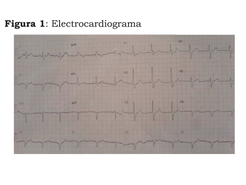
Patient's History: chronic high blood pressure managed with enalapril 20 mg every 12 hr; heavy smoker until seven years ago, maxillary basal cell carcinoma treated with primary resection on several occasions, relapses and need to perform a maxillectomy, resection of the orbital floor, turbinates, lip, gum and palate. Additionally, 10 sessions of radiotherapy were administered. Therapeutic management had been stopped one year ago. The pre-operative paraclinical tests were as follows: hemoglobin 9 gr/dl, platelets 120,000; tromboplastin time 15.4/10.4. EKG: right ventricle hypertrophy (Figure 1).
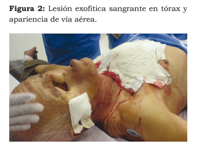
Physical examination with the patient awake-BP: 90/50, HR: 86 min y RR: 18 min. Marked facial deformity with soft tisue discontinuity in the right maxillary area, limited mouth opening of 1.5 cm. 10 cm x 10 cm exophytic chest lesion with moderate active bleeding (Figure 2).
INDUCTION OF ANESTHESIA
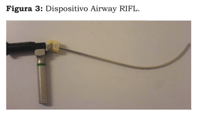
Application of topical anesthesia with 2 % lidocaine in the mouth and oropharinx, followed by remifentanil infusion 0.1 mcg/kg/min, maintaining spontaneous breathing. Direct laryngoscopy was not an option due to the limited mouth opening. Several blind nasal intubation attempts failed so the decision was to do use the Airway RIFL (rigid intubation fiberoptic laryngoscope) to attempt oral intubation (Figure 3).
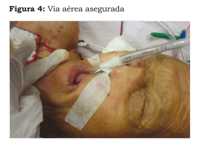
The device was prepared with lidocaine gel and a 7.5 tube; traction of the lower jaw was performed for a mid-line approach, maintaining the vision on the proximal optic of the device. Upon visualizing the epiglottis and entering the glottis, the endotracheal tube is advanced up to the trachea, the Airway RIFL is removed and the endotracheal tube is fixed (Figure 4), followed by the immediate administration of 50 mg of propo-fol I.V.
No hemodynamic instability or hypoxia episodes occurred during the procedure. The anesthesia for the surgical procedure was maintained with 2 % sevofluorane and remifentanil 0.15 mcg/kg/min.
Once the surgery is complete, the remifentanil and sevoflourane infusion is suspended, the patient recovers the spontaneous ventilation, the awareness and airway reflexes. At this point, the orotracheal tube is removed. The patient is observed for two additional hours free of complications.
DISCUSSION
Endotracheal intubation is an incredible achievement in the history of anesthesia. Late in the 18th Century, the London Royal Humane Society used endotracheal intubation to resuscitate drowned people. Almost one hundred years later, McEwan (1) reported one case of tracheal digital intubation in a patient awake under general anesthesia with chloroform to resect a tongue tumor. Then in 1928, Magill (2) adopts blind nasal tracheal intubation as part of the anesthesia procedure.
However, it was Alfred Kirstein in 1895 that disseminated the use of tracheal intubation to ventilate patients unable to maintain the patency of the airway on their own. The introduction of direct laryngoscopy increased the success rate of tracheal intubation, making it more effective both in surgical and non-surgical events. Later on, the introduction of the de Miller and Macintosh blades in the forties, expanded the acceptance and use of these devices, strengthening the techniques used until then (3).
Currently other airway management devices have evolved, using the same patency and visualization principles, enabling the physician to deal with different scenarios that were unsuccessfully managed in the past.
However, due to the large number of devices available for managing the airway, careful consideration should be given to their individual advantages and disadvantages; therefore, there is a need to choose the most appropriate device for each particular patient (4). But making the right choice is not enough; the anesthesiologist must be trained to use each one of them (5).
Consequently, the final choice of a particular device should also be based on the following considerations: frequency of use at the institution, easy installation and care of the device (6).
The development of intubation techniques using optical devices has resulted in the design of a large number of these devices; however, the difficulty to develop the necessary skills, their high cost, cleansing and disinfection care and the difficulty to visualize the laryngeal structures due to the presence of secretions or blood, have all limited their use (7). For instance, only 59 % of the anesthesiologist in the U.S. reported having the skills to intubate with fiberoptic devices (8).
Rigid, indirect vision devices have been developed as an alternative to flexible bronchoscope intubation and direct laryngoscopy, with favorable success rates reported in normal and difficult airway intubation (9,10). There are however few reports on their usefulness in emergency cases.
Some algorithms for managing the difficult airway recommend intubating with the patient awake, as a safe method to ensure a patent airway in difficult airway patients, whether established or suspected. This procedure is usually accomplished through direct laryngoscopy and there is a rare need to use additional devices to complete the tracheal intubation (11).
In rare occasions, when the patency of the airway with the patient awake and direct laryngoscopy is not achieved and the nature of the emergency calls for immediate intervention, additional techniques should be used, including surgical methods or a combination of non-surgical techniques for managing the airway (11).
Some of the non-surgical techniques for managing the difficult airway include the rigid and semi-rigid stylets. The former devices allow for indirect visualization of the glottis through an image transmitted to an optical display, using a package containing a light beam and fiberoptics travelling through the instrument and allows for pre-loading the endotracheal tube so that it moves forward as soon as the entrance to the airway is visualized.
The device has a curved shape and a 70° and 90° distal angle, designed to scan around the tongue and hypopharynx and to visualize the laryngeal structures. (12). The curved shape is rigid and cannot be modified. Alignment of the three axes is not required for their use, which is particularly advantageous in situations when any movement may give rise to additional complications (trauma, for instance). The advantages include: easy to adapt to the laryngeal anatomy, passes onto the oropharynx through smaller mouth openings and less hemodynamic response to the procedure (13). These devices have proven to be fast and atraumatic for the management of the difficult airway under diverse clinical conditions (14,15).
Two of the rigid stylets are the bonfils retromolar intubation fiberscope and the Airway RIFL (rigid intubation fiberoptic laryngoscope) (16). Managing the difficult airway with a rigid fiberscope in a patient who is awake requires concomitant sedation and the use of local anesthesia techniques; however, the patient's cooperation is critical and the informed consent should be obtained.
Whenever other strategies have failed, the rigid fiberscope may indeed be used for awake intubation of the difficult airway patient in an emergency situation.
REFERENCIAS
1. McEwan W. Clinical observations on the introduction of tracheal tubes by the mouth instead of performing tracheotomy or laryngotomy. BMJ. 1880;2(1022):163-5.
2. Magill IW. Endotracheal anaesthesia. Proc R Soc Med. 1928;22(2):83-8.
3. Macintosh RR. A new laryngoscope. Lancet. 1943;1:205.
4. Davis L, Cook-Sather S, Schreiner M. Lighted stylet tracheal intubation: a review. Anesth Analg. 2000;90(3):745-56.
5. Marco CA, Marco AP. Airway adjuncts. Emerg Med Clin N Am. 2008;26(4):1015-27.
6. Levitan RM, Kush S, Hollander JE. Devices for difficult airway management in academic emergency departments: results of a national survey. Ann Emerg Med. 1999;33(6):694-8.
7. Reeder TJ, Brown CK, Norris DL. Managing the difficult airway: a survey of residency directors and a call for change. J Emerg Med. 2005;28(4):473-8.
8. Ezri T, Szmuk P, Warters RD, Katz J, Hagberg CA. Difficult airway management practice patterns among anesthesiologists practicing in the United States: have we made any progress? J Clin Anesth. 2003;15(6):418-22.
9. Liem EB, Bjoraker DG, Gravenstein D. New options for airway management: intubating fibreoptic stylets. Br J Anaesth. 2003;91(3):408-18.
10. Pfitzner L, Cooper MG, Ho D. The Shikani Seeing Stylet for difficult intubation in children: initial experience. Anaesth Intensive Care. 2002;30(4):462-6.
11. Gempeler F, Devis A, Pedraza P. Intubación con paciente despierto con fibroscopio retromolar de Bonfils bajo sedación con dexmedetomidina. Reporte de casos. Rev Col Anest. 2009;37:49-56.
12. Kovacs G, Law A, Petrie D. Awake fiberoptic intubation using an optical stylet in ananticipated dificult airway. Ann Emerg Med. 2007;49(1):81-3.
13. Brimacombe J, Keller C, Künzel KH, Gaber O, Boehler M, Pühringer F. Cervical spine motion during airway management:a cinefluoroscopic study of the posteriorly destabilized third vertebrae in human cadavers. Anesth Analg. 2000;91(5):1274-8.
14. Rudolph C, Schlender M. Clinical experiences with fiber optic intubation with the Bonfils intubation fiberscope. Anaesthesiol Reanim. 1996;21(5):127-30.
15. Hagberg CA. Special devices and techniques. Anesthesiology Clin N Am. 2002;20(4):907-32.
16. Hung O, Murphy M. Management of the difficult and failed airway. New York: McGraw Hill; 2007.
Conflicto de intereses: ninguno declarado.
1. McEwan W. Clinical observations on the introduction of tracheal tubes by the mouth instead of performing tracheotomy or laryngotomy. BMJ. 1880;2(1022):163-5. [ Links ]
2. Magill IW. Endotracheal anaesthesia. Proc R Soc Med. 1928;22(2):83-8. [ Links ]
3. Macintosh RR. A new laryngoscope. Lancet. 1943;1:205. [ Links ]
4. Davis L, Cook-Sather S, Schreiner M. Lighted stylet tracheal intubation: a review. Anesth Analg. 2000;90(3):745-56. [ Links ]
5. Marco CA, Marco AP. Airway adjuncts. Emerg Med Clin N Am. 2008;26(4):1015-27. [ Links ]
6. Levitan RM, Kush S, Hollander JE. Devices for difficult airway management in academic emergency departments: results of a national survey. Ann Emerg Med. 1999;33(6):694-8. [ Links ]
7. Reeder TJ, Brown CK, Norris DL. Managing the difficult airway: a survey of residency directors and a call for change. J Emerg Med. 2005;28(4):473-8. [ Links ]
8. Ezri T, Szmuk P, Warters RD, Katz J, Hagberg CA. Difficult airway management practice patterns among anesthesiologists practicing in the United States: have we made any progress? J Clin Anesth. 2003;15(6):418-22. [ Links ]
9. Liem EB, Bjoraker DG, Gravenstein D. New options for airway management: intubating fibreoptic stylets. Br J Anaesth. 2003;91(3):408-18. [ Links ]
10. Pfitzner L, Cooper MG, Ho D. The Shikani Seeing Stylet for difficult intubation in children: initial experience. Anaesth Intensive Care. 2002;30(4):462-6. [ Links ]
11. Gempeler F, Devis A, Pedraza P. Intubación con paciente despierto con fibroscopio retromolar de Bonfils bajo sedación con dexmedetomidina. Reporte de casos. Rev Col Anest. 2009;37:49-56. [ Links ]
12. Kovacs G, Law A, Petrie D. Awake fiberoptic intubation using an optical stylet in ananticipated dificult airway. Ann Emerg Med. 2007;49(1):81-3. [ Links ]
13. Brimacombe J, Keller C, Künzel KH, Gaber O, Boehler M, Pühringer F. Cervical spine motion during airway management:a cinefluoroscopic study of the posteriorly destabilized third vertebrae in human cadavers. Anesth Analg. 2000;91(5):1274-8. [ Links ]
14. Rudolph C, Schlender M. Clinical experiences with fiber optic intubation with the Bonfils intubation fiberscope. Anaesthesiol Reanim. 1996;21(5):127-30. [ Links ]
15. Hagberg CA. Special devices and techniques. Anesthesiology Clin N Am. 2002;20(4):907-32. [ Links ]
16. Hung O, Murphy M. Management of the difficult and failed airway. New York: McGraw Hill; 2007. [ Links ]
