


 text in
  English (pdf)
 Article in xml format
 Article references
 Send this article by e-mail
 Cited by SciELO
  Cited by Google
  Similars in
SciELO
Similars in
SciELO  Similars in Google
 Permalink
INTRODUCTION
Microorganisms are the main etiologic agent for pulp and periapical disease; therefore, one of the main objectives of the endodontic therapy is the three-dimensional sealing of the root canal with biocompatible materials1,2,3 to prevent the entry of new microorganisms or their reproduction in the canal system. The success of the endodontic treatment depends largely on the sealing of all routes of filtration of microbial irritators, preventing reinfection within the canal system.1 It has been reported that 40% to 60% of endodontic failures are due to poor obturations that cause microleakage.4
Various obturation techniques are currently available to ensure the effective sealing of root canals. However, there has been controversy around traditional techniques such as lateral compaction and warm vertical compaction, since they do not provide an airtight sealing but offer predictable outcomes.1 Lateral compaction does not meet the requirement of providing homogeneous sealing of the root canal, unlike the warm vertical compaction technique, which provides homogeneous sealing allowing adaptation of material to the walls, and even sealing root canal irregularities.5,6 The cold single-cone technique, on the contrary, is being widely used nowadays due to its simplicity and low cost, even though it does not provide a three- dimensional sealing of the root canal, in contrast to the evidence, which has demonstrated that this technique has high failure rates due to poor sealing.7,8,9,10 Dye penetration and microbial filtration are the two most common methods to evaluate microleakage of endodontic obturations,11 but microbial filtration is the most relevant method, since it fits clinical conditions. This study used the dual chamber technique, a method that was first introduced by Torabinejad et al12 Figure 1.

Figure 1 Assembly of tube (upper and lower chamber) and marker of bacterial presence (phenol red) in lower chamber
Enterococcus faecalis is a Gram+ facultative anaerobe microorganism that has been widely studied in endodontics. This microorganism is linked to endodontic failure due to its morphological characteristics and virulence which allow it to survive in extreme conditions within the root canal.13,14
The purpose of this study was to evaluate bacterial microleakage of E. faecalis in three endodontic obturation techniques: lateral compaction, warm vertical compaction (System B and Obtura II), and single cone, searching for the technique that provides patients with the best possible root canal sealing.
MATERIALS AND METHODS
Preparation of samples
Thirty caries-free premolars extracted for orthodontic reasons were used in this study. The teeth were immersed in 5.25% sodium hypochlorite (NaOCL) for 15 min to remove organic tissue from the root surface, and any remnants were removed with sterile gauze pads. The teeth were later rinsed and stored in sterile saline solution; digital x-rays were taken (Radiovisiografo Gendex, VixWin Platinium) to verify that the teeth were single- rooted and free from internal or external resorptions or calcifications. The external surface of roots was observed with a microscope to verify the absence of fractures (Medical Zumax, Jiangsu, China).
The teeth’s crowns were removed with diamond discs (Miltex, Germany) and roots were standardized to a length of 10 mm. The working length was defined as 1 mm shorter with respect to the point in which the 10 K-file (Dentsply, Maillefer, Ballaigues, Switzerland) appeared through the apical foramen.
Preparation and obturation
All teeth were prepared with the ProTaper Universal (Dentsply, Maillefer, Ballaigues, Switzerland) corono-apical rotary technique up to file F3. During preparation, the samples were irrigated between one file and the other with 2 mL of 5.25% NaOCL. Next, the samples were irrigated with 2 mL of 17% EDTA (MDCleanser™, MetaBiomed Co. Ltd., Cheongju City, Chungbuk, Korea) for 3 minutes, followed by 5 mL of 5.25% NaOCL to remove smear layer. The root canals were finally rinsed with 5 mL of sterile saline solution and dried with sterile paper tips ProTaper F3 (Dentsply, Maillefer, Ballaigues, Switzerland). All preparations and obturations were performed by a single operator (an endodontist).
The teeth were randomly divided into three experimental groups (n = 8 each), three negative and three positive control groups (n = 6). The obturations were made in a laminar flow cabinet, under aseptic conditions, and the same sealing cement was used in all the experimental groups (Top Seal - Dentsply, Maillefer, Ballaigues, Switzerland).
Lateral compaction technique
Teeth from the first group were filled using the lateral compaction technique. A ProTaper F3 cone was used as master cone and brought to the working length with sealing cement Top Seal. Lateral compaction was made using standardized digital spacers (Dentsply, Maillefer, Ballaigues, Switzerland), corresponding to 2% taper guttapercha cones (Dentsply, Maillefer, Ballaigues, Switzerland). Obturations were considered complete when the spacer did not enter the root canal area (10 mm).
Vertical compaction technique
The System B (Analytic Endodontics) and Obtura II (Obtura II; Obtura Corp., Fenton, MO) warm vertical compaction technique was used. A ProTaper F3 cone was used as master cone and brought to the working length with sealing cement. Then, System B was used for obturation of the 3 apical millimeters, removing the heat source and conducting vertical compaction with a manual compactor (Machtou’s heat-carrier pluggers; Dentsply Maillefer, Ballaigues, Switzerland). Finally, obturation of the middle and coronal third was completed with the guttapercha injection system Obtura II up to 10 mm of root length.
Single-cone technique
A ProTaper F3 cone was used as master cone and brought to the working length with sealing cement; it was sectioned at 10 mm, by the site of root end. Three teeth were used for the negative control, which were prepared but did not receive any type of obturation neither were inoculated with the microorganism; three teeth were also used for the positive control, and these were prepared but did not receive any type of obturation and were inoculated with the microorganism.
All teeth were stored at 37 °C and 100% humidity for 8 hours as recommended by the manufacturer to achieve hardening of the Top Seal sealing cement prior to the experiment.
Microleakage test
Preparation of the sample
The dual chamber technique was used for microleakage testing. Plastic tubes were selected for this model. The plastic tubes were chemically sterilized with Alkacime for 30 min and ultraviolet light for 30 min. They were later thoroughly rinsed with sterile saline solution to avoid remnants of Alkacime. In addition, the teeth were sterilized in an autoclave, in individual packages at 121 °C for 50 min. Epoxy resin was used to seal the periphery of teeth, except for the last 3 mm, to avoid microleakage through sites other than the apical third.
Bacterial filtration technique
Teeth were placed in plastic tubes, using the lower and upper chamber method Figure 1. The periphery of the union of tooth and vial was sealed with acrylic to prevent microfiltration. Before inserting each tooth, the vial’s lower chamber was filled with 4.8 ml of BHI broth (Merck Eurolab, Darmstadt, Germany), which contained phenol red marker and covered the 3 apical millimeters of samples. An ATCC 29212 E faecalis strain was used as microleakage marker. The strain was isolated in Tryptic Soy Agar (TSA), preparing suspensions in BHI, which were adjusted by spectrophotometer to 0.5 McFarland (1.5 X 10⁸ UFC/mL). Based on this suspension, and after 5 hours, to get a microorganism in its most active phase and verify sterility of vials and teeth, 2 ml were inoculated in the upper chamber and teeth were incubated at 37 °C. The culture medium of the upper chamber was changed every 48 hours until showing signs of microfiltration, so samples were evaluated visually every 24 hours.
Microleakage was assessed based on color change of the medium in the lower chamber from red to yellow. The entire process of culture mediums assembly and union of teeth to vials was conducted in a laminar flow cabinet to ensure aseptic conditions.
This project was submitted to the Universidad Cooperativa de Colombia Bioethics Committee, which considered it a low-risk project according to Resolution 8430 of 1993, also because this School is equipped with microbiology laboratories that comply with the technical standards for the management of the type of microorganisms used in the study.
RESULTS
The samples were observed for 21 days. All positive control samples showed filtration by the second day of incubation. The negative control samples showed no filtration throughout the experimental period.
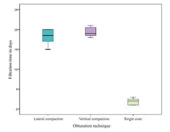
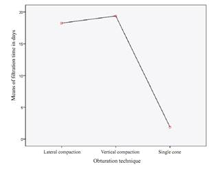
The results were processed using version 21 of the SPSS software. Descriptive statistical analysis of filtration time was conducted, finding out an average of 18.25 microfiltration time for the lateral compaction group, while for the warm vertical compaction group this average was 19.38, and 1.88 for the single cone group Table 1.
Table 1 Summary, average filtration time, means comparison, and multiple comparisons test
| Obturation technique | Microfiltration | 95% confidence interval | |||
|---|---|---|---|---|---|
| Average in days | Lower limit | Upper limit | |||
| Filtration time in days | Lateral compaction | 18.25 | 16.65 | 19.85 | |
| Vertical compaction | 19.38 | 18.38 | 20.37 | ||
| Single cone | 1.88 | 1.18 | 2.57 | ||
| Significance | |||||
| ANOVA test for mean differences | 0.000 | ||||
| Technique | vs. Technique | Significance | |||
| Bonferroni test for multiple comparisons to establish which groups present differences. | Lateral compaction | Single cone | 0.000 | ||
| Vertical compaction | Single cone | 0.000 | |||
| Lateral compaction | Vertical compaction | 0,357 | |||
A Shapiro-Wilk normality test was conducted, finding out that all study groups showed normal distribution. For comparison among groups, a one-way ANOVA test was conducted with a 95% confidence level (p = 0.000) (see table 1 finding out statistically significant differences among groups. A post-test was conducted using Bonferroni and Tukey’s test Table 1 for multiple comparisons, and as a result both the warm vertical compaction group and the lateral compaction group showed statistically significant differences in comparison to the single-cone group. However, there were no statistically significant differences between the warm vertical compaction group and the lateral compaction groups.
The results showed that the single-cone group had a shorter filtration time compared to the other two groups. Figure 2 shows microfiltration distribution time according to technique.
DISCUSSION
Several techniques are currently available to evaluate microleakage in endodontics. Gómez et al15 grouped microfiltration techniques as follows: air and pressure, microbiological studies, studies with radioisotopes, neutron activation analysis, electrochemical studies, studies with electron scanning microscope, studies with chemical markers, and dye penetration studies.
The authors note that air and pressure studies and electrochemical studies are obsolete; other studies are not easily available since they are too sophisticated, such as neutron activation analysis or studies with radioisotopes. Other studies are more similar to clinical conditions, such as microbiological studies and dye penetration studies, which were the most commonly used in the past because of their availability and simplicity. We used the bacterial microleakage technique in our study using the E. faecalis microorganism, which is highly resistant to root canal disinfection processes.
The results of this study are similar to other studies that have concluded that no material can completely seal the root canal and that no obturation technique completely avoids the microfiltration of microorganisms to the periapex.16,17,18 These studies, like ours, observed the samples for a specific period of time, but did not quantify the number of microorganisms that entered the radicular apex.
The single-cone ProTaper technique has reported problems related to gutta-percha adaptation to root canal walls, due to its inability to adapt to irregularities. In our study, this technique was the one with the highest microleakage time, with 1.88 days in average. This result differs from that by Mercês et al,17 who compared microleakage in two obturation techniques (active lateral compaction and single cone), prior preparation with the ProTaper rotary technique, and using ProTaper cones for the obturations, finding out that the samples filled with thesingle-conetech niquehad 67 day sofmicroleakage in average.17 In comparison to the lateral compaction technique, microleakage averaged 87 days, while in our study the average microleakage for the lateral compaction technique was 18.25 days.
In a 2009 microleakage study, Muñoz- Bolaños19 showed, as did the present study, the inability of the vertical compaction and lateral compaction techniques to seal after 25 days, obtaining an average microleakage time of 23.4 days for the lateral compaction technique and 23.1 days for the vertical compaction technique. It should be noted, however, that in our study we evaluated the sealing capacity to E. faecalis, while Muñoz-Bolaños used Streptococcus salivarius.
In 2015, Mathur et al20 assessed microleakage with dye penetration and bacterial microleakage for a period of 21 days and, like in the present study, demonstrated the inability of the lateral compaction and vertical compaction techniques to properly seal against microorganisms. These techniques, as well as in the present study, did not show statistically significant differences; however, the sealing values were more favorable for the vertical compaction technique. It is important to note that the microorganism Mathur et al used in their study was Proteus vulgaris.
In 2010, Ito et al assessed bacterial microleakage in three root canal obturation techniques (lateral compaction, warm vertical compaction, and single cone), finding out that there was no E. faecalis microleakage in 24 samples filled with lateral and vertical compaction, but there were 2 samples out of 14 that showed filtration with the single-cone technique by days 22 and 23.21 These results are inconsistent with our study, but this difference may be due to discrepancies in methodology.22
Pinheiro et al23 used the bacterial microleakage technique to assess the sealing capacity of various cements used in endodontic sealers, concluding that none of the tested cements (AH Plus, Epiphany, Acroseal, Endofill, and Polifi) prevented the microleakage of E. faecalis into the root apex. In the present study, we used the TopSeal resin-based cement, which also failed to prevent microleakage of the microorganism in the samples.
By contrast, Hernández-Valerín et al,24 in a 2009 study comparing microleakage of E. faecalis in root canals filled with gutta-percha using TopSeal cement and the Resilon-Epiphany obturation system, and observing the samples for 60 days, found out that samples filled with gutta-percha and TopSeal cement showed greater resistance to bacterial filtration, with an average filtration time of 12.6 days, but overall failed to block filtration. This result is similar to the one in our study, in which microleakage of the microorganism could not be avoided with any of the three techniques plus the TopSeal cement.
Our study showed a difference in microleakage between warm vertical compaction and lateral compaction, being more favorable to the warm vertical compaction technique, but with no statistically significant differences. This result, slightly favorable to the warm vertical compaction technique, may be due to the fact that-as reported by the literature- thermoplasticized gutta-percha can easily reach the root canal irregularities, which cannot be achieved with lateral compaction, since irregularities and curvatures become a difficulty for this technique. The warm compaction techniques produce greater amount of gutta-percha in comparison to lateral compaction techniques. Therefore, improving gutta-percha homogenicity and adaptation to the surface was a necessity, and thus thermoplasticized obturation was introduced in the clinical practice.25,26
CONCLUSIONS
In conclusion, the present study showed that none of the three obturation techniques used was able to avoid the microleakage of E. faecalis towards the root apex, and the warm vertical compaction and lateral compaction techniques showed the greatest resistance to microleakage, with no statistically significant differences; in numerical terms, however, the vertical compaction technique showed a slightly better performance. In contrast, the single-cone technique showed an almost complete inability to prevent microleakage, which happened in less than two days.
Due to different methodologies described in the literature to evaluate microleakage, and the disparity in results, it is suggested to perform new studies to try to establish a universal methodology for the study of microleakage, in order to make comparisons that allow to evaluate more rigorously used in endodontic obturation techniques.

