


 English (pdf)
 Article in xml format
 Article references
 Send this article by e-mail
 Cited by SciELO
  Cited by Google
  Similars in
SciELO
Similars in
SciELO  Similars in Google
 Permalink
INTRODUCTION
Angiolymphoid hyperplasia with eosinophilia (ALHE), also known as epithelioid hemangioma or histiocytoid hemangioma, is a rare inflammatory vascular alteration of unknown origin, with a benign evolution and with periods of recurrence after surgical treatment.1 It is unclear whether ALHE occurs more often in men or women, and reports in the medical literature are contradictory.2
Its pathogenesis is unknown, but it is considered to be a reactive disorder induced by various stimuli, including vascular malformations, trauma, and pregnancy.3 The presence of high serum estrogen levels (also the case in the use of oral contraceptives) is one of the hypotheses; however, currently the most accepted theory is that of a reactional vascular hyperplasia caused by different stimuli.4 A case of dermal ALHE was found to harbor a mutation in the TEK gene, which encodes the Tie-2 endothelial cell tyrosine kinase receptor, showing that certain molecular alterations may contribute to the pathogenesis of this condition.5
Clinically, there can be erythematous or brownish papules of about 0.5-1 cm in diameter, and subcutaneous plaques or lesions that mainly affect the head and neck, especially in the periauricular region. It can also appear in maxillary bones, salivary glands, muscle tissues, and skin.6 The oral mucosa is a rare site, with few reports in the literature, but when it occurs, the most affected sites are the lips, the jugal mucosa, and the tongue.4 ALHE can be asymptomatic or produce pruritus, pain, or spontaneous bleeding; however, little is known about how often symptoms occur.2
Histologically, it is characterized by well-formed but often immature vessels, which are delineated by an endothelium of epithelioid form cells associated with a prominent inflammatory infiltrate including lymphocytes, mastocytes, eosinophils, and sometimes lymphoid follicles.7 Two main histological components are found: one inflammatory and one vascular. The degree of infiltration by eosinophils varies, but the lack of these puts the diagnosis of ALHE into question.8
The misdiagnosis of this condition is related to the high recurrence of the disease after initial treatment. Dermatoscopy and histopathological examination are necessary to diagnose this entity, providing a better healing rate.9 Panoramic, periapical, and occlusal x-rays usually show no alteration.10
The main differential diagnosis is Kimura disease, which is most common in Asian patients and occurs as deep nodules or subcutaneous lesions that typically affect the major salivary glands and regional lymph nodes; this is associated with the elevation of peripheral blood eosinophil count and IgE levels. Other diseases that should be considered in differential diagnosis include Kaposi sarcoma, angiosarcoma, and hemangioendothelioma.3 ALHE is a pathology of low prevalence and it rarely occurs in the oral cavity; when it happens, differential diagnosis includes benign neoplasms of salivary glands (mainly pleomorphic adenoma) and of other tissues such as leiomyoma, neurofibromas, and lipomas.
Various treatments have been reported, but due to its low incidence, there is no standardized treatment.12 Treatment options include cryosurgery, pulse dye laser, topical tacrolimus, surgery, curettage, and oral retinoids.8 The variety of attempted treatment options probably reflects a knowledge gap in the pathogenesis of ALHE.2
As this is a rare condition, the objective of this article is to report the unusual location of angiolymphoid hyperplasia in the oral cavity, which has become a challenge for dental practitioners in terms of diagnosis; in addition, the clinical, radiographic, and histological findings will be described.
CASE REPORT
A 32-year-old female patient consulted the stomatology and oral surgery clinic due to a nodular lesion in gingiva, of approximately 4 months of evolution, asymptomatic, without any kind of treatment. The patient had routine blood tests, reporting hemoglobin 12.9 gr/L, leukocytes 8,300 mm3, eosinophils 700/L, lymphocytes 4,600/mm3, neutrophils 5,000/mm3, and platelet count 280,000 mm3, showing a mild eosinophilia, with no pharmacological history, hypersensitivity reactions, or immune or allergic diseases.
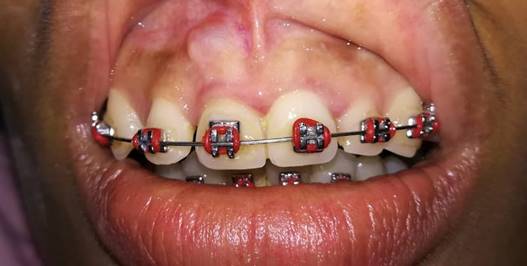
The intraoral examination showed a nodular lesion located at the level of free gingiva of the right upper central incisor, of the same color of adjacent mucosa with a violet central region, which was maintained despite the increase in size. The lesion was approximately 1 cm in diameter, asymptomatic to palpation, of fluctuating consistency, with liquid content, 4 months of evolution, and unknown etiology. Clinically, the lesion did not coincide with infectious processes, and the images showed no bone compromise. The patient had had bimaxillary orthodontic appliance for about 1 year, which was still active (Figure 1).
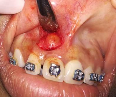
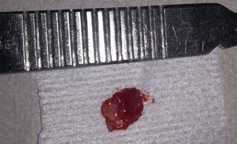
Following asepsis and antisepsis, the surgical approach was performed under local anesthesia by Parstch operation or semilunar incision and partial-thickness flap lift (Figure 2). After the excision, the lesion was sent for histopathological analysis (Figure 3). Three profuse rinses were performed with saline solution, conducting hemostasis, and taking suture points with 4.0 polyglycolic acid. The histological study showed a proliferation of blood capillaries with a thick vascular wall, endothelial cells, partial obliteration of vascular light and an infiltrate with significant infiltration of eosinophils. The definitive anatomopathological report was “angiolymphoid hyperplasia with eosinophilia”.
A follow-up 8 days after surgery showed stitches in place. These were removed, showing good clinical healing and recovery of soft tissues. No relapse has occurred after 10 months of follow up.
DISCUSSION
Angiolymphoid hyperplasia with eosinophilia is an unusual benign pathology clinically characterized by the presence of unique or multiple angiomatous lesions frequently located on the scalp and face, and less frequently in the oral mucosa of young adults.8 In the present case there is an unusual location in the oral cavity, specifically in the free gingiva. In 2013, Singh et al reported no contribution of periapical, panoramic, and occlusal x-rays to diagnose angiolymphoid hyperplasia with eosinophilia because these show no alterations,10 just as in the present case, where no imaging alterations are reported.
In 2016, Da Rocha et al described divisional biopsy under local anesthesia as a diagnostic method, obtaining small fragments of soft tissue, which were fixed in 10% formalin for histopathological analysis. In most cases there was a benign vascular lesion characterized by the proliferation of blood capillaries with thick vascular wall as well as a significant inflammatory component composed of lymphocytes, plasmacytes, and numerous eosinophils.4 All this is consistent with the present case, where the same alterations were found in the histological analysis.
In 2014, Lorente et al reported the presence of eosinophilia in blood tests, with no changes in other lab parameters, which could suggest a diagnosis of angiolymphoid hyperplasia with eosinophilia.5 This is exactly the same in the present case, where mild eosinophilia and other normal values were reported, which helped established the diagnosis once the complementary tests were examined.
In 2016, Adler et al reported 593 cases, documenting specific monotherapy or combination therapy. Surgical excision was the most reported therapeutic intervention for ALHE (44.2%). Treatment failure (defined as incomplete disease resolution or recurrence after treatment) was lower in cases treated with excision (40.8%).2 Such surgical procedure and result were seen in the present case, which was monitored after 10 months with no signs of relapse.
In 2011, Kukreja et al reported that ALHE lesions are superficial and contain blood vessels of different luminal sizes, some of which may not be channeled, coated by a distinctive endothelium of epithelioid appearance. Eosinophilic abscesses are not seen and peripheral eosinophilia is not always present.13 On the other hand, in 2019 Wortsman et al commented that vascular changes are characteristic of ALHE, the vascular component shows proliferation of endothelial cells, including epithelioid or histiocytoid, some vessels are immature or poorly channeled, the light of some vessels may be occluded by the hypertrophy and protrusion of these cells, showing thickening of the wall of blood vessels with concentric rings of fibrosis and edema separating the cubic endothelial cells, some of them sometimes contain vacuolate cytoplasm, it has nuclei with irregular folds, cytoplasm in moderate quantity, strongly eosinophilic and vacuolate, sometimes diffuse or marked infiltrated lymphocytes and plasma cells with or without lymphoid follicles, the location of lymphoid commitment to the periphery is common, except in dermal lesions.14 This is consistent with the present case in which the histological study showed a proliferation of blood capillaries with thick vascular wall, endothelial cells, partial obliteration of vascular light, and an infiltrate with significant infiltration of eosinophils (Figures 4A and 4B).
CONCLUSION
Angiolymphoid hyperplasia is a benign lesion with an unusual location in the oral cavity that presents unspecific symptoms and etiology. However, in this case there were no symptoms, so the diagnosis was done through clinical and histopathological findings only. The treatment of choice was surgical, showing no evidence of recurrence to date. On the other hand, the details found in the x-ray study are of no relevance. Therefore, reporting lesions caused by this entity is considered important in order to contribute to a better understanding not only of its clinical or radiographic behavior, but also its biological and histological manifestations.

