


 English (pdf)
 Article in xml format
 Article references
 Send this article by e-mail
 Cited by SciELO
  Cited by Google
  Similars in
SciELO
Similars in
SciELO  Similars in Google
 Permalink
Introduction
Peste des petits ruminants (PPR) is endemic across the African and Asian continents causing economic and veterinary important disease leading to considerable economic losses (1,2). It is known to be endemic in Nigeria as confirmed by Shamaki et al. (3). It is extremely contagious viral disease of sheep and goats, clinically manifested by pyrexia, purulent ocular and nasal discharges, dyspnoea, sneezing, pneumonia, ulceration of mucous membranes of the mouth, gastroenteritis and severe diarrhoea (4,5,6,7). The disease is caused by an RNA virus, the PPR virus of the Genus Morbilivirus and the Family Paramyxovirus. Genetic characterization of the virus has grouped it into four distinct lineages: three from Africa and one from Asia. One of the African lineages is also found in Asia (8,9,10). The aetiologic virus is easily transmitted by direct contact with the aerosols, secretions and/or excretions of infected animals (2,4,11). PPR is a transboundary animal disease causing enormous economic losses due to the high morbidity and mortality associated with the disease and has therefore been classified among World Organisation for Animal Health (OIE) notifiable diseases (12). It poses a significant threat to the development of livestock production in areas where it is endemic (13,14).
Programs aimed at controlling PPR, have costed farmers, governments and aid agencies so much money. Hence, this disease remains as a major constraint in the productivity of small ruminants and is responsible for severe economic losses according to Ogunsanmi et al. (15) and Libeau et al. (16). In addition to this, drug development provides possible solutions to some tropical health diseases, although pharmaceutical research is very exorbitant and a lack of return on investment has constrained the production of new drugs (17).
Vaccination has been the main method of control but despite it, sporadic outbreaks have also been reported by Şevik and Sait (18). In Nigeria, homologous vaccine for PPR was developed from the attenuation of Nig 75/1 PPRV strain Diallo (19) but recently, Luka et al. (20) reported that Nigerian strains of PPRV demonstrate 93-94% homology with PPRV homologous vaccine. Hence, there is no doubt that it is the vaccine of choice to control the PPR in Nigeria. However, the thermolabile nature of the vaccine may increase the likelihood of low sero-conversion following vaccination wih the vaccine Sen et al. (21).
Hence this research was conducted to use clinical signs, Post-mortem lesions and Monoclonal antibody Blocking ELISA method to serologically diagnose PPR in West African Dwarf goats and to assess the efficacy of different chemotherapeutic regimens including Acyclovir for the treatment of PPR in West African Dwarf goats.
Materials and Methods
Study Animals
Twenty WAD male goats, whose ages ranges from six months to one year with no history of PPR vaccination adjudged by clinical signs and gross lesions to be suffering from natural PPR infection were purchased from the local markets in Ibagwa and Orba, Nsukka and Udenu Local Government Area, Enugu State, Nigeria. The WAD goats were kept together in a well aerated pen in the Department of Veterinary Surgery, University of Nigeria, Nsukka (UNN) in the experimental animal house unit. They were left to acclimatize for 7 days (1 week) prior to the commencement of the study.
During this period of acclimatization, the animals were thoroughly checked for the manifestation of characteristic clinical signs that are suggestive and points to the scope of the study. Also, during this period, the WAD goats were fed on (fresh and green) grasses consisting of Guinea grass (Panicum maximum) and Elephant grass (Pennisetum purpureum), which is the usual practice in this eco-zone. Water was also made available ad libitum.
Experimental Drugs and Administration
Chemotherapeutic drugs used include Acyclovir tablet 200 mg (Virest 200®) manufactured by HOVID Bhd., Malaysia. It was administered orally (Per.Os) to the animals for 5 days. Oxytetracycline Hydrochloride Short acting (Oxy-pet®/SA) manufactured by Shanghai Gongyi Vet Medicine Plant, Shanghai (People’s Republic of. China). It was administered intramuscularly at the dose of 20 mg of the drug per kg of body weight for five days and Ivermectin (Kepromec®) manufactured by KEPRO B.V., Maagdenburgstraat (Holland) was used for treatment at 1ml per 25kg and was administered subcutaneously once in two weeks according to their body weight. These treatments were instituted from the 7th day, post infection and based on confirmatory PPR diagnosis.
Experimental Design
Twenty WAD goats were randomly assigned into five treatment groups - 1, 2, 3, 4 and 5, of four goats per group. Each goat was duly identified using neck tags: Group 1- The goats were treated with Acyclovir, Oxytetracycline and Ivermectin; Group 2- The goats were treated with Acyclovir only; Group 3- The goats were treated with Ivermectin only; Group 4- The goats were treated with Oxytetracycline hydrochloride only; and Group 5- The goats were left untreated.
Experimental Infection
The WAD goats after a period of stabilization and acclimatization were infected by keeping together in a common pen with goats confirmed by haemagglutination inhibition test and Monoclonal antibody based blocking ELISA to be suffering from PPR infection. This infection was induced according to standard procedures as described by Ugochukwu et al. (6). Three to five days after, all the animals came down with the infection, showing classical clinical signs of PPR. These animals were also subjected to Monoclonal antibody-based blocking ELISA and were all positive for PPR infection.
Blood Sample Collection
Whole blood (2ml) was collected from all experimental animals prior to their separation into groups. This procedure was aseptically done via the jugular vein by venipuncture for serology. The blood samples collected were put in sample bottles not containing anticoagulant and could clot and later centrifuged at 3000 rpm for 15 minutes. The sera obtained were harvested and stored in a deep freezer prior to the start with the serological analysis.
Physiological Parameters
The major physiological parameters, namely, temperature, pulse rate, heart rate and respiratory rate were obtained using standard procedures as described by Chakrabarti (22) and Ugochukwu (6). These physiological parameters were obtained in the mornings throughout the experiment.
Serological Diagnosis
Monoclonal Antibody-Based Blocking Enzyme-Linked Immunosorbent Assay was done by the following procedure and using commercially available Enzyme-linked immunosorbent assay (ELISA) kit (CIRAD-EMVT, Montpellier, France) for PPRV. This diagnostic kit detects antibodies directed against the nucleoprotein of PPRV and was developed by a FAO reference laboratory, CIRAD-EMVT, Montpellier, France. The reaction was stopped after 10 minutes with 50 μl of 1 M sulphuric acid. Results were read on an ELISA reader at 492 nm. The absorbance was converted to percentage inhibition (PI). Sera showing PI greater than 50% were regarded as positive. This procedure was done according to standard procedures as described by Saliki et al. (23) and Munir et al. (24).
Survivability and Clinical Signs
The goats were observed once daily throughout the experimental period for clinical signs of the disease (e.g., palpable lymph node, body condition/weight changes, appetite, mucus membrane colour, ocular discharges, nasal discharges, etc.) as well as mortality and these were recorded.
Results
Mortality Rate
Table 1 shows the percentage mortality rate of the different groups. Percentage mortality per group was: Group 1, 25%; Group 2, 75%; Group 3, 75%; Group 4, 75%; and Group 5, 100%. Additionally, Groups 2, 3 and 4 had comparable mean mortality rates across the groups.
Table 1 Survivability and mortality of WAD goats infected naturally with PPR and treated with different chemotherapeutic regimens
| DAY | GROUP 1 | GROUP 2 | GROUP 3 | GROUP 4 | GROUP 5 |
|---|---|---|---|---|---|
| 0 | 0/4 | 0/4 | 0/4 | 0/4 | 0/4 |
| 7 | 0/4 | 0/4 | 0/4 | 0/4 | 2/4 |
| 11 | 0/4 | 2/4 | 0/4 | 0/4 | 3/4 |
| 13 | 0/4 | 2/4 | 3/4 | 0/4 | 3/4 |
| 15 | 0/4 | 2/4 | 3/4 | 2/4 | 3/4 |
| 17 | 0/4 | 3/4 | 3/4 | 2/4 | 3/4 |
| 18 | 1/4 | 3/4 | 3/4 | 2/4 | 4/4 |
| 19 | 1/4 | 3/4 | 3/4 | 3/4 | 4/4 |
Figures represent number of mortality/number of animals in the group
Source: own work
Temperature
Table 2 shows the mean rectal temperature of the different groups. There was no significant difference (P>0.05) across the groups in week 0 and week 1. In weeks 2 and 3, Groups 1, 2, 3 and 4 had a comparable mean temperature and the mean temperatures were significantly lower than (P<0.05) Group 5 (untreated) in week 2.
Table 2 Mean±SD of temperature (OC) inWAD goats infected naturally with PPR and treated with different chemotherapeutic regimens
| Week | Group 1 (combined) | Group 2 (Acyclovir) | Group 3 (Ivermectin) | Group 4 (Oxytetracycline) | Group 5 (Untreated) |
|---|---|---|---|---|---|
| 0 | 38.33 ± 0.25a | 38.23 ± 0.32a | 38.42 ± 0.30a | 37.84 ± 0.16a | 38.28 ± 0.15a |
| 1 | 38.58 ± 0.26a | 38.07 ± 0.32a | 37.86 ± 0.20a | 38.44 ± 0.16a | 38.29 ± 0.31a |
| 2 | 38.31 ± 0.20a | 38.88 ± 0.45a | 38.89 ± 0.23a | 38.58 ± 0.27a | 40.84 ± 0.47b |
| 3 | 37.65 ± 0.31b | 39.60 ± 0.15b | 38.50 ± 0.31b | 38.25 ± 0.18b | 0.00 ± 0.00a |
Superscripta and b represent the order of significance.
Source: own work
It was observed that the rectal temperatures inthe WAD goats were within physiological range (38{40oC) in Groups 1, 2, 3 and 4 throughout the experimental period. Also, Group 5, in weeks 0 and 1, had temperatures within the physiologic range, but, in week 2, there was an increase in the temperature value suggesting pyrexia.
Pulse Rate
The mean pulse rates of the different groups are shown in Table 3. There was no significant differences (P>0.05) across all the groups in week 0 and week 1. In week 2, Groups 1, 2, 3 and 4 had a comparable mean pulse rate and were significantly (P<0.05) higher than the mean pulse rate for Group 5 (untreated).
In week 3, the mean pulse rate for Group 2 (treated with Acyclovir only) was significantly (P<0.05) higher than Group 5 (untreated). Also, there was a significant difference (P<0.05) in Groups 1, 2 and 3 although they had a comparable mean pulse rate across the groups.
Table 3 Mean ± SD of pulse rates (beats/minute) inWAD goats infected naturally with PPR and treated with different chemotherapeutic regimens
| Week | Group 1 (combined) | Group 2 (Acyclovir) | Group 3 (Ivermectin) | Group 4 (Oxytetracycline) | Group 5 (Untreated) |
|---|---|---|---|---|---|
| 0 | 87.71 ± 5.10a | 93.71 ± 5.10a | 92.57 ± 5.97a | 89.14 ± 5.08a | 83.35 ± 5.66a |
| 1 | 84.86 ± 5.56a | 76.86 ± 2.98a | 87.24 ± 4.77a | 84.66 ± 3.30a | 85.43 ± 8.30a |
| 2 | 95.14 ± 6.47b | 93.71 ± 4.77b | 94.86 ± 5.07b | 103.71 ± 4.91b | 66.67 ± 10.06a |
| 3 | 102.50 ± 3.59ab | 123.00 ± 18.00b | 108.00 ± 8.04ab | 112.00 ± 21.17ab | 0.00 ± 0.00a |
Superscripts a and b represent the order of significance.
Source: own work
At weeks 0 and 1, the pulse rates across the groups were within the physiologic range (70-90), except for Groups 2 and 3, in week 0, which had slight increase, but, from week 2 throughout the experimental period, the pulse rates were higher than normal in Groups 1, 2, 3 and 4. Group 5 showed pulse rate within the normal range in week 1, but decreased drastically throughout the experiment.
Heart Rate
Table 4 shows the mean heart rate of the different groups. There was no significant difference (P>0.05) across the groups in week 0 and week 1. In week 2, there was a significant increase (P<0.05) in Groups 1, 2, 3 and 4 when compared with Group 5 (untreated) groups. In week 3, Group 2 (treated with Acyclovir only) was significantly higher than (P<0.05) Group 5 (untreated). Also, there was a significant difference (P<0.05) in Groups 1, 2, 3 and 4 and had a comparable mean heart rate across the groups.
Table 4 Mean±SD of the heart rates (beats/minute) inWAD goats infected naturally with PPR and treated with different chemotherapeutic regimens
| Week | Group 1 (combined) | Group 2 (Acyclovir) | Group 3 (Ivermectin) | Group 4 (Oxytetracycline) | Group 5 (Untreated) |
|---|---|---|---|---|---|
| 0 | 95.85 ± 5.20a | 106.28 ± 5.31a | 108.57 ± 6.70a | 99.62 ± 4.40a | 101.81 ± 5.57a |
| 1 | 97.71 ± 4.37a | 98.28 ± 5.63a | 100.19 ± 5.94a | 100.14 ± 5.12a | 86.57 ± 2.92a |
| 2 | 107.05 ± 10.12b | 119.71 ± 8.94b | 126.29 ± 9.64b | 117.71 ± 4.29b | 81.33 ± 4.81a |
| 3 | 113.00 ± 9.31ab | 144.00 ± 6.73b | 125.00 ± 5.51ab | 120.00 ± 4.32ab | 0.00 ± 0.00a |
Superscripts a and b represent the order of significance.
Source: own work
Respiratory Rate
It was observed that the respiratory rates across the groups were higher than normal. Normal respiratory rate ranges from 15cpmto 20cpm. Table 5 shows the mean respiratory rate in the different groups. In week 0, Group 5 (untreated) was significantly higher than (P<0.05) Group 2 (treated with Acyclovir only). Also, Groups 1, 3 and 4 had a comparable mean respiratory rate across the groups. In week 1, Group 2 (treated with Acyclovir only) was significantly higher than (P<0.05) Group 5 (untreated). Also, Groups 1, 3 and 4 had a comparable mean respiratory rate across the groups. In week 2, Groups 1, 2, 3 and 4 was significantly higher than (P<0.05) Group 5 (untreated). In week 3, Groups 1, 2 and 3 was significantly higher than (P<0.05) Group 5 (untreated).
Table5 Mean±SD of the respiratory rates (cycles/minute) inWAD goats infected naturally with PPR and treated with different chemotherapeutic regimens
| Week | Group 1 (combined) | Group 2 (Acyclovir) | Group 3 (Ivermectin) | Group 4 (Oxytetracycline) | Group 5 (Untreated) |
|---|---|---|---|---|---|
| 0 | 029.90 ± 1.40ab | 28.19 ± 1.35a | 32.76 ± 1.67ab | 31.24 ± 1.45ab | 33.52 ± 1.81b |
| 1 | 31.43 ± 2.63ab | 35.14 ± 1.65b | 31.90 ± 2.16ab | 33.23 ± 1.46ab | 28.57 ± 2.03a |
| 2 | 31.19 ± 2.14a | 30.29 ± 2.29a | 37.14 ± 4.16a | 27.57 ± 2.40a | 13.14 ± 6.69a |
| 3 | 32.50 ± 1.71b | 30.50 ± 2.99b | 34.00 ± 2.58ab | 25.00 ± 1.00ab | 0.00 ± 0.00a |
Superscripts a and b represent the order of significance.
Source: own work
Clinical Signs and Post-Mortem Lesions
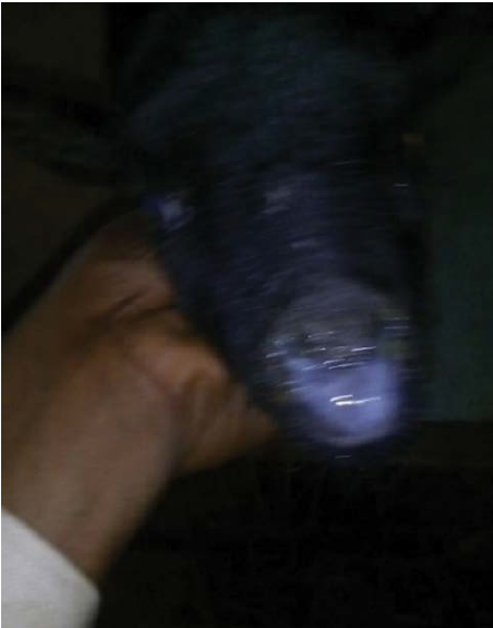
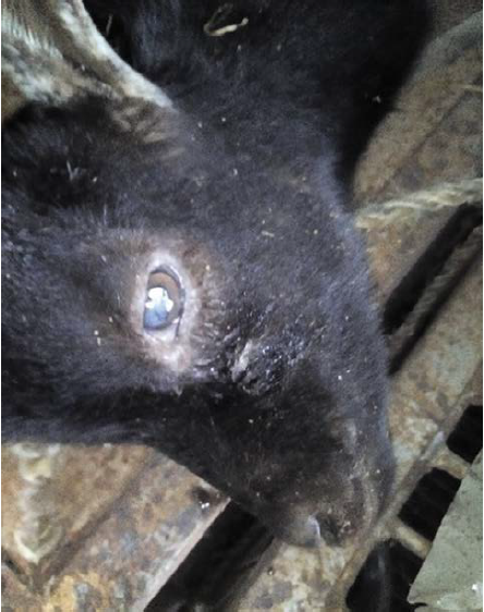
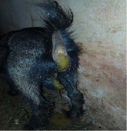
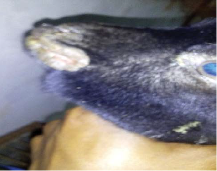
During the experiment, other clinical signs of PPR were observed. The clinical signs initially observed were respiratory distress, ocular and nasal discharges as shown in Figure 1 and 2, lacrimation, pyrexia, dehydration, anorexia and terminally, diarrhoea as shown in Figure 3. Meanwhile, other clinical signs later developed including ulcerative stomatitis, gingivitis and labial Orf-like lesions as shown in Figure 4.
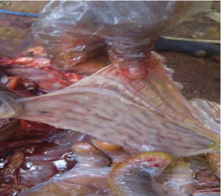
Furthermore, some Post-mortem lesions were observed including oral ulcerative lesions, gastro-enteritis and the characteristic discontinuous streaks of congestion, also called Zebra-markings, in the colon and rectum as shown in Figure 5.
Discussion
The results from this experiment showed that the mortality rate of West African Dwarf (WAD) goats infected with Peste des petits ruminants without supportive therapy and/or treatment was high, which is in agreement with the reports by Kitching, (25) and Jaisree et al. (26). Clinically, the affected animals showed signs of pyrexia, anorexia, emaciation, catarrhal nasal discharge, severe lacrimal and ocular discharges, conjunctival encrustation in the medial canthus ulcerative stomatitis and severe diarrhoea. These symptoms agree with those described by the FAO (27), Wolhsein and Saliki (28), Islam et al. (29), Abubakar and Irfan (5) and Ugochukwu et al. (30) in goats with this disease.
It is also worth noting that the findings from this study is consistent with the report of Blood et al. (31), who observed that the disease was very fatal in young goats (60-80%) especially in the age group of 7-12 months. This occurrence is observed in Groups 2, 3, 4 and 5, since these groups showed a mortality rate range between 75%-100%, with the untreated infected PPR goats having 100% mortality rate. This fact agrees with the findings by Dhar et al. (1) and Das et al. (32).
The findings of this experiment are in tandem with the report by Gupta et al. (33), who reported that broad spectrum antibiotics (especially Oxytetracycline and Sulphadimidine) when administered parenterally, have therapeutic action against PPR infection, which suggests taking preventive measures in terms of vaccination and supportive therapy with oxytetracycline will help to effectively control the disease.
According to Jackson and Cockcroft (34) and Radostits et al. (35) the per-rectum temperature was within normal reference values. This is in accordance with the initial findings of this work but, however, later on, there was a significant increase in the mean temperature in the untreated group, suggesting pyrexia associated with the disease as reported by Lefevre and Diallo (36). It can thus be deduced that the chemotherapeutic agents (Acyclovir, Oxytetracycline short-acrting Hydrochloride and Ivermectin) had effects on the treated groups by stabilizing the temperature to remain within the normal range.
The heart rate, pulse rate and respiratory rate were also high, deferring from the normal reference values reported by Jackson and Cockcroft (34), Radostits et al., (35), but this occurrence agrees with the report by Naznin et al. (37), who reported that the heart rate, pulse rate and respiratory rate in PPR affected goats are high. It could be postulated, also, that the significant increase in the respiratory rate was due to harsh environmental and weather conditions as well as the presence of the virus.
Ivermectin, a veterinary drug, was incorporated into the combined treatment regimen to kill internal helminth parasites that were observed in some of the goats suffering from PPR used in this experiment. It agrees with findings by Crump and Omura (38). They stated the efficacy of ivermectin,’’wonder drug’’ against external and internal parasites.
Naznin et al. (37) reported that the recovery rate from clinical signs was higher in goats treated with parenteral administration of oxytetracycline but as seen in the findings of this work, it was observed that the combined therapy using Acyclovir, Oxytetracycline and Ivermectin, had a better efficacy than the other groups treated singly. The recovery rate of the combined therapy and the single-Oxytetracycline treated groups were comparable one week post-treatment. It can be deduced that the Oxytetracycline effects were against bacteria becoming opportunistic due to the immunosuppressive effects of the virus. It was also postulated that the combined therapy group also symptomatically combated opportunistic bacterial infection (Oxytetracycline) as well as the PPR virus (Acyclovir) and parasitic infections (Ivermectin) that may complicate the disease condition. It provides a platform for rationale for survivability and lower mortality rate in this group.
It was reported by Nawathe (39) that the use of chemotherapeutic agents combined with hygiene, sanitation, isolation, and quarantine of the infected goats had positive effects in managing the disease. The present findings of this work are in agreement with this report. Also, this work is in tandem with the report by Islam et al. (2012) and Bupasha et al. (40) who investigated and reported the efficacy of Oxytetracycline in managing PPR infection.
Conclusions
Acyclovir has not been widely used in PPR cases, but it is proven in this work that it produced positive efficacy when combined with Oxytetracycline and Ivermectin as palliative or supportive therapy. This in turn, may allow for immune responses and antibody build up which might lead to recovery and resistance of the goats against the virus.
Combination of Acyclovir, Oxytetracycline and Ivermectin is effective in achieving recovery from clinical signs when compared to the use of these chemotherapeutic agents singly. The presence of the virus had a significant effect on the temperature, pulse rate and respiratory rate. It also caused high mortality in untreated PPR infected goats.
A prolonged study should be done to determine the point of full recovery post-treatment. Although not specific for Paramyxoviruses, it is recommended that more investigations and assessments should be done on the use and efficacy of Acyclovir in the treatment of PPR.

